SLAC மற்றும் SNAC மணிக்கட்டு
Patients › Wrist
SLAC/SNAC wrist – understanding pain from arthritis at the wrist, often after injury.
நீங்கள் என்ன உணர்கிறீர்கள்
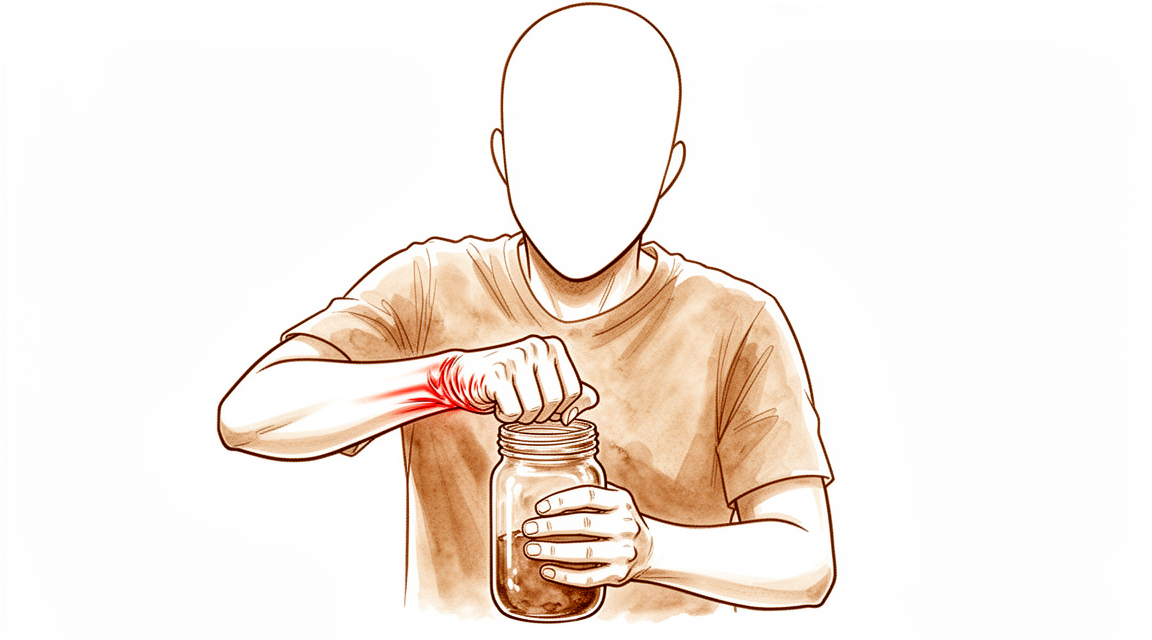
பழைய காயத்திற்குப் பிறகு உங்கள் மணிக்கட்டில் வலி தொடங்குகிறது. இது உடைந்துபோகும் மூட்டுவலி என்று அழைக்கப்படுகிறது. உங்கள் மணிக்கட்டில் உள்ள எலும்புகள் இடத்திலிருந்து நகரும்போது இது நிகழ்கிறது. உங்கள் மணிக்கட்டின் மேல் பகுதியில் ஆழமான வலியை நீங்கள் உணரலாம். நீங்கள் எதையாவது அழுத்தும்போது அல்லது உங்கள் கையைத் திருப்பும்போது வலி பெரும்பாலும் மோசமடைகிறது. ஒரு ஜாடி திறப்பது அல்லது கதவு கைப்பிடியை திருப்புவது போன்ற எளிய பணிகள் கடினமாகிவிடும்.
நீங்கள் முதலில் எழுந்திருக்கும்போது விறைப்புத்தன்மையை நீங்கள் கவனிக்கலாம். இது வழக்கமாக நீங்கள் சிறிது நேரம் நகர்ந்த பிறகு மறைந்துவிடும். இருப்பினும், நீங்கள் நாள் முழுவதும் உங்கள் கையைப் பயன்படுத்திய பிறகு வலி மீண்டும் வரலாம். பல நோயாளிகள் கையை ஓய்வெடுப்பது உதவுகிறது என்று கருதுகின்றனர். சிலருக்கு இரவில் வலி ஏற்படுகிறது, குறிப்பாக அவர்கள் அந்த பக்கத்தில் தூங்கினால். இது உங்கள் தூக்கத்தை சீர்குலைத்து உங்களை சோர்வாக உணர வைக்கும்.
அன்றாட நடவடிக்கைகள் முன்னெப்போதையும் விட கடினமாக உணரலாம். உங்கள் முதுகுக்குப் பின்னால் ஒரு பிராவைப் பிணைப்பது வேதனையாக இருக்கலாம். சட்டைக்குள் புகுத்துவதற்கு உங்கள் கையை வலிக்கும் வகையில் உயர்த்த வேண்டியிருக்கலாம். பொருள்களை உயர்த்துவது, இலகுவானவை கூட, கூர்மையான அச om கரியத்தை ஏற்படுத்தும். வலியைத் தூண்டும் என்ற பயத்தில் உங்கள் கையைப் பயன்படுத்துவதைத் தவிர்க்கலாம். இது எளிய நடைமுறைகளை ஒரு சவாலாக உணரக்கூடும்.
இந்த நிலை உங்கள் மணிக்கட்டில் எலும்புகள் எவ்வாறு நகர்கின்றன என்பதை பாதிக்கிறது. முக்கிய எலும்புகள் இனி சரியாக சீரமைக்கப்படாமல் போகலாம். இந்த தவறான சீரமைப்பு மூட்டுகளில் கூடுதல் தேய்மானத்தை ஏற்படுத்துகிறது. உங்கள் மணிக்கட்டை நகர்த்தும்போது ஒரு அரைக்கும் உணர்வை நீங்கள் உணரலாம் அல்லது கிளிக் செய்யும் ஒலியைக் கேட்கலாம். இது எப்போதும் வேதனையானது அல்ல, ஆனால் அது எரிச்சலூட்டும்.
எல்லா மணிக்கட்டு காயங்களும் இந்த பிரச்சினைக்கு வழிவகுக்காது என்பதை அறிந்து கொள்வது முக்கியம். கடந்த காலத்தில் உங்களுக்கு ஒரு இழைக் கிழிவு ஏற்பட்டிருந்தால், ஆனால் உங்கள் எக்ஸ்-கதிர்கள் சாதாரணமாகத் தோன்றினால், நீங்கள் இந்த மூட்டுவலிக்கு ஆளாகாமல் இருக்கலாம். இருப்பினும், உங்களுக்கு நீண்டகால நிலையற்ற தன்மை இருந்தால், ஆபத்து அதிகமாக இருக்கும். உங்கள் அறுவை சிகிச்சையாளர் உங்கள் குறிப்பிட்ட வரலாறு மற்றும் அறிகுறிகளைப் பார்ப்பார். உங்கள் வலி இந்த வடிவத்துடன் பொருந்துகிறதா என்பதை அவர்கள் தீர்மானிப்பார்கள். நீங்கள் என்ன உணர்கிறீர்கள் என்பதைப் புரிந்துகொள்வது உங்கள் அறுவை சிகிச்சையாளருக்கு உங்கள் வலியைக் குறைப்பதற்கும் உங்கள் செயல்பாட்டை மேம்படுத்துவதற்கும் சரியான சிகிச்சையைத் தேர்வுசெய்ய உதவுகிறது.
உண்மையில் என்ன நடக்கிறது
உங்கள் மணிக்கட்டில் எட்டு சிறிய எலும்புகள் உள்ளன. SLAC மற்றும் SNAC மணிக்கட்டுகளில், இந்த எலும்புகளுக்கு இடையேயான இயல்பான சறுக்கல் இயக்கம் உடைகிறது. இது பொதுவாக எலும்புகளை ஒன்றாக வைத்திருக்கும் இழைகளுக்கு சேதம் ஏற்படுவதன் மூலம் அல்லது எலும்பு முறிவுக்குப் பிறகு குணமடையாத ஒரு எலும்புடன் தொடங்குகிறது. இந்த ஆதரவுகள் பலவீனமடையும்போது, எலும்புகள் அவற்றின் சரியான சீரமைப்பிலிருந்து விலகிச் செல்கின்றன.
உங்கள் மணிக்கட்டை நன்கு எண்ணெய் பூசப்பட்ட கீல் போல சிந்தியுங்கள். எலும்புகளின் முனைகளில் உள்ள மென்மையான பூச்சு, அவை சிராய்ப்பு இல்லாமல் சறுக்குவதற்கு உதவுகிறது. எலும்புகள் தவறாக சீரமைக்கப்படும்போது, இந்த பூச்சு சீராக அணியாது. இந்த அணியக்கூடிய மூட்டுவலி எலும்பு-எலும்பு தொடர்பை உருவாக்குகிறது. இதன் விளைவாக வலி, கடினத்தன்மை மற்றும் உங்கள் கையை நகர்த்தும்போது அரைக்கும் உணர்வு ஏற்படுகிறது. மூட்டு சுற்றி ஒரு ஸ்லீவ் போல செயல்படும் கூட்டு காப்ஸ்யூலும் அழற்சி மற்றும் இறுக்கமாக மாறக்கூடும்.
இந்த தவறான சீரமைப்பு உங்கள் கை வழியாக வலிமை எவ்வாறு பயணிக்கிறது என்பதை மாற்றுகிறது. சாதாரணமாக, சுமை சமமாக விநியோகிக்கப்படுகிறது. இப்போது, சில புள்ளிகள் அதிக அழுத்தத்தை எடுத்துக்கொள்கின்றன. இது சேதத்தை துரிதப்படுத்துகிறது மற்றும் நீங்கள் உணரும் குறிப்பிட்ட அறிகுறிகளுக்கு வழிவகுக்கிறது. உங்கள் அறுவை சிகிச்சையாளர் இந்த மாற்றங்களை படத்தில் காணலாம். எலும்புகள் இனி மென்மையான, ஒருங்கிணைந்த வழியில் நகரவில்லை என்பதற்கான அறிகுறிகளை அவர்கள் தேடுகிறார்கள்.
இந்த அசாதாரண இயக்கத்தை நிறுத்துவதே சிகிச்சையின் குறிக்கோள். எலும்புகளை உறுதிப்படுத்துவதன் மூலமோ அல்லது சேதமடைந்த பகுதிகளை அகற்றுவதன் மூலமோ, இயக்கத்திற்கான மென்மையான பாதையை மீட்டெடுப்பதை நாங்கள் நோக்கமாகக் கொண்டுள்ளோம். இது அரைப்பதைக் குறைக்கிறது மற்றும் மூட்டுகளில் உள்ள உணர்திறன் நரம்பு முனைகளில் இருந்து அழுத்தத்தை எடுக்கிறது. மூட்டு அழற்சியை முழுவதுமாக மாற்ற முடியாது என்றாலும், உங்கள் மணிக்கட்டு எவ்வாறு செயல்படுகிறது என்பதை நாங்கள் கணிசமாக மேம்படுத்தலாம் மற்றும் உங்கள் வலியைக் குறைக்கலாம்.
நாம் என்ன செய்ய முடியும்
உங்கள் அறுவை சிகிச்சையாளர் சுய மேலாண்மை மற்றும் இயற்பியல் சிகிச்சையுடன் தொடங்குவார். இந்த அணுகுமுறையானது வலியைக் குறைப்பதையும் உங்கள் மணிக்கட்டை நகர்த்துவதையும் நோக்கமாகக் கொண்டுள்ளது. உங்கள் மணிக்கட்டைச் சுற்றியுள்ள தசைகளை வலுப்படுத்த மென்மையான பயிற்சிகளை நீங்கள் கற்றுக்கொள்வீர்கள். இந்த இயக்கங்கள் மூட்டுக்கு ஆதரவளிக்கவும் தினசரி செயல்பாட்டை மேம்படுத்தவும் உதவுகின்றன. இந்த கன்சர்வேடிவ் பராமரிப்புக்கு வேலை செய்ய ஒரு நியாயமான வாய்ப்பை வழங்க வேண்டும். பெரும்பாலான நோயாளிகள் அறுவை சிகிச்சை இல்லாமல் அறிகுறிகள் மேம்படுகிறதா என்று பார்க்க முதலில் இந்த அறுவை சிகிச்சை அல்லாத விருப்பங்களை முயற்சி செய்கிறார்கள்.
எளிய உடற்பயிற்சிகள் போதுமானதாக இல்லாவிட்டால், உங்கள் அறுவை சிகிச்சை நிபுணர் மருத்துவ மேலாண்மையைப் பற்றி விவாதிக்கலாம். இது பொதுவாக வலி மருந்துகள் மற்றும் அழற்சி எதிர்ப்பு மருந்துகளை உள்ளடக்கியது. இது உங்களுக்கு வசதியாக உணர உதவுகிறது. உங்கள் அறுவை சிகிச்சை நிபுணர் ஊசிகளையும் பரிந்துரைக்கலாம். கோர்டிசோன் ஊசிகள் வீக்கம் மற்றும் வலியை ஒரு குறிப்பிட்ட காலத்திற்கு குறைக்கலாம். ஹைலூரோனிக் அமிலம் அல்லது பிளேட்லெட்-பணக்கார பிளாஸ்மா (பிஆர்பி) ஊசிகள் மூட்டுகளை மெருகூட்ட அல்லது குணப்படுத்துவதற்கு விருப்பமாக இருக்கலாம். இந்த சிகிச்சைகள் மூட்டுச்சலையை குணப்படுத்தாது, ஆனால் நீங்கள் உங்கள் அன்றாட நடவடிக்கைகளைத் தொடரும்போது அவை நிவாரணத்தை அளிக்கலாம். நிவாரணத்தின் காலம் நபருக்கு நபர் மாறுபடும்.
தற்காப்பு பராமரிப்பு இனி உங்கள் வலியைக் கட்டுப்படுத்தாதபோது, அறுவை சிகிச்சை பரிசீலிக்கப்படலாம். உங்கள் வாழ்க்கைத் தரம் கணிசமாக பாதிக்கப்படும்போது இது பொதுவாக பரிந்துரைக்கப்படுகிறது. அறுவை சிகிச்சை விருப்பங்கள் உங்கள் கீல்வாதம் மற்றும் உங்கள் மணிக்கட்டு எலும்புகளின் நிலை ஆகியவற்றைப் பொறுத்தது. ஆரம்ப கட்ட சோர்வு மற்றும் கண்ணீருக்காக, உங்கள் அறுவை சிகிச்சையாளர் ஒரு வரிசையில் சிறிய எலும்புகளை அகற்றலாம் (நெடுநிலை வரிசை கார்பெக்டோமி) அல்லது ஸ்காஃபோய்டை அகற்றிய பிறகு நான்கு குறிப்பிட்ட எலும்புகளை ஒன்றாக இணைக்கலாம் (நான்கு மூலை இணைப்பு). இந்த நடைமுறைகள் முடிந்தவரை மணிக்கட்டு இயக்கத்தை பாதுகாக்கும்போது வலியைக் குறைப்பதை நோக்கமாகக் கொண்டுள்ளன. சில சந்தர்ப்பங்களில், மூட்டு இயக்கம் கொண்டிருக்கும்போது வலி சமிக்ஞைகளைக் குறைக்க மணிக்கட்டு டெனர்வேஷன் என்று அழைக்கப்படும் ஒரு நடைமுறை பயன்படுத்தப்படலாம். உங்கள் அறுவை சிகிச்சையாளர் உங்கள் குறிப்பிட்ட முறை மற்றும் காய இலக்குகளின் அடிப்படையில் சிறந்த விருப்பத்தைத் தேர்ந்தெடுவார்.
எதிர்பார்ப்பது என்ன
உங்கள் மணிக்கட்டு வலி மற்றும் இறுக்கம் சிகிச்சை இல்லாமல் நீடிக்கும். இந்த நிலைமைகள் ஒரு பழைய காயத்திற்குப் பிறகு உடைந்து போகும் மூட்டு அழற்சியால் ஏற்படுகின்றன. சேதம் பொதுவாக காலப்போக்கில் முன்னேறுகிறது. தலையீடு இல்லாமல், நீங்கள் தொடர்ச்சியான அச om கரியத்தையும் தினசரி பணிகளுக்கு உங்கள் கையை பயன்படுத்துவதற்கான திறனைக் குறைப்பதையும் அனுபவிக்கலாம்.
சரியான சிகிச்சையுடன், நீங்கள் கணிசமான நிவாரணத்தை எதிர்பார்க்கலாம். உங்கள் வலி மற்றும் ஒட்டுமொத்த மணிக்கட்டு செயல்பாட்டை மேம்படுத்த அறுவை சிகிச்சை விருப்பங்கள் வடிவமைக்கப்பட்டுள்ளன. பெரும்பாலான நோயாளிகள் இந்த நடைமுறைகளுக்குப் பிறகு சிறந்த அகநிலை முடிவுகளையும் குறைவான வலியையும் தெரிவிக்கின்றனர். உங்கள் அறுவை சிகிச்சையாளர் உங்கள் மூட்டு அழற்சியின் குறிப்பிட்ட கட்டம் மற்றும் உங்கள் மூட்டு மேற்பரப்புகளின் நிலை ஆகியவற்றின் அடிப்படையில் சிறந்த விருப்பத்தைத் தேர்ந்தெடுப்பார்.
நீண்ட காலமாக வலி குணமடைவதற்கும், நல்ல செயல்பாட்டை அடைவதற்கும் உங்களுக்கு ஒன்றுக்கு மேற்பட்ட அறுவை சிகிச்சை தேவைப்படலாம். இரண்டாவது அல்லது மூன்றாவது அறுவை சிகிச்சை இன்னும் நீடித்த நன்மைகளைத் தரும், இது வேலைக்கும் அன்றாட நடவடிக்கைகளுக்கும் திரும்புவதற்கு உங்களை அனுமதிக்கிறது.
சில நோயாளிகள் சில சரிசெய்தல் அறுவை சிகிச்சைகளுக்குப் பிறகு லேசான அல்லது மிதமான மூட்டுவலி அறிகுறிகளை அனுபவிக்கக்கூடும் என்றாலும், பெரும்பாலானவை ஏற்றுக்கொள்ளக்கூடிய நீண்ட கால செயல்பாட்டைப் பராமரிக்கின்றன.
உங்கள் விரல்களின் இயல்புகள் உங்கள் காயத்திற்கு முன்பு இருந்ததைப் போலவே இருக்காது. இருப்பினும், நீங்கள் ஒரு நீடித்த முடிவை எதிர்பார்க்கலாம், இது அன்றாட பணிகளை குறைந்த வலியுடன் செய்ய அனுமதிக்கிறது. உங்கள் அறுவை சிகிச்சை நிபுணர் இந்த பயணத்தின் மூலம் உங்களுக்கு வழிகாட்டுவார், உங்கள் குறிப்பிட்ட சூழ்நிலைக்கு முடிந்தவரை இயக்கம் மற்றும் வலிமையைப் பாதுகாப்பதில் கவனம் செலுத்துவார்.
யாரையாவது எப்போது பார்க்க வேண்டும்
உங்கள் மண்டை வலி தொடர்ச்சியாக இருந்தால் ஒரு நிபுணரின் மதிப்பாய்வைக் கேளுங்கள். ஓய்வெடுப்பதன் மூலம் மேம்படாத வலி. நீங்கள் பலவீனம், நிலையற்ற தன்மை அல்லது பூட்டுதல் அல்லது வழிவகுக்கும் உணர்வைக் கண்டால் கவனிப்பை நாடுங்கள். இந்த அறிகுறிகள் உங்கள் தூக்கம் அல்லது வேலையில் தலையிடக்கூடும். உங்கள் நிலை திடீரென்று மோசமடைவதை நீங்கள் சந்தித்தால் உங்கள் மருத்துவரைத் தொடர்பு கொள்ளுங்கள். நீங்கள் ஆண் மற்றும் மண்டை காயம் ஏற்பட்டால் இது மிகவும் முக்கியமானது. முந்தைய காயம் தொடர்பான உடைந்துபோகும் கீல் அழற்சி உங்களுக்கு இருக்கிறதா என்பதை ஆரம்ப மதிப்பீடு தீர்மானிக்க உதவுகிறது. உங்கள் மண்டை இயக்கம் மற்றும் செயல்பாட்டைப் பாதுகாக்க எளிய ஓய்வு போதுமானதா அல்லது கூடுதல் சிகிச்சை தேவைப்படுகிறதா என்பதை உங்கள் அறுவை சிகிச்சையாளர் மதிப்பீடு செய்யலாம்.
Evidence & references
Overview
- Proximal row carpectomy and four-corner fusion with scaphoid bone excision are the most widely used surgical procedures for stage II wrist osteoarthritis secondary to SLAC or SNAC wrist [6].
- Both proximal row carpectomy and four-corner fusion provide improvements in pain and subjective outcome measures for patients with symptomatic and appropriately staged SLAC or SNAC wrists [1].
- Scaphoid excision and four-corner fusion is an increasingly popular treatment option for patients with stage II or III SLAC/SNAC arthritis that preserves wrist motion through the radiolunate and ulnocarpal joints [3].
- Scaphoid excision and four-corner fusion are effective treatments for SLAC and SNAC patterns of wrist arthritis [8].
- For stage II SLAC wrist with a preserved capitolunate joint, proximal row carpectomy is preferred because it is technically less demanding and yields durable results [10].
- The authors prefer proximal row carpectomy for SLAC wrists with preserved capitate head cartilage due to socio-economic benefits, lower complication rates, and procedural ease [46].
- In patients with SLAC/SNAC II, proximal row carpectomy might be favourable to a midcarpal arthrodesis solely based on better flexion-extension range of motion of the radiocarpal joint after proximal row carpectomy [4].
- Wrist denervation is a viable salvage option for patients with symptomatic SLAC wrist osteoarthritis to preserve motion, decrease pain, and increase function with a low absolute failure rate at mid- to long-term follow-up [13].
- Arthroscopic wrist debridement and radial styloidectomy may have advantages in relieving pain while preserving wrist motion for SLAC stage 2 or 3 disease [23].
- It is mostly indicated as a palliative procedure in elderly patients with posttraumatic SNAC or SLAC wrist with limited functional demands [18].
- A second and even a third operation can result in long-term pain improvement, good function and capacity for work, and re-operation is recommended in symptomatic cases with minor osteoarthritis of the wrist (SNAC stage 0 or 1) [11].
- Further data in a larger cohort with longer follow-up is required to determine the effect on SLAC-wrist deterioration [2].
Anatomy & Pathophysiology
- Surgical treatments for scapholunate advanced collapse (SLAC) wrist result in decreased wrist kinematic motion and functional performance compared with individuals with normal wrists [24].
- Lunate morphology affects the 3-dimensional kinematics of the carpus during wrist flexion and extension [25].
- Three-corner fusions produce motion that is smoother and more closely replicates the normal axis and functional motion of the wrist compared with four-corner fusions [26].
- Computed fiber elongations of the dorsal carpal ligaments vary linearly with wrist position [27].
- Four-dimensional computed tomography (4DCT) is a non-invasive and affordable method to assess and quantify wrist kinematics by incorporating the temporal dimension [28].
- During simple unresisted wrist motions, force in the scapholunate interosseous ligament does not exceed 20 N [29].
- Kinematic changes in scapholunate instability may predict the development of radioscaphoid arthritis and help identify a kinematically abnormal wrist [30].
- Scaphoid nonunions have a dramatic impact on carpal kinematics by partially uncoupling the proximal and distal carpal rows [31].
- Dynamic imaging allows for the derivation of standardized protocols for mapping carpal motion that are clinically applicable and reproducible [32].
- Computer-aided CT analysis provides guidelines for measuring and quantifying carpal alignment three-dimensionally and establishes a database for normal values [33].
- More than half the motion of the carpus when the wrist is loaded in extension occurs at the midcarpal joint [34].
- Radioscapholunate fusion shows the most biomechanically similar behavior to the healthy wrist among three compared fusion types [35].
- Contact areas between the scaphoid and distal radius are maximized during full extension of the wrist, which helps stabilize the radiocarpal joint [36].
- Tendon ball arthroplasty and proximal carpal stabilization with tendon graft restore the integrity of the proximal carpal row [37].
- In unstable wrists, dorsally applied polylactic acid (PLA) plates restore carpal kinematics for 1,000 cycles of motion when fixation is not compromised by carpal size or osteoporosis [38].
- Electrogoniometric and radiologic evaluation characterizes the modification of the wrist center of rotation during flexion and extension [39].
- Wrist arthrodesis may only compromise select wrist functions [40].
- The 'dart thrower's motion' of the wrist, from radial extension to ulnar flexion, may be a unifying concept of functional wrist motion that is uniquely human [41].
- Correction of scapholunate dissociation via modified Brunelli technique or Blatt capsulodesis might correlate with improved carpal dynamics and improved clinical outcomes [42].
Classification
- SLAC wrist diagnosis should be reserved for patients in whom traumatic disruption of the proximal carpal row initiates the defined sequence of arthritic change outlined by Watson and Ballet [5].
- Radiographic classification of SLAC wrist has moderate reliability and reproducibility [14].
- Radiographic classification of SNAC wrist has limited reliability [14].
- SNAC wrists differ from SLAC wrists by exhibiting a decreased sagittal lunotriquetral angle, indicating a distinct pathomechanism of carpal instability [7].
- Bone density is greater at the capitolunate joint, the radial styloid, and the radiolunate joint in SNAC wrists compared to controls [9].
- Carpal malalignment in SLAC wrists affects the radio- and midcarpal joints and extends to the third carpometacarpal joint, with malalignment evident in both sagittal and coronal planes [16].
- Patients with SLAC wrist are more likely to be male and have a history of trauma compared to patients with first CMC OA [12].
Clinical Presentation
- SLAC wrist diagnosis is reserved for patients in whom traumatic disruption of the proximal carpal row initiates a defined sequence of arthritic changes [5].
- Patients with SLAC wrist are more likely to be male and have a history of trauma compared to patients with first CMC OA [12].
- SNAC wrists differ from SLAC wrists by exhibiting a decreased sagittal lunotriquetral angle, indicating a distinct pathomechanism of carpal instability [7].
- Bone density is greater at the capitolunate joint, the radial styloid, and the radiolunate joint in SNAC wrists compared to controls [9].
- Carpal malalignment in SLAC wrists affects the radio- and midcarpal joints and extends to the third carpometacarpal joint, with malalignment evident in both sagittal and coronal planes [16].
- Distal row pronation and translation and radiolunate arthritis are demonstrated in the SNAC wrist via quantitative 3-D CT [9].
- Radiographic classification of SLAC wrist has moderate reliability and reproducibility [14].
- Radiographic classification of SNAC wrist has limited reliability [14].
- There is room for improvement in the assessment of patients with SNAC wrists [45].
- A scapholunate ligament injury visualized arthroscopically, without static x-ray changes, does not inevitably lead to SLAC wrist [15].
- Isolated lunocapitate osteoarthritis presents differently from SLAC wrist and is not related to scapholunate instability [20].
- Bilateral scapholunate widening may have a nontraumatic aetiology and progress to carpal instability and osteoarthritis with advancing age [44].
- Patients without carpal instability or osteoarthritis were younger than those with bilateral SLAC wrists [44].
- There is no absolute evidence to confirm that bilateral wide gaps inexorably progress to carpal instability and osteoarthritis [44].
Investigations
- The diagnosis of SLAC wrist should be reserved for patients in whom a traumatic disruption of the proximal carpal row has initiated the defined sequence of arthritic change outlined by Watson and Ballet [5].
- There is currently no scientific evidence that a scapholunate ligament injury visualized arthroscopically, without static x-ray changes, inevitably leads to SLAC wrist [15].
- Radiographic classification of SLAC wrist has moderate reliability and reproducibility [14].
- Classification of SNAC wrist has limited reliability [14].
- SNAC wrists differ from SLAC wrists in exhibiting a decreased sagittal lunotriquetral angle, indicating a distinct pathomechanism of carpal instability [7].
- Bone density is greater at the capitolunate joint, the radial styloid, and the radiolunate joint in SNAC wrists compared to controls [9].
- Carpal malalignment in SLAC wrists affects the radio- and midcarpal joints and extends to the third carpometacarpal joint, with malalignment evident in both the sagittal and coronal planes [16].
- Patients with SLAC wrist are more likely to be male and have a history of trauma compared to patients with first CMC OA [12].
- Isolated lunocapitate osteoarthritis presents differently from SLAC wrist and is not related to scapholunate instability [20].
- Cineradiography has a high diagnostic value for diagnosing scapholunate dissociations [49].
- Dynamic CT scan of the wrist is a user-friendly way of measuring the scapholunate distance, which is minimal in the normal wrist below 40 years of age [51].
Treatment
- Proximal row carpectomy and four-corner fusion with scaphoid bone excision are the most widely used surgical procedures for stage II wrist osteoarthritis secondary to SLAC or SNAC wrist [6].
- Both proximal row carpectomy and four-corner fusion provide improvements in pain and subjective outcome measures for patients with symptomatic and appropriately staged SLAC or SNAC wrists [1].
- Scaphoid excision and four-corner fusion is an increasingly popular treatment option for patients with stage II or III SLAC/SNAC arthritis that preserves wrist motion through the radiolunate and ulnocarpal joints [3].
- Scaphoid excision and four-corner fusion are effective treatments for SLAC and SNAC patterns of wrist arthritis [8].
- For stage II SLAC wrist with a preserved capitolunate joint, proximal row carpectomy is preferred because it is technically less demanding and yields durable results [10].
- In patients with SLAC/SNAC II, proximal row carpectomy might be favourable to a midcarpal arthrodesis solely based on better flexion-extension range of motion of the radiocarpal joint after proximal row carpectomy [4].
- The LCF (lunate-capitate arthrodesis) is not less efficient than the 4CF (four-corner fusion) in the treatment of SNAC II and III wrist injuries [19].
- Wrist denervation is a viable salvage option for patients with symptomatic SLAC wrist osteoarthritis to preserve motion, decrease pain, and increase function with a low absolute failure rate at mid- to long-term follow-up [13].
- Arthroscopic wrist debridement and radial styloidectomy may have advantages in relieving pain while preserving wrist motion for SLAC stage 2 or 3 disease [23].
- Motion-preserving procedures of the wrist can obtain good long-term results if indications are accurately respected and the technique is well performed to prevent complications [43].
- Proximal row carpectomy is mostly indicated as a palliative procedure in elderly patients with posttraumatic SNAC or SLAC wrist with limited functional demands [18].
- A second and even a third operation can result in long-term pain improvement, good function and capacity for work, and re-operation is recommended in symptomatic cases with minor osteoarthritis of the wrist (SNAC stage 0 or 1) [11].
- Further data in a larger cohort with longer follow-up is required to determine the effect of treatments on SLAC-wrist deterioration [2].
Complications
- Proximal row carpectomy and four-corner fusion provide improvements in pain and subjective outcome measures for patients with symptomatic and appropriately staged SLAC or SNAC wrists [1].
- Further data in a larger cohort with longer follow-up is required to determine the effect of surgical interventions on SLAC-wrist deterioration [2].
- Scaphoid excision and four-corner fusion preserves wrist motion through the radiolunate and ulnocarpal joints [3].
- Proximal row carpectomy may be favourable to midcarpal arthrodesis in patients with SLAC/SNAC II based on better flexion-extension range of motion of the radiocarpal joint after proximal row carpectomy [4].
- The diagnosis of SLAC should be reserved for patients in whom a traumatic disruption of the proximal carpal row has initiated the defined sequence of arthritic change outlined by Watson and Ballet [5].
- Proximal row carpectomy and four-corner fusion with scaphoid bone excision are the most widely used surgical procedures for stage II wrist osteoarthritis secondary to SLAC or SNAC wrist [6].
- SNAC wrists differ from SLAC wrists in exhibiting a decreased sagittal lunotriquetral angle, indicating a distinct pathomechanism of carpal instability [7].
- Bone density was greater at the capitolunate joint, the radial styloid, and the radiolunate joint in SNAC wrists compared to controls [9].
- For stage II SLAC wrist with a preserved capitolunate joint, proximal row carpectomy is preferred because it is technically less demanding and yields durable results [10].
- A second and even a third operation can result in long-term pain improvement, good function and capacity for work, and re-operation is recommended in symptomatic cases with minor osteoarthritis of the wrist (SNAC stage 0 or 1) [11].
- Patients with SLAC wrist were more likely to be male and have a history of trauma compared to patients with first CMC OA [12].
- Wrist denervation was a viable salvage option for patients with symptomatic SLAC wrist osteoarthritis to preserve motion, decrease pain, and increase function with a low absolute failure rate at mid- to long-term follow-up [13].
- There is currently no scientific evidence that a scapholunate ligament injury visualized arthroscopically, without static x-ray changes, inevitably leads to SLAC wrist [15].
- Although ongoing scapholunate instability resulted in early arthritic degeneration after dorsal intercarpal ligament capsulodesis, most patients had acceptable long-term function of the wrist [21].
- Scapholunate ligament reconstruction using a part of the extensor carpi radialis brevis tendon through a dorsal approach resulted in long-term, improved outcomes compared with other techniques, even in scapholunate advanced collapse type I wrists [22].
- Distal scaphoid resection arthroplasty produced favorable, long-term clinical results and did not result in noteworthy wrist collapse [52].
Recovery
- Proximal row carpectomy and four-corner fusion both provide improvements in pain and subjective outcome measures for patients with symptomatic and appropriately staged SLAC or SNAC wrists [1].
- Further data in a larger cohort with longer follow-up is required to determine the effect of interventions on SLAC-wrist deterioration [2].
- Scaphoid excision and four-corner fusion preserves wrist motion through the radiolunate and ulnocarpal joints in patients with stage II or III SLAC/SNAC arthritis [3].
- In patients with SLAC/SNAC II, proximal row carpectomy might be favourable to midcarpal arthrodesis based on better flexion-extension range of motion of the radiocarpal joint after proximal row carpectomy [4].
- For stage II SLAC wrist with a preserved capitolunate joint, proximal row carpectomy is preferred because it is technically less demanding and yields durable results [10].
- A second and even a third operation can result in long-term pain improvement, good function, and capacity for work, supporting re-operation in symptomatic cases with minor osteoarthritis of the wrist (SNAC stage 0 or 1) [11].
- Wrist denervation is a viable salvage option for patients with symptomatic SLAC wrist osteoarthritis to preserve motion, decrease pain, and increase function with a low absolute failure rate at mid- to long-term follow-up [13].
- Although ongoing scapholunate instability resulted in early arthritic degeneration, most patients had acceptable long-term function of the wrist after dorsal intercarpal ligament capsulodesis [21].
- Scapholunate ligament reconstruction using a part of the extensor carpi radialis brevis tendon resulted in long-term, improved outcomes compared with other techniques, even in scapholunate advanced collapse type I wrists [22].
- Residual scaphoid deformity has no relevant negative impact on mid-term wrist function after scaphoid nonunion surgery [53].
- Wrist alignment was maintained over time after corrective osteotomy for distal radius malunion, but 13 patients presented mild to moderate symptomatic wrist arthritis [54].
- Carpal collapse recurred within a short time after dorsal intercarpal ligament capsulodesis because the procedure cannot withstand large and repetitive forces [55].
- The reduction and association of the scaphoid and lunate procedure should be abandoned due to early radiographic failure in the short term, despite relatively low outcomes measures scores [56].
Key Evidence
- [L4] Both procedures provide improvements in pain and subjective outcome measures for patients with symptomatic and appropriately staged SLAC or SNAC wrists. [1] (10.1177/1753193408100954)
- [Paper] Scaphoid excision and four-corner fusion is an increasingly popular treatment option for patients with stage II or III SLAC/SNAC arthritis that preserves wrist motion through the radiolunate and ulnocarpal joints. [3] (10.1016/j.hcl.2005.08.012)
- [L1] In patients with SLAC/SNAC II, proximal row carpectomy might be favourable to a midcarpal arthrodesis solely based on better FE ROM of the radiocarpal joint after proximal row carpectomy. [4] (10.1186/s12891-024-07527-6)
- [L5] The diagnosis of SLAC should be reserved for patients in whom a traumatic disruption of the proximal carpal row has initiated the defined sequence of arthritic change outlined by Watson and Ballet. [5] (10.1016/j.jhsa.2015.06.110)
- [L4] Proximal row carpectomy and four-corner fusion with scaphoid bone excision are the most widely used surgical procedures for stage II wrist osteoarthritis secondary to SLAC or SNAC wrist. [6] (10.1016/j.otsr.2014.06.025)
- [L4] SNAC wrists differ from SLAC wrists in exhibiting a decreased sagittal lunotriquetral angle, indicating a distinct pathomechanism of carpal instability. [7] (10.1186/s12891-025-08652-6)
- [L3] Scaphoid excision and four-corner fusion are effective treatments for SLAC and SNAC patterns of wrist arthritis. [8] (10.1016/s0363-5023(09)60084-8)
- [L3] Bone density was greater at the capitolunate joint, the radial styloid, and the radiolunate joint in SNAC wrists compared to controls. [9] (10.2106/jbjs.22.01350)
- [L4] For stage II SLAC wrist with a preserved capitolunate joint, proximal row carpectomy is preferred because it is technically less demanding and yields durable results. [10] (10.5435/00124635-200307000-00007)
- [L4] A second and even a third operation can result in long-term pain improvement, good function and capacity for work, and we recommend re-operation in symptomatic cases with minor osteoarthritis of the wrist (SNAC stage 0 or 1). [11] (10.1177/1753193409346093)
- [L3] Patients with SLAC wrist were more likely to be male and have a history of trauma compared to patients with first CMC OA. [12] (10.1177/1558944718788672)
- [L4] This method of wrist denervation was a viable salvage option for patients with symptomatic SLAC wrist osteoarthritis to preserve motion, decrease pain, and increase function with a low absolute failure rate at mid- to long-term follow-up. [13] (10.1016/j.jhsa.2021.02.023)
- [L4] Radiographic classification of SLAC wrist has moderate reliability and reproducibility, whereas classification of SNAC wrist has limited reliability. [14] (10.1177/1753193413484629)
- [L5] There is currently no scientific evidence that a scapholunate ligament injury visualized arthroscopically, without static x-ray changes, inevitably leads to SLAC wrist. [15] (10.1016/j.jhsa.2011.01.018)
- [L3] Carpal malalignment in SLAC wrists not only affects the radio- and midcarpal joints, but also extends to the third carpometacarpal joint, with malalignment evident in both the sagittal and coronal planes. [16] (10.1016/j.jhsa.2024.09.021)
- [L4] The LCF is not less efficient than the 4CF in the treatment of SNAC II and III wrist injuries. [19] (10.1186/s12891-024-07755-w)
- [L3] Although the consequent ongoing scapholunate instability resulted in early arthritic degeneration, most patients had acceptable long-term function of the wrist. [21] (10.1302/0301-620x.94b12.30007)
- [L4] This technique, even in scapholunate advanced collapse type I wrists, resulted in long-term, improved outcomes compared with other techniques. [22] (10.1177/17531934221143679)
- [L4] The procedure studied may have advantages in relieving pain while preserving wrist motion for SLAC stage 2 or 3 disease. [23] (10.1177/1558944717725383)
- [L2] Both surgical groups demonstrated decreased wrist kinematic motion and functional performance compared with individuals with normal wrists. [24] (10.1016/j.jhsa.2015.04.035)
- [L5] This study describes the effect of lunate morphology on 3-dimensional carpal kinematics during wrist flexion and extension. [25] (10.1016/j.jhsa.2014.09.019)
- [L3] Motion was smoother and more closely replicated the normal axis and functional motion of the wrist. [26] (10.1016/j.jhsa.2015.02.027)
- [L5] Despite complex carpal bone anatomy and kinematics, computed fiber elongations were found to vary linearly with wrist position. [27] (10.1016/j.jhsa.2012.04.025)
- [L5] Four-dimensional computed tomography (4DCT) is a promising, non-invasive, and affordable method to assess and quantify wrist kinematics, extending conventional CT by incorporating the temporal dimension. [28] (10.1177/17531934251326028)
- [L5] However, during simple unresisted wrist motions, the force did not exceed 20 N. [29] (10.1016/j.jhsa.2015.04.007)
- [L3] These kinematic changes may predict the development of radioscaphoid arthritis and help identify a kinematically abnormal wrist. [30] (10.1177/17531934241242676)
- [L4] Scaphoid nonunions have a dramatic impact on carpal kinematics, partially uncoupling the proximal and distal carpal rows. [31] (10.1016/j.jhsa.2008.03.008)
- [L4] With the increased focus on dynamic imaging for wrist motion, it may be possible to derive a standardized protocol for mapping the carpal motion that is clinically applicable and reproducible. [32] (10.1016/j.jhsg.2022.10.001)
- [L4] This study provides guidelines on how to measure and quantify carpal alignment three-dimensionally and establishes a database for normal values, which may be useful when analysing various wrist pathologies and kinematics. [33] (10.1177/17531934231160100)
- [L4] More than half the motion of the carpus when the wrist was loaded in extension occurred at the midcarpal joint. [34] (10.1016/j.jhsa.2012.10.035)
- [L5] The contact areas between the scaphoid and distal radius are maximized during full extension of the wrist, which helps stabilize the radiocarpal joint and potentially reduces the risk of injury to the carpus and the distal radius. [36] (10.1177/1753193413507810)
- [L4] The technique demonstrated reduced wrist pain and improved wrist motion and grip strength while restoring the integrity of the proximal carpal row. [37] (10.1177/17531934241238939)
- [L5] The study shows that in the unstable wrist, following ligament sectioning, where fixation is not compromised by carpal size or osteoporosis, a dorsally applied PLA plate does restore carpal kinematics for 1,000 cycles of motion. [38] (10.1016/j.jhsa.2008.01.016)
- [L4] The study also characterized the modification of the wrist CoR during flexion and extension, noting that stability is considered more important than mobility in clinical conditions. [39] (10.1016/s0749-0712(03)00008-8)
- [L4] Our findings suggest that wrist arthrodesis may only compromise select wrist functions. [40] (10.1177/1558944715626930)
- [L5] The 'dart thrower's motion' of the wrist, from radial extension to ulnar flexion, may be a unifying concept of functional wrist motion that is uniquely human. [41] (10.5435/00124635-201001000-00007)
- [L5] This correction might correlate with improved carpal dynamics and improved clinical outcomes. [42] (10.1016/j.jhsa.2010.06.029)
- [L4] While bilateral SLAC wrists were not exceptional and patients without carpal instability or osteoarthritis were younger, there is no absolute evidence to confirm that bilateral wide gaps inexorably progress to carpal instability and osteoarthritis. [44] (10.1177/1753193418819653)
- [L4] There is room for improvement in the way we assess patients with SNAC wrists. [45] (10.1177/1558944716677541)
- [L3] The authors prefer proximal row carpectomy for SLAC wrists with preserved capitate head cartilage due to socio-economic benefits, lower complication rates, and procedural ease. [46] (10.1177/1753193408087116)
- [L3] Cineradiography has a high diagnostic value for diagnosing scapholunate dissociations. [49] (10.1177/1753193413489056)
- [L4] This novel dynamic CT scan of the wrist is a user-friendly way of measuring the scapholunate distance, which is minimal in the normal wrist below 40 years of age. [51] (10.1177/1558944717726372)
- [L4] Distal scaphoid resection arthroplasty produced favorable, long-term clinical results and did not result in noteworthy wrist collapse. [52] (10.1016/j.jhsa.2014.05.031)
- [L4] Residual scaphoid deformity has no relevant negative impact on mid-term wrist function. [53] (10.1177/17531934221125355)
- [L4] Wrist alignment was maintained over time but 13 patients presented mild to moderate symptomatic wrist arthritis. [54] (10.1177/1753193409357373)
- [L4] With a majority of patients experiencing early radiographic failure of the procedure in the short term, our experience suggests that the reduction and association of the scaphoid and lunate procedure should be abandoned despite the relatively low outcomes measures scores. [56] (10.1016/j.jhsa.2014.07.014)
References
[1] Proximal Row Carpectomy vs Four Corner Fusion for Scapholunate (Slac) or Scaphoid Nonunion Advanced Collapse (Snac) Wrists: A Systematic Review of Outcomes. Journal of Hand Surgery (European Volume). 2009. DOI: 10.1177/1753193408100954 [2] 10.1055-s-0033-1341582. n.d.. [3] Scaphoid Excision with Four-Corner Fusion. Hand Clinics. 2005. DOI: 10.1016/j.hcl.2005.08.012 [4] Patient reported and functional outcome measures after surgical salvage procedures for posttraumatic radiocarpal osteoarthritis – a systematic review. BMC Musculoskeletal Disorders. 2024. DOI: 10.1186/s12891-024-07527-6 [5] Scapholunate Advanced Collapse: Nomenclature and Differential Diagnosis. The Journal of Hand Surgery. 2015. DOI: 10.1016/j.jhsa.2015.06.110 [6] Wrist osteoarthritis. Orthopaedics & Traumatology: Surgery & Research. 2015. DOI: 10.1016/j.otsr.2014.06.025 [7] Computer-aided three-dimensional analysis of carpal alignment in scaphoid nonunion advanced collapse wrists: A comparative study with scapholunate advanced collapse and healthy wrists. BMC Musculoskeletal Disorders. 2025. DOI: 10.1186/s12891-025-08652-6 [8] Four-Corner Fusion: A Retrospective Comparative Study of Fixation with a Circular Plate versus Compression Screws. The Journal of Hand Surgery. 2009. DOI: 10.1016/s0363-5023(09)60084-8 [9] Quantitative 3-D CT Demonstrates Distal Row Pronation and Translation and Radiolunate Arthritis in the SNAC Wrist. Journal of Bone and Joint Surgery. 2023. DOI: 10.2106/jbjs.22.01350 [10] Proximal Row Carpectomy and Intercarpal Arthrodesis for the Management of Wrist Arthritis. Journal of the American Academy of Orthopaedic Surgeons. 2003. DOI: 10.5435/00124635-200307000-00007 [11] Is revision bone grafting worthwhile after failed surgery for scaphoid nonunion? Minimum 8 year follow-up of 18 patients. Journal of Hand Surgery (European Volume). 2009. DOI: 10.1177/1753193409346093 [12] The Epidemiology of Scapholunate Advanced Collapse. HAND. 2018. DOI: 10.1177/1558944718788672 [13] Midterm Patient-Reported Outcomes in Wrist Denervation for Post-Traumatic Arthritis. The Journal of Hand Surgery. 2021. DOI: 10.1016/j.jhsa.2021.02.023 [14] Reproducibility of radiographic classification of scapholunate advanced collapse (SLAC) and scaphoid nonunion advanced collapse (SNAC) wrist. Journal of Hand Surgery (European Volume). 2013. DOI: 10.1177/1753193413484629 [15] Scapholunate Advanced Collapse and Scaphoid Nonunion Advanced Collapse Arthritis—Update on Evaluation and Treatment. The Journal of Hand Surgery. 2011. DOI: 10.1016/j.jhsa.2011.01.018 [16] Comparative Computer-Aided Analysis of Three-Dimensional Carpal Alignment in Scapholunate Advanced Collapse and Healthy Wrists. The Journal of Hand Surgery. 2025. DOI: 10.1016/j.jhsa.2024.09.021 [18] 10.1055-s-0032-1329615. n.d.. [19] Lunate-capitate arthrodesis for scaphoid nonunion: a comparative study. BMC Musculoskeletal Disorders. 2024. DOI: 10.1186/s12891-024-07755-w [20] 10.1055-s-0034-1372515. n.d.. [21] Long-term results of dorsal intercarpal ligament capsulodesis for the treatment of chronic scapholunate instability. The Journal of Bone and Joint Surgery. British volume. 2012. DOI: 10.1302/0301-620x.94b12.30007 [22] Scapholunate ligament reconstruction using a part of the extensor carpi radialis brevis tendon through a dorsal approach. Journal of Hand Surgery (European Volume). 2023. DOI: 10.1177/17531934221143679 [23] Arthroscopic Wrist Debridement and Radial Styloidectomy for Advanced Scapholunate Advanced Collapse Wrist: Long-term Follow-up. HAND. 2017. DOI: 10.1177/1558944717725383 [24] Surgical Treatments for Scapholunate Advanced Collapse Wrist: Kinematics and Functional Performance. The Journal of Hand Surgery. 2015. DOI: 10.1016/j.jhsa.2015.04.035 [25] The Effect of Lunate Morphology on the 3-Dimensional Kinematics of the Carpus. The Journal of Hand Surgery. 2015. DOI: 10.1016/j.jhsa.2014.09.019 [26] Comparison of the Clinical and Functional Outcomes Following 3- and 4-Corner Fusions. The Journal of Hand Surgery. 2015. DOI: 10.1016/j.jhsa.2015.02.027 [27] Elongation of the Dorsal Carpal Ligaments: A Computational Study of In Vivo Carpal Kinematics. The Journal of Hand Surgery. 2012. DOI: 10.1016/j.jhsa.2012.04.025 [28] Dynamic wrist imaging: How it works and how to assess kinematic changes in wrists with scapholunate instability. Journal of Hand Surgery (European Volume). 2025. DOI: 10.1177/17531934251326028 [29] Force in the Scapholunate Interosseous Ligament During Active Wrist Motion. The Journal of Hand Surgery. 2015. DOI: 10.1016/j.jhsa.2015.04.007 [30] Radiocarpal and midcarpal kinematics in scapholunate instability: a four-dimensional CT study in vivo. Journal of Hand Surgery (European Volume). 2024. DOI: 10.1177/17531934241242676 [31] Interfragmentary Motion in Patients With Scaphoid Nonunion. The Journal of Hand Surgery. 2008. DOI: 10.1016/j.jhsa.2008.03.008 [32] Radiographic Evaluation of Carpal Mechanics and the Scapholunate Angle in a Clenched Fist with Dynamic Computed Tomography Imaging. Journal of Hand Surgery Global Online. 2023. DOI: 10.1016/j.jhsg.2022.10.001 [33] Three-dimensional carpal alignment: computer-aided CT analysis of carpal axes and normal ranges. Journal of Hand Surgery (European Volume). 2023. DOI: 10.1177/17531934231160100 [34] In Vivo Kinematics of the Scaphoid, Lunate, Capitate, and Third Metacarpal in Extreme Wrist Flexion and Extension. The Journal of Hand Surgery. 2013. DOI: 10.1016/j.jhsa.2012.10.035 [35] Load_transfer_through_the_radiocarpal_joint_and_the_effects_of_partial_wrist_art_1753193412441761. 1934. [36] Contact areas of the scaphoid and lunate with the distal radius in neutral and extension: correlation of falling strategies and distal radial anatomy. Journal of Hand Surgery (European Volume). 2013. DOI: 10.1177/1753193413507810 [37] Tendon ball arthroplasty and proximal carpal stabilization with tendon graft for advanced Kienböck’s disease. Journal of Hand Surgery (European Volume). 2024. DOI: 10.1177/17531934241238939 [38] Treatment of Scapholunate Dissociation With a Bioresorbable Polymer Plate: A Biomechanical Study. The Journal of Hand Surgery. 2008. DOI: 10.1016/j.jhsa.2008.01.016 [39] Electrogoniometric and radiologic evaluation of scapho-trapezo-trapezoid arthrodesis. Hand Clinics. 2003. DOI: 10.1016/s0749-0712(03)00008-8 [40] Assessment of Wrist Function After Simulated Total Wrist Arthrodesis. HAND. 2016. DOI: 10.1177/1558944715626930 [41] The Advantage of Throwing the First Stone: How Understanding the Evolutionary Demands of Homo sapiens Is Helping Us Understand Carpal Motion. Journal of the American Academy of Orthopaedic Surgeons. 2010. DOI: 10.5435/00124635-201001000-00007 [42] Radiographic Evaluation of the Modified Brunelli Technique Versus the Blatt Capsulodesis for Scapholunate Dissociation in a Cadaver Model. The Journal of Hand Surgery. 2010. DOI: 10.1016/j.jhsa.2010.06.029 [43] 10.1055-s-0032-1330070. n.d.. [44] Bilateral scapholunate widening may have a nontraumatic aetiology and progress to carpal instability and osteoarthritis with advancing age. Journal of Hand Surgery (European Volume). 2019. DOI: 10.1177/1753193418819653 [45] Does a Comparison View Improve the Reliability of Staging Wrist Osteoarthritis?. HAND. 2016. DOI: 10.1177/1558944716677541 [46] Proximal Row Carpectomy Versus Four-Corner Arthrodesis as a Treatment for SLAC (Scapholunate Advanced Collapse) Wrist. Journal of Hand Surgery (European Volume). 2008. DOI: 10.1177/1753193408087116 [49] The diagnostic accuracy of wrist cineradiography in diagnosing scapholunate dissociation. Journal of Hand Surgery (European Volume). 2013. DOI: 10.1177/1753193413489056 [51] Dynamic CT Scan of the Normal Scapholunate Joint in a Clenched Fist and Radial and Ulnar Deviation. HAND. 2017. DOI: 10.1177/1558944717726372 [52] Distal Scaphoid Resection for Degenerative Arthritis Secondary to Scaphoid Nonunion: A 20-Year Experience. The Journal of Hand Surgery. 2014. DOI: 10.1016/j.jhsa.2014.05.031 [53] Residual flexion deformity after scaphoid nonunion surgery: 7-year follow-up study. Journal of Hand Surgery (European Volume). 2022. DOI: 10.1177/17531934221125355 [54] Long-Term Outcomes of Corrective Osteotomy for the Treatment of Distal Radius Malunion. Journal of Hand Surgery (European Volume). 2010. DOI: 10.1177/1753193409357373 [55] 10.1055-s-0033-1343079. n.d.. [56] Reduction and Association of the Scaphoid and Lunate Procedure: Short-Term Clinical and Radiographic Outcomes. The Journal of Hand Surgery. 2014. DOI: 10.1016/j.jhsa.2014.07.014


