SLAC và SNAC cổ tay
Patients › Wrist
SLAC/SNAC wrist – understanding pain from arthritis at the wrist, often after injury.
Những gì bạn đang cảm nhận
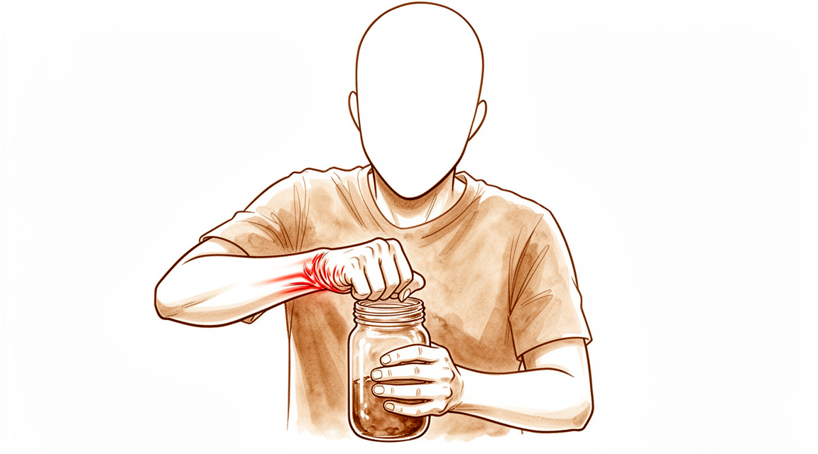
Cơn đau của bạn có khả năng bắt đầu từ cổ tay sau một chấn thương cũ. Tình trạng này được gọi là viêm xương khớp do hao mòn. Nó xảy ra khi các xương trong cổ tay của bạn bị lệch khỏi vị trí bình thường. Bạn có thể cảm thấy một cơn đau âm ỉ ở mặt trên của cổ tay. Cơn đau thường trở nên nghiêm trọng hơn khi bạn đẩy vào một vật hoặc xoay bàn tay. Các hoạt động đơn giản như mở nắp lọ hoặc xoay tay nắm cửa có thể trở nên khó khăn.
Bạn có thể nhận thấy cảm giác cứng khớp khi vừa thức dậy. Điều này thường biến mất sau khi bạn di chuyển một chút. Tuy nhiên, cơn đau có thể quay trở lại sau khi bạn sử dụng tay cả ngày. Nhiều bệnh nhân nhận thấy rằng nghỉ ngơi bàn tay giúp giảm bớt. Một số người cảm thấy đau vào ban đêm, đặc biệt nếu họ ngủ nghiêng về phía đó. Điều này có thể làm gián đoạn giấc ngủ và khiến bạn cảm thấy mệt mỏi.
Các hoạt động hàng ngày có thể trở nên khó khăn hơn trước. Với tay ra sau lưng để cài áo ngực có thể gây đau. Nhét áo vào quần có thể đòi hỏi bạn phải nâng cánh tay theo một cách gây đau. Nhấc các vật, ngay cả những vật nhẹ, có thể gây khó chịu nhói lên. Bạn có thể tránh sử dụng tay vì sợ kích hoạt cơn đau. Điều này có thể khiến các thói quen đơn giản trở thành một thách thức.
Tình trạng này ảnh hưởng đến cách các xương trong cổ tay di chuyển. Các xương chính có thể không còn căn chỉnh đúng vị trí nữa. Sự lệch lạc này gây ra sự hao mòn thêm cho các khớp. Bạn có thể cảm thấy một cảm giác mài mòn hoặc nghe thấy tiếng lách cách khi bạn di chuyển cổ tay. Điều này không phải lúc nào cũng gây đau, nhưng nó có thể gây khó chịu.
Điều quan trọng là phải biết rằng không phải tất cả các chấn thương cổ tay đều dẫn đến vấn đề này. Nếu bạn từng bị rách dây chằng trong quá khứ nhưng kết quả chụp X-quang của bạn bình thường, bạn có thể không phát triển viêm xương khớp này. Tuy nhiên, nếu bạn đã có tình trạng mất ổn định kéo dài, nguy cơ sẽ cao hơn. Bác sĩ phẫu thuật của bạn sẽ xem xét tiền sử và triệu chứng cụ thể của bạn. Họ sẽ xác định xem cơn đau của bạn có phù hợp với mô hình này hay không. Hiểu rõ những gì bạn đang cảm nhận giúp bác sĩ phẫu thuật lựa chọn phương pháp điều trị đúng đắn để giảm đau và cải thiện chức năng của bạn.
Những gì thực sự đang xảy ra
Cổ tay của bạn là một cụm phức tạp gồm tám xương nhỏ được gọi là xương cổ tay. Trong cổ tay SLAC và SNAC, chuyển động trượt bình thường giữa các xương này bị suy giảm. Điều này thường bắt đầu với tổn thương các dây chằng giữ các xương lại với nhau hoặc với một xương không lành sau khi gãy xương. Khi các cấu trúc hỗ trợ này suy yếu, các xương bị lệch khỏi vị trí đúng của chúng.
Hãy tưởng tượng cổ tay của bạn như một bản lề được bôi trơn tốt. Sụn là lớp phủ trơn tru trên đầu các xương cho phép chúng trượt mà không có ma sát. Khi các xương bị lệch, lớp phủ này bị mòn không đều. Viêm xương khớp do hao mòn này tạo ra sự tiếp xúc xương trên xương. Kết quả là đau, cứng và cảm giác mài mòn khi bạn di chuyển bàn tay. Bao hoạt dịch khớp, hoạt động như một ống bọc quanh khớp, cũng có thể bị viêm và co cứng.
Sự lệch vị trí này thay đổi cách lực truyền qua bàn tay của bạn. Bình thường, tải trọng được chia sẻ đều đặn. Bây giờ, một số vị trí chịu quá nhiều áp lực. Điều này làm tăng tốc độ tổn thương và dẫn đến các triệu chứng cụ thể mà bạn cảm nhận được. Bác sĩ phẫu thuật của bạn có thể thấy những thay đổi này trên hình ảnh chẩn đoán. Họ tìm kiếm các dấu hiệu cho thấy các xương không còn di chuyển theo cách trơn tru, phối hợp nữa.
Mục tiêu của điều trị là ngăn chặn chuyển động bất thường này. Bằng cách ổn định các xương hoặc loại bỏ các phần bị tổn thương, chúng tôi nhằm mục đích khôi phục một đường đi chuyển động trơn tru hơn. Điều này làm giảm cảm giác mài mòn và giảm áp lực lên các đầu dây thần kinh nhạy cảm trong khớp. Mặc dù chúng tôi không thể đảo ngược hoàn toàn viêm xương khớp, chúng tôi có thể cải thiện đáng kể chức năng của cổ tay và giảm đau cho bạn.
Những gì chúng tôi có thể làm về vấn đề này
Bác sĩ phẫu thuật của bạn có thể sẽ bắt đầu bằng việc tự chăm sóc và vật lý trị liệu. Phương pháp này nhằm mục đích giảm đau và duy trì khả năng vận động của cổ tay. Bạn sẽ được học các bài tập nhẹ nhàng để tăng cường sức mạnh cho các cơ xung quanh cổ tay. Những chuyển động này giúp hỗ trợ khớp và cải thiện chức năng trong sinh hoạt hàng ngày. Bạn nên dành cơ hội thích đáng cho phương pháp điều trị bảo tồn này phát huy tác dụng. Hầu hết bệnh nhân đều thử các lựa chọn không phẫu thuật trước tiên để xem liệu các triệu chứng có cải thiện mà không cần phẫu thuật hay không.
Nếu các bài tập đơn giản không đủ hiệu quả, bác sĩ phẫu thuật của bạn có thể thảo luận về quản lý bằng thuốc. Điều này thường bao gồm thuốc giảm đau và thuốc chống viêm để giúp bạn cảm thấy dễ chịu hơn. Bác sĩ phẫu thuật của bạn cũng có thể đề xuất các loại tiêm. Tiêm cortisone có thể giảm sưng và đau trong một khoảng thời gian. Tiêm axit hyaluronic hoặc huyết tương giàu tiểu cầu (PRP) cũng có thể là các lựa chọn để bôi trơn khớp hoặc thúc đẩy quá trình lành thương. Những phương pháp điều trị này không chữa khỏi bệnh viêm xương khớp, nhưng chúng có thể giảm bớt triệu chứng trong khi bạn tiếp tục các hoạt động hàng ngày. Thời gian giảm đau thay đổi tùy theo từng người.
Khi phương pháp điều trị bảo tồn không còn kiểm soát được cơn đau của bạn, phẫu thuật có thể được xem xét. Điều này thường được khuyến nghị khi chất lượng cuộc sống của bạn bị ảnh hưởng đáng kể. Các lựa chọn phẫu thuật phụ thuộc vào giai đoạn viêm xương khớp của bạn và tình trạng của các xương cổ tay. Đối với tình trạng mòn khớp ở giai đoạn sớm, bác sĩ phẫu thuật của bạn có thể cắt bỏ một hàng xương nhỏ (cắt bỏ hàng xương cổ tay gần) hoặc cố định bốn xương cụ thể lại với nhau sau khi cắt bỏ xương thuyền (cố định bốn góc). Các thủ thuật này nhằm mục đích giảm đau trong khi bảo tồn tối đa khả năng vận động của cổ tay. Trong một số trường hợp, một thủ thuật được gọi là cắt dây thần kinh cổ tay có thể được sử dụng để giảm các tín hiệu đau trong khi vẫn giữ cho khớp linh hoạt. Bác sĩ phẫu thuật của bạn sẽ chọn lựa chọn tốt nhất dựa trên mô hình chấn thương cụ thể và mục tiêu của bạn.
Những điều cần biết
Đau và cứng cổ tay của bạn có khả năng sẽ tiếp tục tồn tại nếu không được điều trị. Các tình trạng này do viêm xương khớp do hao mòn gây ra sau một chấn thương cũ. Tổn thương thường tiến triển theo thời gian. Nếu không can thiệp, bạn có thể gặp phải tình trạng khó chịu kéo dài và giảm khả năng sử dụng bàn tay cho các hoạt động hàng ngày.
Với phương pháp điều trị phù hợp, bạn có thể mong đợi sự giảm đau đáng kể. Các lựa chọn phẫu thuật như cắt bỏ hàng xương cổ tay gần hoặc hợp nhất bốn xương góc được thiết kế để cải thiện tình trạng đau và chức năng tổng thể của cổ tay. Hầu hết bệnh nhân báo cáo kết quả chủ quan tốt hơn và ít đau hơn sau các thủ thuật này. Bác sĩ phẫu thuật của bạn sẽ lựa chọn phương án tốt nhất dựa trên giai đoạn cụ thể của viêm xương khớp và tình trạng của bề mặt khớp.
Quá trình hồi phục là một tiến trình, không phải là giải pháp tức thì. Bạn có thể cần nhiều hơn một cuộc phẫu thuật để đạt được sự cải thiện đau lâu dài và chức năng tốt. Phẫu thuật lần thứ hai hoặc thậm chí lần thứ ba vẫn có thể mang lại những lợi ích bền vững, cho phép bạn trở lại làm việc và các hoạt động hàng ngày. Điều này đặc biệt đúng nếu viêm xương khớp của bạn đang ở giai đoạn sớm.
Ngay cả khi bạn cần một thủ thuật cứu cánh sau này, chẳng hạn như cắt thần kinh cổ tay, bạn vẫn có thể duy trì khả năng vận động và giảm đau. Các lựa chọn này có tỷ lệ thất bại thấp trong trung và dài hạn. Trong khi một số bệnh nhân có thể gặp phải các triệu chứng viêm xương khớp từ nhẹ đến trung bình sau một số phẫu thuật chỉnh hình nhất định, phần lớn vẫn duy trì được chức năng lâu dài chấp nhận được.
Điều quan trọng là phải có những kỳ vọng thực tế. Bạn sẽ không lấy lại chính xác cùng cơ học cổ tay như trước khi bị chấn thương. Tuy nhiên, bạn có thể mong đợi một kết quả bền vững cho phép bạn thực hiện các nhiệm vụ hàng ngày với ít đau đớn hơn. Bác sĩ phẫu thuật của bạn sẽ hướng dẫn bạn trong hành trình này, tập trung vào việc bảo tồn nhiều khả năng vận động và sức mạnh nhất có thể cho tình trạng cụ thể của bạn.
Khi nào cần gặp bác sĩ
Hãy yêu cầu đánh giá bởi chuyên gia nếu bạn có đau cổ tay dai dẳng không cải thiện khi nghỉ ngơi. Hãy tìm kiếm chăm sóc y tế nếu bạn nhận thấy yếu cơ, mất ổn định, hoặc cảm giác kẹt cứng hoặc sập khớp. Những triệu chứng này có thể ảnh hưởng đến giấc ngủ hoặc công việc của bạn. Hãy liên hệ với bác sĩ nếu bạn trải qua tình trạng xấu đi đột ngột của bệnh. Điều này đặc biệt quan trọng nếu bạn là nam giới và có tiền sử chấn thương cổ tay. Đánh giá sớm giúp xác định xem bạn có bị viêm xương khớp do hao mòn liên quan đến chấn thương trước đó hay không. Bác sĩ phẫu thuật của bạn có thể đánh giá liệu việc nghỉ ngơi đơn thuần có đủ hay cần điều trị thêm để duy trì tầm vận động và chức năng của cổ tay.
Evidence & references
Overview
- Proximal row carpectomy and four-corner fusion with scaphoid bone excision are the most widely used surgical procedures for stage II wrist osteoarthritis secondary to SLAC or SNAC wrist [6].
- Both proximal row carpectomy and four-corner fusion provide improvements in pain and subjective outcome measures for patients with symptomatic and appropriately staged SLAC or SNAC wrists [1].
- Scaphoid excision and four-corner fusion is an increasingly popular treatment option for patients with stage II or III SLAC/SNAC arthritis that preserves wrist motion through the radiolunate and ulnocarpal joints [3].
- Scaphoid excision and four-corner fusion are effective treatments for SLAC and SNAC patterns of wrist arthritis [8].
- For stage II SLAC wrist with a preserved capitolunate joint, proximal row carpectomy is preferred because it is technically less demanding and yields durable results [10].
- The authors prefer proximal row carpectomy for SLAC wrists with preserved capitate head cartilage due to socio-economic benefits, lower complication rates, and procedural ease [46].
- In patients with SLAC/SNAC II, proximal row carpectomy might be favourable to a midcarpal arthrodesis solely based on better flexion-extension range of motion of the radiocarpal joint after proximal row carpectomy [4].
- Wrist denervation is a viable salvage option for patients with symptomatic SLAC wrist osteoarthritis to preserve motion, decrease pain, and increase function with a low absolute failure rate at mid- to long-term follow-up [13].
- Arthroscopic wrist debridement and radial styloidectomy may have advantages in relieving pain while preserving wrist motion for SLAC stage 2 or 3 disease [23].
- It is mostly indicated as a palliative procedure in elderly patients with posttraumatic SNAC or SLAC wrist with limited functional demands [18].
- A second and even a third operation can result in long-term pain improvement, good function and capacity for work, and re-operation is recommended in symptomatic cases with minor osteoarthritis of the wrist (SNAC stage 0 or 1) [11].
- Further data in a larger cohort with longer follow-up is required to determine the effect on SLAC-wrist deterioration [2].
Anatomy & Pathophysiology
- Surgical treatments for scapholunate advanced collapse (SLAC) wrist result in decreased wrist kinematic motion and functional performance compared with individuals with normal wrists [24].
- Lunate morphology affects the 3-dimensional kinematics of the carpus during wrist flexion and extension [25].
- Three-corner fusions produce motion that is smoother and more closely replicates the normal axis and functional motion of the wrist compared with four-corner fusions [26].
- Computed fiber elongations of the dorsal carpal ligaments vary linearly with wrist position [27].
- Four-dimensional computed tomography (4DCT) is a non-invasive and affordable method to assess and quantify wrist kinematics by incorporating the temporal dimension [28].
- During simple unresisted wrist motions, force in the scapholunate interosseous ligament does not exceed 20 N [29].
- Kinematic changes in scapholunate instability may predict the development of radioscaphoid arthritis and help identify a kinematically abnormal wrist [30].
- Scaphoid nonunions have a dramatic impact on carpal kinematics by partially uncoupling the proximal and distal carpal rows [31].
- Dynamic imaging allows for the derivation of standardized protocols for mapping carpal motion that are clinically applicable and reproducible [32].
- Computer-aided CT analysis provides guidelines for measuring and quantifying carpal alignment three-dimensionally and establishes a database for normal values [33].
- More than half the motion of the carpus when the wrist is loaded in extension occurs at the midcarpal joint [34].
- Radioscapholunate fusion shows the most biomechanically similar behavior to the healthy wrist among three compared fusion types [35].
- Contact areas between the scaphoid and distal radius are maximized during full extension of the wrist, which helps stabilize the radiocarpal joint [36].
- Tendon ball arthroplasty and proximal carpal stabilization with tendon graft restore the integrity of the proximal carpal row [37].
- In unstable wrists, dorsally applied polylactic acid (PLA) plates restore carpal kinematics for 1,000 cycles of motion when fixation is not compromised by carpal size or osteoporosis [38].
- Electrogoniometric and radiologic evaluation characterizes the modification of the wrist center of rotation during flexion and extension [39].
- Wrist arthrodesis may only compromise select wrist functions [40].
- The 'dart thrower's motion' of the wrist, from radial extension to ulnar flexion, may be a unifying concept of functional wrist motion that is uniquely human [41].
- Correction of scapholunate dissociation via modified Brunelli technique or Blatt capsulodesis might correlate with improved carpal dynamics and improved clinical outcomes [42].
Classification
- SLAC wrist diagnosis should be reserved for patients in whom traumatic disruption of the proximal carpal row initiates the defined sequence of arthritic change outlined by Watson and Ballet [5].
- Radiographic classification of SLAC wrist has moderate reliability and reproducibility [14].
- Radiographic classification of SNAC wrist has limited reliability [14].
- SNAC wrists differ from SLAC wrists by exhibiting a decreased sagittal lunotriquetral angle, indicating a distinct pathomechanism of carpal instability [7].
- Bone density is greater at the capitolunate joint, the radial styloid, and the radiolunate joint in SNAC wrists compared to controls [9].
- Carpal malalignment in SLAC wrists affects the radio- and midcarpal joints and extends to the third carpometacarpal joint, with malalignment evident in both sagittal and coronal planes [16].
- Patients with SLAC wrist are more likely to be male and have a history of trauma compared to patients with first CMC OA [12].
Clinical Presentation
- SLAC wrist diagnosis is reserved for patients in whom traumatic disruption of the proximal carpal row initiates a defined sequence of arthritic changes [5].
- Patients with SLAC wrist are more likely to be male and have a history of trauma compared to patients with first CMC OA [12].
- SNAC wrists differ from SLAC wrists by exhibiting a decreased sagittal lunotriquetral angle, indicating a distinct pathomechanism of carpal instability [7].
- Bone density is greater at the capitolunate joint, the radial styloid, and the radiolunate joint in SNAC wrists compared to controls [9].
- Carpal malalignment in SLAC wrists affects the radio- and midcarpal joints and extends to the third carpometacarpal joint, with malalignment evident in both sagittal and coronal planes [16].
- Distal row pronation and translation and radiolunate arthritis are demonstrated in the SNAC wrist via quantitative 3-D CT [9].
- Radiographic classification of SLAC wrist has moderate reliability and reproducibility [14].
- Radiographic classification of SNAC wrist has limited reliability [14].
- There is room for improvement in the assessment of patients with SNAC wrists [45].
- A scapholunate ligament injury visualized arthroscopically, without static x-ray changes, does not inevitably lead to SLAC wrist [15].
- Isolated lunocapitate osteoarthritis presents differently from SLAC wrist and is not related to scapholunate instability [20].
- Bilateral scapholunate widening may have a nontraumatic aetiology and progress to carpal instability and osteoarthritis with advancing age [44].
- Patients without carpal instability or osteoarthritis were younger than those with bilateral SLAC wrists [44].
- There is no absolute evidence to confirm that bilateral wide gaps inexorably progress to carpal instability and osteoarthritis [44].
Investigations
- The diagnosis of SLAC wrist should be reserved for patients in whom a traumatic disruption of the proximal carpal row has initiated the defined sequence of arthritic change outlined by Watson and Ballet [5].
- There is currently no scientific evidence that a scapholunate ligament injury visualized arthroscopically, without static x-ray changes, inevitably leads to SLAC wrist [15].
- Radiographic classification of SLAC wrist has moderate reliability and reproducibility [14].
- Classification of SNAC wrist has limited reliability [14].
- SNAC wrists differ from SLAC wrists in exhibiting a decreased sagittal lunotriquetral angle, indicating a distinct pathomechanism of carpal instability [7].
- Bone density is greater at the capitolunate joint, the radial styloid, and the radiolunate joint in SNAC wrists compared to controls [9].
- Carpal malalignment in SLAC wrists affects the radio- and midcarpal joints and extends to the third carpometacarpal joint, with malalignment evident in both the sagittal and coronal planes [16].
- Patients with SLAC wrist are more likely to be male and have a history of trauma compared to patients with first CMC OA [12].
- Isolated lunocapitate osteoarthritis presents differently from SLAC wrist and is not related to scapholunate instability [20].
- Cineradiography has a high diagnostic value for diagnosing scapholunate dissociations [49].
- Dynamic CT scan of the wrist is a user-friendly way of measuring the scapholunate distance, which is minimal in the normal wrist below 40 years of age [51].
Treatment
- Proximal row carpectomy and four-corner fusion with scaphoid bone excision are the most widely used surgical procedures for stage II wrist osteoarthritis secondary to SLAC or SNAC wrist [6].
- Both proximal row carpectomy and four-corner fusion provide improvements in pain and subjective outcome measures for patients with symptomatic and appropriately staged SLAC or SNAC wrists [1].
- Scaphoid excision and four-corner fusion is an increasingly popular treatment option for patients with stage II or III SLAC/SNAC arthritis that preserves wrist motion through the radiolunate and ulnocarpal joints [3].
- Scaphoid excision and four-corner fusion are effective treatments for SLAC and SNAC patterns of wrist arthritis [8].
- For stage II SLAC wrist with a preserved capitolunate joint, proximal row carpectomy is preferred because it is technically less demanding and yields durable results [10].
- In patients with SLAC/SNAC II, proximal row carpectomy might be favourable to a midcarpal arthrodesis solely based on better flexion-extension range of motion of the radiocarpal joint after proximal row carpectomy [4].
- The LCF (lunate-capitate arthrodesis) is not less efficient than the 4CF (four-corner fusion) in the treatment of SNAC II and III wrist injuries [19].
- Wrist denervation is a viable salvage option for patients with symptomatic SLAC wrist osteoarthritis to preserve motion, decrease pain, and increase function with a low absolute failure rate at mid- to long-term follow-up [13].
- Arthroscopic wrist debridement and radial styloidectomy may have advantages in relieving pain while preserving wrist motion for SLAC stage 2 or 3 disease [23].
- Motion-preserving procedures of the wrist can obtain good long-term results if indications are accurately respected and the technique is well performed to prevent complications [43].
- Proximal row carpectomy is mostly indicated as a palliative procedure in elderly patients with posttraumatic SNAC or SLAC wrist with limited functional demands [18].
- A second and even a third operation can result in long-term pain improvement, good function and capacity for work, and re-operation is recommended in symptomatic cases with minor osteoarthritis of the wrist (SNAC stage 0 or 1) [11].
- Further data in a larger cohort with longer follow-up is required to determine the effect of treatments on SLAC-wrist deterioration [2].
Complications
- Proximal row carpectomy and four-corner fusion provide improvements in pain and subjective outcome measures for patients with symptomatic and appropriately staged SLAC or SNAC wrists [1].
- Further data in a larger cohort with longer follow-up is required to determine the effect of surgical interventions on SLAC-wrist deterioration [2].
- Scaphoid excision and four-corner fusion preserves wrist motion through the radiolunate and ulnocarpal joints [3].
- Proximal row carpectomy may be favourable to midcarpal arthrodesis in patients with SLAC/SNAC II based on better flexion-extension range of motion of the radiocarpal joint after proximal row carpectomy [4].
- The diagnosis of SLAC should be reserved for patients in whom a traumatic disruption of the proximal carpal row has initiated the defined sequence of arthritic change outlined by Watson and Ballet [5].
- Proximal row carpectomy and four-corner fusion with scaphoid bone excision are the most widely used surgical procedures for stage II wrist osteoarthritis secondary to SLAC or SNAC wrist [6].
- SNAC wrists differ from SLAC wrists in exhibiting a decreased sagittal lunotriquetral angle, indicating a distinct pathomechanism of carpal instability [7].
- Bone density was greater at the capitolunate joint, the radial styloid, and the radiolunate joint in SNAC wrists compared to controls [9].
- For stage II SLAC wrist with a preserved capitolunate joint, proximal row carpectomy is preferred because it is technically less demanding and yields durable results [10].
- A second and even a third operation can result in long-term pain improvement, good function and capacity for work, and re-operation is recommended in symptomatic cases with minor osteoarthritis of the wrist (SNAC stage 0 or 1) [11].
- Patients with SLAC wrist were more likely to be male and have a history of trauma compared to patients with first CMC OA [12].
- Wrist denervation was a viable salvage option for patients with symptomatic SLAC wrist osteoarthritis to preserve motion, decrease pain, and increase function with a low absolute failure rate at mid- to long-term follow-up [13].
- There is currently no scientific evidence that a scapholunate ligament injury visualized arthroscopically, without static x-ray changes, inevitably leads to SLAC wrist [15].
- Although ongoing scapholunate instability resulted in early arthritic degeneration after dorsal intercarpal ligament capsulodesis, most patients had acceptable long-term function of the wrist [21].
- Scapholunate ligament reconstruction using a part of the extensor carpi radialis brevis tendon through a dorsal approach resulted in long-term, improved outcomes compared with other techniques, even in scapholunate advanced collapse type I wrists [22].
- Distal scaphoid resection arthroplasty produced favorable, long-term clinical results and did not result in noteworthy wrist collapse [52].
Recovery
- Proximal row carpectomy and four-corner fusion both provide improvements in pain and subjective outcome measures for patients with symptomatic and appropriately staged SLAC or SNAC wrists [1].
- Further data in a larger cohort with longer follow-up is required to determine the effect of interventions on SLAC-wrist deterioration [2].
- Scaphoid excision and four-corner fusion preserves wrist motion through the radiolunate and ulnocarpal joints in patients with stage II or III SLAC/SNAC arthritis [3].
- In patients with SLAC/SNAC II, proximal row carpectomy might be favourable to midcarpal arthrodesis based on better flexion-extension range of motion of the radiocarpal joint after proximal row carpectomy [4].
- For stage II SLAC wrist with a preserved capitolunate joint, proximal row carpectomy is preferred because it is technically less demanding and yields durable results [10].
- A second and even a third operation can result in long-term pain improvement, good function, and capacity for work, supporting re-operation in symptomatic cases with minor osteoarthritis of the wrist (SNAC stage 0 or 1) [11].
- Wrist denervation is a viable salvage option for patients with symptomatic SLAC wrist osteoarthritis to preserve motion, decrease pain, and increase function with a low absolute failure rate at mid- to long-term follow-up [13].
- Although ongoing scapholunate instability resulted in early arthritic degeneration, most patients had acceptable long-term function of the wrist after dorsal intercarpal ligament capsulodesis [21].
- Scapholunate ligament reconstruction using a part of the extensor carpi radialis brevis tendon resulted in long-term, improved outcomes compared with other techniques, even in scapholunate advanced collapse type I wrists [22].
- Residual scaphoid deformity has no relevant negative impact on mid-term wrist function after scaphoid nonunion surgery [53].
- Wrist alignment was maintained over time after corrective osteotomy for distal radius malunion, but 13 patients presented mild to moderate symptomatic wrist arthritis [54].
- Carpal collapse recurred within a short time after dorsal intercarpal ligament capsulodesis because the procedure cannot withstand large and repetitive forces [55].
- The reduction and association of the scaphoid and lunate procedure should be abandoned due to early radiographic failure in the short term, despite relatively low outcomes measures scores [56].
Key Evidence
- [L4] Both procedures provide improvements in pain and subjective outcome measures for patients with symptomatic and appropriately staged SLAC or SNAC wrists. [1] (10.1177/1753193408100954)
- [Paper] Scaphoid excision and four-corner fusion is an increasingly popular treatment option for patients with stage II or III SLAC/SNAC arthritis that preserves wrist motion through the radiolunate and ulnocarpal joints. [3] (10.1016/j.hcl.2005.08.012)
- [L1] In patients with SLAC/SNAC II, proximal row carpectomy might be favourable to a midcarpal arthrodesis solely based on better FE ROM of the radiocarpal joint after proximal row carpectomy. [4] (10.1186/s12891-024-07527-6)
- [L5] The diagnosis of SLAC should be reserved for patients in whom a traumatic disruption of the proximal carpal row has initiated the defined sequence of arthritic change outlined by Watson and Ballet. [5] (10.1016/j.jhsa.2015.06.110)
- [L4] Proximal row carpectomy and four-corner fusion with scaphoid bone excision are the most widely used surgical procedures for stage II wrist osteoarthritis secondary to SLAC or SNAC wrist. [6] (10.1016/j.otsr.2014.06.025)
- [L4] SNAC wrists differ from SLAC wrists in exhibiting a decreased sagittal lunotriquetral angle, indicating a distinct pathomechanism of carpal instability. [7] (10.1186/s12891-025-08652-6)
- [L3] Scaphoid excision and four-corner fusion are effective treatments for SLAC and SNAC patterns of wrist arthritis. [8] (10.1016/s0363-5023(09)60084-8)
- [L3] Bone density was greater at the capitolunate joint, the radial styloid, and the radiolunate joint in SNAC wrists compared to controls. [9] (10.2106/jbjs.22.01350)
- [L4] For stage II SLAC wrist with a preserved capitolunate joint, proximal row carpectomy is preferred because it is technically less demanding and yields durable results. [10] (10.5435/00124635-200307000-00007)
- [L4] A second and even a third operation can result in long-term pain improvement, good function and capacity for work, and we recommend re-operation in symptomatic cases with minor osteoarthritis of the wrist (SNAC stage 0 or 1). [11] (10.1177/1753193409346093)
- [L3] Patients with SLAC wrist were more likely to be male and have a history of trauma compared to patients with first CMC OA. [12] (10.1177/1558944718788672)
- [L4] This method of wrist denervation was a viable salvage option for patients with symptomatic SLAC wrist osteoarthritis to preserve motion, decrease pain, and increase function with a low absolute failure rate at mid- to long-term follow-up. [13] (10.1016/j.jhsa.2021.02.023)
- [L4] Radiographic classification of SLAC wrist has moderate reliability and reproducibility, whereas classification of SNAC wrist has limited reliability. [14] (10.1177/1753193413484629)
- [L5] There is currently no scientific evidence that a scapholunate ligament injury visualized arthroscopically, without static x-ray changes, inevitably leads to SLAC wrist. [15] (10.1016/j.jhsa.2011.01.018)
- [L3] Carpal malalignment in SLAC wrists not only affects the radio- and midcarpal joints, but also extends to the third carpometacarpal joint, with malalignment evident in both the sagittal and coronal planes. [16] (10.1016/j.jhsa.2024.09.021)
- [L4] The LCF is not less efficient than the 4CF in the treatment of SNAC II and III wrist injuries. [19] (10.1186/s12891-024-07755-w)
- [L3] Although the consequent ongoing scapholunate instability resulted in early arthritic degeneration, most patients had acceptable long-term function of the wrist. [21] (10.1302/0301-620x.94b12.30007)
- [L4] This technique, even in scapholunate advanced collapse type I wrists, resulted in long-term, improved outcomes compared with other techniques. [22] (10.1177/17531934221143679)
- [L4] The procedure studied may have advantages in relieving pain while preserving wrist motion for SLAC stage 2 or 3 disease. [23] (10.1177/1558944717725383)
- [L2] Both surgical groups demonstrated decreased wrist kinematic motion and functional performance compared with individuals with normal wrists. [24] (10.1016/j.jhsa.2015.04.035)
- [L5] This study describes the effect of lunate morphology on 3-dimensional carpal kinematics during wrist flexion and extension. [25] (10.1016/j.jhsa.2014.09.019)
- [L3] Motion was smoother and more closely replicated the normal axis and functional motion of the wrist. [26] (10.1016/j.jhsa.2015.02.027)
- [L5] Despite complex carpal bone anatomy and kinematics, computed fiber elongations were found to vary linearly with wrist position. [27] (10.1016/j.jhsa.2012.04.025)
- [L5] Four-dimensional computed tomography (4DCT) is a promising, non-invasive, and affordable method to assess and quantify wrist kinematics, extending conventional CT by incorporating the temporal dimension. [28] (10.1177/17531934251326028)
- [L5] However, during simple unresisted wrist motions, the force did not exceed 20 N. [29] (10.1016/j.jhsa.2015.04.007)
- [L3] These kinematic changes may predict the development of radioscaphoid arthritis and help identify a kinematically abnormal wrist. [30] (10.1177/17531934241242676)
- [L4] Scaphoid nonunions have a dramatic impact on carpal kinematics, partially uncoupling the proximal and distal carpal rows. [31] (10.1016/j.jhsa.2008.03.008)
- [L4] With the increased focus on dynamic imaging for wrist motion, it may be possible to derive a standardized protocol for mapping the carpal motion that is clinically applicable and reproducible. [32] (10.1016/j.jhsg.2022.10.001)
- [L4] This study provides guidelines on how to measure and quantify carpal alignment three-dimensionally and establishes a database for normal values, which may be useful when analysing various wrist pathologies and kinematics. [33] (10.1177/17531934231160100)
- [L4] More than half the motion of the carpus when the wrist was loaded in extension occurred at the midcarpal joint. [34] (10.1016/j.jhsa.2012.10.035)
- [L5] The contact areas between the scaphoid and distal radius are maximized during full extension of the wrist, which helps stabilize the radiocarpal joint and potentially reduces the risk of injury to the carpus and the distal radius. [36] (10.1177/1753193413507810)
- [L4] The technique demonstrated reduced wrist pain and improved wrist motion and grip strength while restoring the integrity of the proximal carpal row. [37] (10.1177/17531934241238939)
- [L5] The study shows that in the unstable wrist, following ligament sectioning, where fixation is not compromised by carpal size or osteoporosis, a dorsally applied PLA plate does restore carpal kinematics for 1,000 cycles of motion. [38] (10.1016/j.jhsa.2008.01.016)
- [L4] The study also characterized the modification of the wrist CoR during flexion and extension, noting that stability is considered more important than mobility in clinical conditions. [39] (10.1016/s0749-0712(03)00008-8)
- [L4] Our findings suggest that wrist arthrodesis may only compromise select wrist functions. [40] (10.1177/1558944715626930)
- [L5] The 'dart thrower's motion' of the wrist, from radial extension to ulnar flexion, may be a unifying concept of functional wrist motion that is uniquely human. [41] (10.5435/00124635-201001000-00007)
- [L5] This correction might correlate with improved carpal dynamics and improved clinical outcomes. [42] (10.1016/j.jhsa.2010.06.029)
- [L4] While bilateral SLAC wrists were not exceptional and patients without carpal instability or osteoarthritis were younger, there is no absolute evidence to confirm that bilateral wide gaps inexorably progress to carpal instability and osteoarthritis. [44] (10.1177/1753193418819653)
- [L4] There is room for improvement in the way we assess patients with SNAC wrists. [45] (10.1177/1558944716677541)
- [L3] The authors prefer proximal row carpectomy for SLAC wrists with preserved capitate head cartilage due to socio-economic benefits, lower complication rates, and procedural ease. [46] (10.1177/1753193408087116)
- [L3] Cineradiography has a high diagnostic value for diagnosing scapholunate dissociations. [49] (10.1177/1753193413489056)
- [L4] This novel dynamic CT scan of the wrist is a user-friendly way of measuring the scapholunate distance, which is minimal in the normal wrist below 40 years of age. [51] (10.1177/1558944717726372)
- [L4] Distal scaphoid resection arthroplasty produced favorable, long-term clinical results and did not result in noteworthy wrist collapse. [52] (10.1016/j.jhsa.2014.05.031)
- [L4] Residual scaphoid deformity has no relevant negative impact on mid-term wrist function. [53] (10.1177/17531934221125355)
- [L4] Wrist alignment was maintained over time but 13 patients presented mild to moderate symptomatic wrist arthritis. [54] (10.1177/1753193409357373)
- [L4] With a majority of patients experiencing early radiographic failure of the procedure in the short term, our experience suggests that the reduction and association of the scaphoid and lunate procedure should be abandoned despite the relatively low outcomes measures scores. [56] (10.1016/j.jhsa.2014.07.014)
References
[1] Proximal Row Carpectomy vs Four Corner Fusion for Scapholunate (Slac) or Scaphoid Nonunion Advanced Collapse (Snac) Wrists: A Systematic Review of Outcomes. Journal of Hand Surgery (European Volume). 2009. DOI: 10.1177/1753193408100954 [2] 10.1055-s-0033-1341582. n.d.. [3] Scaphoid Excision with Four-Corner Fusion. Hand Clinics. 2005. DOI: 10.1016/j.hcl.2005.08.012 [4] Patient reported and functional outcome measures after surgical salvage procedures for posttraumatic radiocarpal osteoarthritis – a systematic review. BMC Musculoskeletal Disorders. 2024. DOI: 10.1186/s12891-024-07527-6 [5] Scapholunate Advanced Collapse: Nomenclature and Differential Diagnosis. The Journal of Hand Surgery. 2015. DOI: 10.1016/j.jhsa.2015.06.110 [6] Wrist osteoarthritis. Orthopaedics & Traumatology: Surgery & Research. 2015. DOI: 10.1016/j.otsr.2014.06.025 [7] Computer-aided three-dimensional analysis of carpal alignment in scaphoid nonunion advanced collapse wrists: A comparative study with scapholunate advanced collapse and healthy wrists. BMC Musculoskeletal Disorders. 2025. DOI: 10.1186/s12891-025-08652-6 [8] Four-Corner Fusion: A Retrospective Comparative Study of Fixation with a Circular Plate versus Compression Screws. The Journal of Hand Surgery. 2009. DOI: 10.1016/s0363-5023(09)60084-8 [9] Quantitative 3-D CT Demonstrates Distal Row Pronation and Translation and Radiolunate Arthritis in the SNAC Wrist. Journal of Bone and Joint Surgery. 2023. DOI: 10.2106/jbjs.22.01350 [10] Proximal Row Carpectomy and Intercarpal Arthrodesis for the Management of Wrist Arthritis. Journal of the American Academy of Orthopaedic Surgeons. 2003. DOI: 10.5435/00124635-200307000-00007 [11] Is revision bone grafting worthwhile after failed surgery for scaphoid nonunion? Minimum 8 year follow-up of 18 patients. Journal of Hand Surgery (European Volume). 2009. DOI: 10.1177/1753193409346093 [12] The Epidemiology of Scapholunate Advanced Collapse. HAND. 2018. DOI: 10.1177/1558944718788672 [13] Midterm Patient-Reported Outcomes in Wrist Denervation for Post-Traumatic Arthritis. The Journal of Hand Surgery. 2021. DOI: 10.1016/j.jhsa.2021.02.023 [14] Reproducibility of radiographic classification of scapholunate advanced collapse (SLAC) and scaphoid nonunion advanced collapse (SNAC) wrist. Journal of Hand Surgery (European Volume). 2013. DOI: 10.1177/1753193413484629 [15] Scapholunate Advanced Collapse and Scaphoid Nonunion Advanced Collapse Arthritis—Update on Evaluation and Treatment. The Journal of Hand Surgery. 2011. DOI: 10.1016/j.jhsa.2011.01.018 [16] Comparative Computer-Aided Analysis of Three-Dimensional Carpal Alignment in Scapholunate Advanced Collapse and Healthy Wrists. The Journal of Hand Surgery. 2025. DOI: 10.1016/j.jhsa.2024.09.021 [18] 10.1055-s-0032-1329615. n.d.. [19] Lunate-capitate arthrodesis for scaphoid nonunion: a comparative study. BMC Musculoskeletal Disorders. 2024. DOI: 10.1186/s12891-024-07755-w [20] 10.1055-s-0034-1372515. n.d.. [21] Long-term results of dorsal intercarpal ligament capsulodesis for the treatment of chronic scapholunate instability. The Journal of Bone and Joint Surgery. British volume. 2012. DOI: 10.1302/0301-620x.94b12.30007 [22] Scapholunate ligament reconstruction using a part of the extensor carpi radialis brevis tendon through a dorsal approach. Journal of Hand Surgery (European Volume). 2023. DOI: 10.1177/17531934221143679 [23] Arthroscopic Wrist Debridement and Radial Styloidectomy for Advanced Scapholunate Advanced Collapse Wrist: Long-term Follow-up. HAND. 2017. DOI: 10.1177/1558944717725383 [24] Surgical Treatments for Scapholunate Advanced Collapse Wrist: Kinematics and Functional Performance. The Journal of Hand Surgery. 2015. DOI: 10.1016/j.jhsa.2015.04.035 [25] The Effect of Lunate Morphology on the 3-Dimensional Kinematics of the Carpus. The Journal of Hand Surgery. 2015. DOI: 10.1016/j.jhsa.2014.09.019 [26] Comparison of the Clinical and Functional Outcomes Following 3- and 4-Corner Fusions. The Journal of Hand Surgery. 2015. DOI: 10.1016/j.jhsa.2015.02.027 [27] Elongation of the Dorsal Carpal Ligaments: A Computational Study of In Vivo Carpal Kinematics. The Journal of Hand Surgery. 2012. DOI: 10.1016/j.jhsa.2012.04.025 [28] Dynamic wrist imaging: How it works and how to assess kinematic changes in wrists with scapholunate instability. Journal of Hand Surgery (European Volume). 2025. DOI: 10.1177/17531934251326028 [29] Force in the Scapholunate Interosseous Ligament During Active Wrist Motion. The Journal of Hand Surgery. 2015. DOI: 10.1016/j.jhsa.2015.04.007 [30] Radiocarpal and midcarpal kinematics in scapholunate instability: a four-dimensional CT study in vivo. Journal of Hand Surgery (European Volume). 2024. DOI: 10.1177/17531934241242676 [31] Interfragmentary Motion in Patients With Scaphoid Nonunion. The Journal of Hand Surgery. 2008. DOI: 10.1016/j.jhsa.2008.03.008 [32] Radiographic Evaluation of Carpal Mechanics and the Scapholunate Angle in a Clenched Fist with Dynamic Computed Tomography Imaging. Journal of Hand Surgery Global Online. 2023. DOI: 10.1016/j.jhsg.2022.10.001 [33] Three-dimensional carpal alignment: computer-aided CT analysis of carpal axes and normal ranges. Journal of Hand Surgery (European Volume). 2023. DOI: 10.1177/17531934231160100 [34] In Vivo Kinematics of the Scaphoid, Lunate, Capitate, and Third Metacarpal in Extreme Wrist Flexion and Extension. The Journal of Hand Surgery. 2013. DOI: 10.1016/j.jhsa.2012.10.035 [35] Load_transfer_through_the_radiocarpal_joint_and_the_effects_of_partial_wrist_art_1753193412441761. 1934. [36] Contact areas of the scaphoid and lunate with the distal radius in neutral and extension: correlation of falling strategies and distal radial anatomy. Journal of Hand Surgery (European Volume). 2013. DOI: 10.1177/1753193413507810 [37] Tendon ball arthroplasty and proximal carpal stabilization with tendon graft for advanced Kienböck’s disease. Journal of Hand Surgery (European Volume). 2024. DOI: 10.1177/17531934241238939 [38] Treatment of Scapholunate Dissociation With a Bioresorbable Polymer Plate: A Biomechanical Study. The Journal of Hand Surgery. 2008. DOI: 10.1016/j.jhsa.2008.01.016 [39] Electrogoniometric and radiologic evaluation of scapho-trapezo-trapezoid arthrodesis. Hand Clinics. 2003. DOI: 10.1016/s0749-0712(03)00008-8 [40] Assessment of Wrist Function After Simulated Total Wrist Arthrodesis. HAND. 2016. DOI: 10.1177/1558944715626930 [41] The Advantage of Throwing the First Stone: How Understanding the Evolutionary Demands of Homo sapiens Is Helping Us Understand Carpal Motion. Journal of the American Academy of Orthopaedic Surgeons. 2010. DOI: 10.5435/00124635-201001000-00007 [42] Radiographic Evaluation of the Modified Brunelli Technique Versus the Blatt Capsulodesis for Scapholunate Dissociation in a Cadaver Model. The Journal of Hand Surgery. 2010. DOI: 10.1016/j.jhsa.2010.06.029 [43] 10.1055-s-0032-1330070. n.d.. [44] Bilateral scapholunate widening may have a nontraumatic aetiology and progress to carpal instability and osteoarthritis with advancing age. Journal of Hand Surgery (European Volume). 2019. DOI: 10.1177/1753193418819653 [45] Does a Comparison View Improve the Reliability of Staging Wrist Osteoarthritis?. HAND. 2016. DOI: 10.1177/1558944716677541 [46] Proximal Row Carpectomy Versus Four-Corner Arthrodesis as a Treatment for SLAC (Scapholunate Advanced Collapse) Wrist. Journal of Hand Surgery (European Volume). 2008. DOI: 10.1177/1753193408087116 [49] The diagnostic accuracy of wrist cineradiography in diagnosing scapholunate dissociation. Journal of Hand Surgery (European Volume). 2013. DOI: 10.1177/1753193413489056 [51] Dynamic CT Scan of the Normal Scapholunate Joint in a Clenched Fist and Radial and Ulnar Deviation. HAND. 2017. DOI: 10.1177/1558944717726372 [52] Distal Scaphoid Resection for Degenerative Arthritis Secondary to Scaphoid Nonunion: A 20-Year Experience. The Journal of Hand Surgery. 2014. DOI: 10.1016/j.jhsa.2014.05.031 [53] Residual flexion deformity after scaphoid nonunion surgery: 7-year follow-up study. Journal of Hand Surgery (European Volume). 2022. DOI: 10.1177/17531934221125355 [54] Long-Term Outcomes of Corrective Osteotomy for the Treatment of Distal Radius Malunion. Journal of Hand Surgery (European Volume). 2010. DOI: 10.1177/1753193409357373 [55] 10.1055-s-0033-1343079. n.d.. [56] Reduction and Association of the Scaphoid and Lunate Procedure: Short-Term Clinical and Radiographic Outcomes. The Journal of Hand Surgery. 2014. DOI: 10.1016/j.jhsa.2014.07.014


