Gãy khuỷu tay
Patients › Elbow
Olecranon fractures — patterns, non-operative care, and tension-band or plate fixation.
Những gì bạn đang cảm thấy
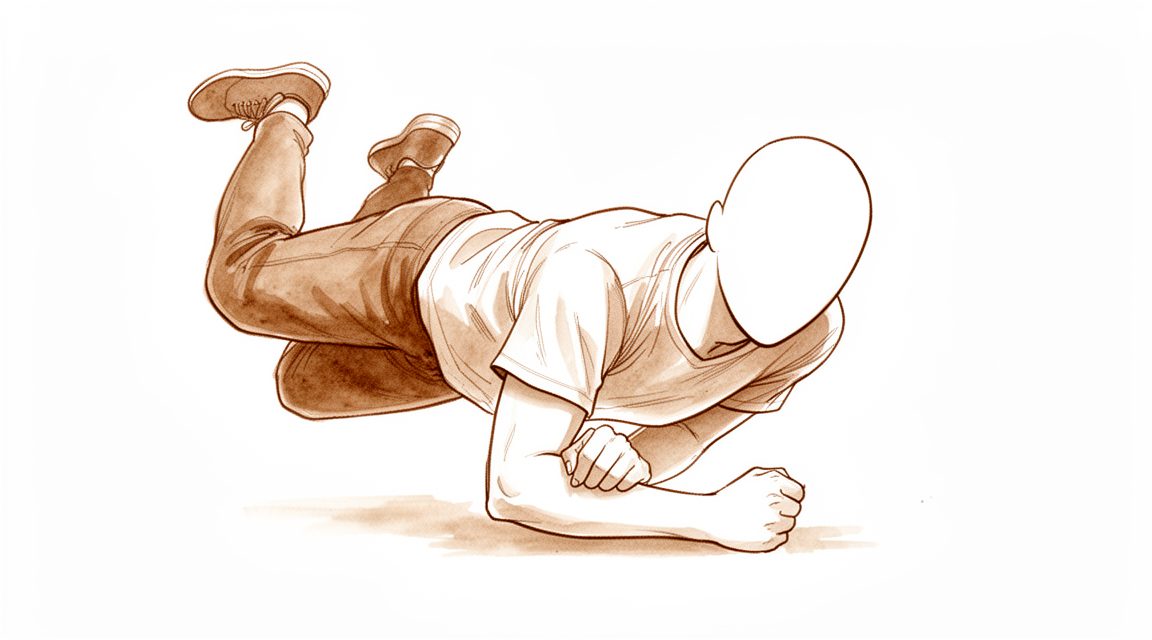
Bạn sẽ cảm thấy đau nhói ở phía sau khuỷu tay. Đây là nơi xương nhọn của xương cánh tay trên gặp xương cẳng tay. Cơn đau thường bùng phát khi bạn cố gắng duỗi thẳng cánh tay chống lại lực cản. Bạn có thể nhận thấy sưng và bầm tím xung quanh khớp. Bạn có thể gặp khó khăn khi nâng bất kỳ vật gì nặng hơn một tách cà phê. Những công việc đơn giản như với tay ra sau lưng để cài áo ngực hoặc nhét áo vào quần trở nên rất khó khăn.
Khuỷu tay của bạn có thể cảm thấy cứng, đặc biệt là khi bạn vừa thức dậy vào buổi sáng. Di chuyển khớp qua toàn bộ phạm vi chuyển động có thể gây đau. Bạn có thể thấy khó ngủ ở bên bị thương. Cơn đau cũng có thể trở nên trầm trọng hơn sau khi bạn đã sử dụng cánh tay cho các hoạt động hàng ngày. Nghỉ ngơi với cánh tay được nâng đỡ thường mang lại một số sự giảm đau. Tuy nhiên, giữ cánh tay hoàn toàn bất động trong quá lâu có thể làm tình trạng cứng khớp tồi tệ hơn.
Bởi vì vết gãy liên quan đến bề mặt khớp, bạn có thể cảm thấy có cảm giác mài mòn hoặc nghe thấy tiếng lách cách khi bạn di chuyển. Điều này là do các xương cọ xát vào nhau nơi lẽ ra chúng phải trượt mượt mà. Ở người lớn tuổi, viêm xương khớp do hao mòn này ảnh hưởng đến khoảng 19% bệnh nhân trong vòng vài năm. Bạn có thể trải qua những cơn đau âm ỉ thỉnh thoảng kéo dài lâu sau khi vết thương ban đầu đã lành. Các triệu chứng này có thể đến và đi, thường được kích hoạt bởi những thay đổi về thời tiết hoặc sử dụng nhiều.
Nếu bạn trên 70 tuổi, bác sĩ phẫu thuật của bạn có thể thảo luận về các lựa chọn không phẫu thuật. Những phương pháp tiếp cận này tập trung vào việc kiểm soát cơn đau và duy trì chức năng thay vì sự căn chỉnh xương hoàn hảo. Nhiều bệnh nhân báo cáo sự hài lòng cao với phương pháp này, ngay cả khi xương không lành trong một vị trí hoàn hảo. Mục tiêu là giúp bạn thực hiện các nhiệm vụ hàng ngày với sự khó chịu tối thiểu. Bác sĩ phẫu thuật của bạn sẽ điều chỉnh kế hoạch dựa trên nhu cầu cụ thể và mức độ hoạt động của bạn.
Những gì thực sự đang xảy ra
Mỏm khuỷu là phần xương nhô ra ở khuỷu tay, nơi bạn thường rests khuỷu tay lên bàn. Đây là một phần của xương trụ, một trong hai xương của cẳng tay. Khi xương này bị gãy, nó thường làm đứt gân ba đầu cánh tay (triceps), hoạt động như một sợi dây thừng chắc chắn gắn vào xương. Mối liên kết này cho phép bạn duỗi thẳng cánh tay chống lại trọng lực. Nếu vết gãy bị lệch, sợi dây này có thể kéo rời khỏi mảnh xương, khiến bạn khó hoặc không thể nâng tay hoặc giữ các vật thể lên.
Mục tiêu chính của bác sĩ phẫu thuật là khôi phục bề mặt trơn tru nơi xương cánh tay trên gặp xương cẳng tay. Bề mặt khớp này phải bằng phẳng để các xương có thể trượt qua nhau mà không bị ma sát. Nếu vết gãy không được cố định chắc chắn, khuỷu tay của bạn có thể bị cứng. Vận động sớm là yếu tố then chốt để ngăn ngừa tình trạng cứng khớp. Bác sĩ phẫu thuật sẽ chọn phương pháp cố định giữ cho xương ổn định đủ để bạn có thể vận động cánh tay sớm sau phẫu thuật, đồng thời vẫn bảo vệ xương đang lành.
Đôi khi, vết gãy quá phức tạp hoặc chất lượng xương quá kém để sửa chữa bằng phương pháp tiêu chuẩn. Trong những trường hợp này, bác sĩ phẫu thuật có thể loại bỏ hoàn toàn mảnh xương gãy và tái gắn gân ba đầu cánh tay trực tiếp vào xương cẳng tay. Phương pháp này tránh các biến chứng liên quan đến thiết bị cố định và thường dẫn đến chức năng tốt hơn với ít đau đớn hơn. Đối với bệnh nhân lớn tuổi có nhu cầu vận động thấp, điều trị không phẫu thuật cũng có thể là một lựa chọn an toàn và hiệu quả.
Ngay cả khi điều trị thành công, viêm xương khớp do hao mòn có thể phát triển trong khớp khuỷu theo thời gian. Dữ liệu cho thấy 19% bệnh nhân phát triển tình trạng này, với thời gian theo dõi trung vị là 41 tháng. Điều này có nghĩa là đối với một số người, lớp sụn bao phủ đầu xương bị mòn đi, có thể gây đau hoặc cứng khớp sau này. Tuy nhiên, hầu hết bệnh nhân đạt được chức năng và sự hài lòng tốt về lâu dài, bất kể họ có trải qua phẫu thuật hay quản lý bảo thủ.
Những gì chúng tôi có thể làm về vấn đề này
Đối với nhiều bệnh nhân, đặc biệt là người lớn tuổi hoặc những người có nhu cầu vận động thấp, điều trị không phẫu thuật là một lựa chọn an toàn và hiệu quả. Bác sĩ phẫu thuật của bạn có thể khuyên nghỉ ngơi, chườm đá và đeo nẹp để giữ khuỷu tay bất động trong khi xương lành lại. Phương pháp này tập trung vào sự thoải mái và cho phép quá trình lành thương tự nhiên mà không cần phẫu thuật. Các nghiên cứu cho thấy rằng các gãy xương rời lệch ở người cao tuổi thường mang lại kết quả ngắn hạn và dài hạn thỏa đáng với phương pháp này. Bạn có thể mong đợi duy trì phạm vi vận động chức năng và trải nghiệm ít đau đớn. Ngay cả khi xương không liền hoàn toàn (liền xương kém), nhiều bệnh nhân vẫn đạt được chức năng khuỷu tay hợp lý và hiếm khi yêu cầu phẫu thuật sau này. Đối với bệnh nhân trẻ tuổi hoặc những trường hợp có độ lệch đáng kể, phẫu thuật thường là tiêu chuẩn để khôi phục sự ổn định.
Kiểm soát đau là một phần quan trọng trong quá trình hồi phục của bạn. Bác sĩ phẫu thuật có thể kê đơn thuốc giảm đau hoặc thuốc chống viêm để giúp bạn duy trì sự thoải mái trong quá trình lành thương. Trong khi các loại tiêm như cortisone, axit hyaluronic hoặc PRP là phổ biến cho đau khớp, bằng chứng về gãy mỏm khuỷu chủ yếu tập trung vào việc lành thương cấu trúc hơn là các loại tiêm cụ thể này. Mục tiêu là kiểm soát đau để bạn có thể bắt đầu vận động nhẹ nhàng ngay khi an toàn. Vận động sớm là rất quan trọng để ngăn ngừa khớp khuỷu tay bị cứng. Nếu phẫu thuật được thực hiện, cố định phải đủ chắc chắn để cho phép vận động sớm này. Hầu hết bệnh nhân giữ lại các thiết bị cấy ghép sau phẫu thuật, và chỉ có 3% gặp phải tình trạng di chuyển thiết bị cấy ghép. Các yếu tố kỹ thuật của thiết bị cấy ghép ít quan trọng hơn các yếu tố cá nhân trong việc quyết định có cần phẫu thuật lần hai để tháo bỏ hay không.
Phẫu thuật được xem xét khi chăm sóc bảo thủ không phù hợp hoặc đã thất bại. Điều này thường gặp ở các gãy xương lệch ở bệnh nhân trẻ, năng động hoặc những người có mô hình chấn thương phức tạp. Phẫu thuật nhằm mục đích giữ các mảnh xương tại chỗ để chúng có thể lành lại đúng cách. Bác sĩ phẫu thuật của bạn sẽ chọn phương pháp phù hợp nhất với loại gãy xương cụ thể và tình trạng sức khỏe của bạn. Dù sử dụng tấm, chỉ hoặc neo, mục tiêu là khôi phục cơ chế cơ tam đầu và chức năng khuỷu tay. Trong một số trường hợp có tổn thương nghiêm trọng, việc loại bỏ mảnh xương gãy và sửa chữa cơ có thể được ưu tiên để giảm biến chứng. Nếu bạn có các chấn thương khác, nguy cơ hạn chế vận động cao hơn, vì vậy bác sĩ phẫu thuật của bạn sẽ thảo luận điều này một cách cởi mở. Thời điểm phẫu thuật không làm tăng đáng kể các biến chứng sớm, vì vậy bạn có thể tiến hành khi bạn sẵn sàng.
Những điều cần biết
Tiên lượng của bạn phụ thuộc phần lớn vào độ tuổi, mức độ hoạt động và việc bạn lựa chọn phẫu thuật hay nghỉ ngơi. Đối với người cao tuổi hoặc những người có nhu cầu vận động thấp, điều trị không phẫu thuật thường mang lại kết quả khả quan cả trong ngắn hạn và dài hạn. Bạn có thể mong đợi chức năng khuỷu tay ở mức độ chấp nhận được ngay cả khi xương không liền hoàn toàn. Phần lớn bệnh nhân trong nhóm này không yêu cầu phẫu thuật thêm.
Nếu bạn còn trẻ và năng động, phẫu thuật thường được khuyến nghị để khôi phục sức mạnh và tầm vận động. Cố định phẫu thuật thường mang lại kết quả chức năng xuất sắc. Bạn có thể mong đợi giữ lại các thiết bị cấy ghép; chỉ có 3% bệnh nhân gặp phải tình trạng di chuyển thiết bị cấy ghép. Các yếu tố kỹ thuật ít quan trọng hơn lựa chọn cá nhân khi quyết định có muốn tháo bỏ thiết bị cấy ghép sau này hay không.
Quá trình hồi phục diễn ra từ từ. Bạn có thể nhận thấy cứng khớp hoặc đau nhức khi khớp đang lành. Khoảng 19% bệnh nhân phát triển viêm xương khớp sau chấn thương, một tình trạng do hao mòn, tại thời điểm theo dõi trung bình là 41 tháng. Điều này có nghĩa là bạn có thể cảm thấy khó chịu thỉnh thoảng khi thời tiết thay đổi hoặc khi sử dụng nhiều. Mặc dù có những thay đổi này, chức năng tốt trong dài hạn vẫn có thể đạt được.
Cần lưu ý rằng gãy mỏm khuỷu ở người cao tuổi có tỷ lệ tử vong sau một năm cao hơn mức dự kiến. Nguy cơ này rất quan trọng cần thảo luận với bác sĩ phẫu thuật của bạn khi cân nhắc các lựa chọn điều trị. Nếu bạn chọn điều trị không phẫu thuật cho một ca gãy xương có di lệch, bạn có thể phải đối mặt với tình trạng xương không liền, nhưng nhiều bệnh nhân vẫn hài lòng với kết quả của mình.
Thời điểm phẫu thuật không làm tăng đáng kể biến chứng sớm hoặc nhu cầu phẫu thuật lại. Bạn không cần phải vội vàng vào phòng mổ vì lý do an toàn, mặc dù cố định sớm hơn có thể giúp cải thiện sự thoải mái. Nhìn chung, phần lớn bệnh nhân duy trì thiết bị cấy ghép và đạt được chức năng tốt, bất kể được điều trị bằng phẫu thuật hay nghỉ ngơi cẩn thận.
Khi nào cần gặp bác sĩ
Hãy gặp bác sĩ đa khoa nếu bạn có cơn đau dai dẳng không cải thiện khi nghỉ ngơi. Hãy yêu cầu đánh giá bởi bác sĩ chuyên khoa nếu bạn cảm thấy yếu hoặc mất vững ở khuỷu tay. Hãy chú ý hiện tượng khóa khớp hoặc khớp bị sập. Hãy tìm kiếm chăm sóc y tế nếu các triệu chứng ảnh hưởng đến giấc ngủ hoặc công việc của bạn. Hãy liên hệ với bác sĩ phẫu thuật nếu bạn nhận thấy tình trạng của mình xấu đi đột ngột. Hãy lưu ý rằng tỷ lệ mắc các loại gãy xương này đã tăng 29% trong suốt thời gian nghiên cứu 20 năm. Viêm xương khớp do hao mòn sau chấn thương xảy ra ở 19% các trường hợp với thời gian theo dõi trung bình là 41 tháng. Bệnh nhân cao tuổi có tỷ lệ tử vong trong vòng một năm cao hơn mức dự kiến. Việc đánh giá sớm giúp quản lý các nguy cơ này một cách hiệu quả.
Evidence & references
Overview
- Non-operative treatment of olecranon fractures in patients aged ≥75 years provided excellent functional results at 6 months without associated complications [1].
- Nonoperative management of isolated displaced olecranon fractures in older, lower-demand patients yields satisfactory short-term and long-term outcomes [4].
- Evidence offers valuable data for developing personalized treatment plans for olecranon fractures in patients over 75, though it does not definitively settle the debate on operative versus non-operative management [5].
- The SOFIE trial is a study protocol aiming to test for superiority of operative versus non-operative treatment for displaced olecranon fractures in the elderly by comparing pain and function up to one year after injury, but it does not report results or conclusions [18].
- Low-profile double-plate osteosynthesis is a safe and effective alternative treatment for olecranon fractures with excellent subjective and objective clinical outcome measures [2].
- Both Kirschner wire tension band combined with anatomical locking plate and other operative procedures effectively treat Mayo type II olecranon fractures [6].
- Plating of the olecranon leads to predictable union, although the most common complication was lack of full extension in 39% of patients [8].
- A majority of olecranon fractures heal uneventfully with good/excellent results, with a small loss of motion to be expected [9].
- The timing of fixation for displaced olecranon fractures does not significantly increase the rate of early complications or reoperation [13].
- Tension band wiring (TBW) remains an effective treatment for appropriately selected olecranon fractures and outperformed plate osteosynthesis in the studied cohort [25].
- Suture fixation is the mainstay of treatment for all simple olecranon fractures, with no re-operations or wound complications observed in the series [28].
- Surgical treatment of olecranon fractures is associated with a high rate of complications, and patients undergoing revisions beyond implant removal had poorer functional outcomes [58].
- No significant differences in functional outcomes or secondary operations were found with respect to fracture type, gender, or surgical method in the context of surgical treatment complications [58].
Anatomy & Pathophysiology
- Concomitant injuries in olecranon fractures are associated with a high risk of limited elbow motion [23].
- Understanding relevant elbow anatomy and factors associated with stability allows for systematic treatment algorithms that ensure sufficient stability for early motion, leading to improved outcomes [29].
- The modified rotational formula (MRCF) provides stable and accurate measurements of rotational displacement despite varying elbow rotations, addressing limitations of the previous method (PRCF) [30].
- An anatomic model of terrible triad injury can be created by exerting axial compression on an elbow in 15° flexion and maximal pronation at speeds of 100 and 10 mm/min [33].
- Individuals with elbow degenerative changes have no inferior subjective elbow function compared to those with normal radiographs, except in cases with joint space reduction [34].
- Elbow range of motion and functional use are maintained in the midterm compared to short-term studies following hemiarthroplasty for distal humeral fractures [35].
- The "spin move" is a maneuver that improves exposure of the coronoid process regardless of the degree of elbow instability [36].
- Restoration of joint motion in posttraumatic stiff elbows is a difficult, time-consuming, and costly challenge [37].
- A portion of the anterior lateral trochlear ridge (aLTR) is covered with articular cartilage but is non-articulating throughout the normal elbow range of motion [41].
- Evaluation and management of elbow injuries in young athletes requires knowledge of immature developing anatomy, injury pathophysiology, and established treatment algorithms [44].
- Reconstruction of the anterior capsule and ligamentous structures is important for providing stability to the elbow joint in complex fracture-dislocations [45].
- Good elbow function can be restored in most cases of comminuted intra-articular distal humeral fractures with minor impairments that do not worsen quality of life [46].
- Use of a standard surgical protocol for elbow dislocations with radial head and coronoid fractures restores sufficient stability to allow early postoperative motion, enhancing functional outcomes [47].
- Disruptions in forearm structures may lead to forearm instability with consequences at the remaining structures [48].
- Open fracture-dislocation (OFD) patterns have the worst functional outcomes among complex elbow injury patterns [51].
- Proper treatment of coronoid fractures requires an understanding of the bony and soft tissue anatomy of the elbow and various injury mechanisms [52].
- While range of motion is typically preserved after reoperation for intra-articular proximal ulna fractures, 35% of patients experience subsequent complications [53].
- Orthogonal plate configuration, olecranon osteotomy, and longer operative time are associated with increased odds of dysfunctional elbow stiffness following operative fixation of distal humerus fractures [54].
- Specific patterns of traumatic elbow instability have correspondingly specific coronoid fracture patterns [55].
Classification
- The Mayo classification was designed to simplify categorization of olecranon fractures [31].
- The Mayo classification does not achieve its goal of simplification due to poor reproducibility [31].
- Quantitative 3-dimensional computed tomography analysis clarified the fracture morphology of Mayo type I, II, and III fractures [57].
Clinical Presentation
- Olecranon fractures are commonly seen in orthopedic practice [56].
- Isolated olecranon fractures occur after low-energy trauma, especially in older women (> 65 years) [16].
- Articular impaction is a common feature of geriatric olecranon fractures [19].
- Patients with olecranon fractures have essentially similar demographic characteristics compared to patients with distal radius fractures [7].
- The incidence of olecranon fractures increased by 29% over the 20-year study period (1999–2018) in Denmark [3].
- Olecranon fractures in the elderly have higher than expected 1 year mortality rates [14].
- More precise studies are needed to properly quantify the specific incidence of various subtypes of forearm and olecranon fractures and associated risk factors [12].
Investigations
- The incidence of olecranon fractures increased by 29% over a 20-year study period in Denmark [3].
- More precise studies are needed to properly quantify the specific incidence of various subtypes of forearm and olecranon fractures and associated risk factors [12].
- Isolated fractures of the olecranon occur after low-energy trauma, especially in older women (> 65 years) [16].
- Patients with olecranon fractures have essentially similar demographic characteristics compared to patients with distal radius fractures [7].
- Olecranon fractures in the elderly have higher than expected 1-year mortality rates [14].
- Articular impaction is a common feature of geriatric olecranon fractures [19].
Treatment
Non-Operative Management
- Non-operative treatment of olecranon fractures in patients aged ≥75 years provided excellent functional results at 6 months, without associated complications [1].
- Nonoperative management of isolated displaced olecranon fractures in older, lower-demand patients yields satisfactory short-term and long-term outcomes [4].
- Primary non-operative management is supported for isolated displaced fractures of the olecranon in the elderly [10].
- Non-operative treatment of Mayo Type II olecranon fractures may be successful, extending the age range for which such treatment of displaced olecranon fractures can be considered [21].
- Patients who present with a non-union after a displaced olecranon fracture managed non-operatively have reasonable elbow function and uncommonly request operative treatment [27].
- Nonoperative treatment as a reasonable option is supported for displaced stable olecranon fractures in elderly patients [40].
- Displaced olecranon fractures in patients older than 70 years may be effectively managed with nonoperative measures to produce high satisfaction and functional range of motion [49].
- Aggregate data support the non-operative treatment of isolated undisplaced olecranon fractures with good results [42].
- The literature on the treatment of olecranon fractures in elderly patients is limited [15].
- More precise studies are needed in order to properly quantify the specific incidence of various subtypes of forearm and olecranon fractures and associated risk factors [12].
- While data offer valuable information for personalized treatment plans, it is not definitively settled whether olecranon fractures should be managed nonoperatively in patients over 75 [5].
- The SOFIE study is a protocol testing for superiority of operative versus non-operative treatment and does not report results or conclusions [18].
- Surgical management remains the standard of care for displaced olecranon fractures until more convincing evidence supports nonsurgical treatment [39].
Operative Management
- Low-profile double-plate osteosynthesis is a safe and effective alternative treatment of olecranon fractures with excellent subjective and objective clinical outcome measures [2].
- Both Kirschner wire tension band combined with anatomical locking plate and other operative procedures effectively treat Mayo type II olecranon fractures [6].
- Suture anchor fixation of displaced olecranon fractures resulted in excellent midterm functional outcomes [11].
- In cases with concomitant injuries, the risk of limited elbow motion is high following open reduction and plate osteosynthesis [23].
- Double tension band wiring (DTBW) produced good clinical and radiological outcomes and could be an effective option for the treatment of olecranon fractures by providing additional stability through a second tension band wire [24].
- Tension band wiring (TBW) remains an effective treatment for appropriately selected olecranon fractures and outperformed plate osteosynthesis in the studied cohort [25].
- Both locking-plate osteosynthesis and intramedullary nailing could be appropriate surgical techniques for fixation of selected olecranon fractures and osteotomies [26].
- Plate has better efficacy and safety than tension band wire for Mayo II olecranon fractures [32].
- The Nickel-Titanium olecranon memory connector (OMC) could be an effective alternative to treat olecranon fractures [38].
- Plate fixation of complex olecranon fractures is an effective, reliable method of treatment with low risk of non-union [50].
- No one technique is suitable for the management of all olecranon fractures [17].
Complications
- Non-operative treatment of olecranon fracture in patients aged ≥75 years provided excellent functional results at 6 months without associated complications [1].
- Low-profile double-plate osteosynthesis is a safe and effective alternative treatment of olecranon fractures with excellent subjective and objective clinical outcome measures [2].
- Nonoperative management of isolated displaced olecranon fractures in older, lower-demand patients yields satisfactory short-term and long-term outcomes [4].
- Plating of the olecranon leads to predictable union, though the most common complication was lack of full extension in 39% of patients [8].
- A majority of olecranon fractures heal uneventfully with good/excellent results, with a small loss of motion to be expected [9].
- Suture anchor fixation of displaced olecranon fractures resulted in excellent midterm functional outcomes [11].
- The timing of fixation of displaced olecranon fractures does not significantly increase the rate of early complications or reoperation [13].
- Olecranon fractures in the elderly have higher than expected 1 year mortality rates [14].
- The median incidence of post-traumatic osteoarthritis following isolated olecranon fractures is 19% at a median follow-up of 41 months [20].
- Suture fixation for simple olecranon fractures resulted in no re-operations or wound complications in the studied series [28].
- Patients who have operative fixation of a fracture of the olecranon can be counseled that most patients keep their implants [61].
- Only 3% of patients experience implant migration after operative fixation of a fracture of the olecranon [61].
- Technical factors such as the type or configuration of an implant seem less important than personal factors in determining who requests a second surgery for implant removal after olecranon fracture fixation [61].
Recovery
- Non-operative treatment of olecranon fractures in patients aged ≥75 years provided excellent functional results at 6 months, without associated complications [1].
- Nonoperative management of isolated displaced olecranon fractures in older, lower-demand patients yields satisfactory short-term and long-term outcomes [4].
- Non-operative treatment of Mayo Type II olecranon fractures may be successful, extending the age range for which such treatment of displaced olecranon fractures can be considered [21].
- The literature on the treatment of olecranon fractures in elderly patients is limited [15].
- Low-profile double-plate osteosynthesis is a safe and effective alternative treatment of olecranon fractures with excellent subjective and objective clinical outcome measures [2].
- Plating of the olecranon leads to predictable union, though the most common complication was lack of full extension in 39% of patients [8].
- Suture anchor fixation of displaced olecranon fractures resulted in excellent midterm functional outcomes [11].
- Good functional long-term results are to be expected in patients with complex olecranon fractures treated with open reduction and internal fixation, despite arthritic changes in the elbow joint [43].
- A majority of olecranon fractures heal uneventfully with good/excellent results with a small loss of motion to be expected [9].
- Among active patients with a simple isolated, displaced fracture of the olecranon, no difference was found between tension-band wire (TBW) and plate fixation in the patient-reported outcome at 1 year following surgery [63].
- The timing of fixation of displaced olecranon fractures does not significantly increase the rate of early complications or reoperation [13].
- Patients with olecranon fractures have essentially similar demographic characteristics compared to patients with distal radius fractures [7].
- Olecranon fractures in the elderly have higher than expected 1 year mortality rates [14].
- The incidence of post-traumatic osteoarthritis following isolated olecranon fractures has a median incidence of 19% at a median follow-up of 41 months [20].
Key Evidence
- [L4] Non-operative treatment of olecranon fracture in patients aged ≥75 years provided excellent functional results at 6 months, without associated complications. [1] (10.1016/j.otsr.2017.10.015)
- [L3] Low-profile double-plate osteosynthesis is a safe and effective alternative treatment of olecranon fractures with excellent subjective and objective clinical outcome measures. [2] (10.1016/j.otsr.2019.08.019)
- [L3] The incidence of olecranon fractures increased by 29% over the 20-year study period. [3] (10.1186/s13018-025-05970-2)
- [L4] We found satisfactory short-term and long-term outcomes following the nonoperative management of isolated displaced olecranon fractures in older, lower-demand patients. [4] (10.2106/jbjs.l.01137)
- [L2] While they did not definitively settle the debate about whether we should manage olecranon fractures nonoperatively in patients over 75, they did offer valuable data that surgeons and patients can use to develop personalized treatment plans tailored to each patient's needs. [5] (10.2106/jbjs.24.01097)
- [L3] Both operative procedures effectively treat Mayo type II olecranon fractures. [6] (10.1186/s12891-025-08843-1)
- [L3] Patients with olecranon fractures have essentially similar demographic characteristics compared to patients with distal radius fractures. [7] (10.1177/17585732221124301)
- [L3] Plating of the olecranon leads to predictable union, though the most common complication was lack of full extension in 39% of patients. [8] (10.1016/j.injury.2016.04.015)
- [L4] A majority of olecranon fractures heal uneventfully with good/excellent results with a small loss of motion to be expected. [9] (10.1016/j.hcl.2015.07.003)
- [L1] These data further support the role of primary non-operative management of isolated displaced fractures of the olecranon in the elderly. [10] (10.1302/0301-620x.99b7.bjj-2016-1112.r2)
- [L4] Suture anchor fixation of displaced olecranon fractures resulted in excellent midterm functional outcomes. [11] (10.5397/cise.2023.00528)
- [L3] More precise studies are needed in order to properly quantify the specific incidence of various subtypes of forearm and olecranon fractures and associated risk factors. [12] (10.1186/s12891-023-07162-7)
- [L3] The timing of fixation of displaced olecranon fractures does not significantly increase the rate of early complications or reoperation. [13] (10.1016/j.jhsg.2023.09.002)
- [L3] Olecranon fractures in the elderly have higher than expected 1 year mortality rates. [14] (10.1177/1758573221994860)
- [L4] The literature on the treatment of olecranon fractures in elderly patients is limited. [15] (10.1007/s11678-018-0488-7)
- [L4] Isolated fractures of the olecranon occur after a low-energy trauma, especially in older women (> 65 years). [16] (10.1007/s00068-021-01765-2)
- [Paper] No one technique is suitable for the management of all olecranon fractures. [17] (10.1016/j.injury.2008.12.013)
- [L2] This document is a study protocol and does not report results or conclusions; the study aims to test for superiority of operative treatment versus non-operative treatment for displaced olecranon fractures in the elderly by comparing pain and function up to one year after injury. [18] (10.1186/s12891-015-0789-6)
- [L4] Articular impaction is a common feature of geriatric olecranon fractures. [19] (10.5435/jaaos-d-20-01293)
- [L4] This review identified a median OA incidence of 19% at a median follow-up of 41 months following isolated olecranon fractures. [20] (10.1016/j.jse.2026.02.024)
- [L4] Non-operative treatment of Mayo Type II olecranon fractures may be successful, extending the age range for which such treatment of displaced olecranon fractures can be considered. [21] (10.1177/1758573217711889)
- [L4] In cases with concomitant injuries, the risk of limited elbow motion is high. [23] (10.1016/j.jse.2010.11.023)
- [L4] DTBW produced good clinical and radiological outcomes and could be an effective option for the treatment of olecranon fractures by providing additional stability through a second TBW. [24] (10.1016/j.jhsa.2014.09.020)
- [L4] TBW remains an effective treatment for appropriately selected olecranon fractures and in this cohort outperformed plate osteosynthesis. [25] (10.1007/s00590-015-1724-0)
- [L3] Both implant types could be appropriate surgical techniques for fixation of selected olecranon fractures and osteotomies. [26] (10.1007/s00264-013-1854-0)
- [L4] Patients who present with a non-union after a displaced olecranon fracture managed non-operatively have reasonable elbow function and uncommonly request operative treatment. [27] (10.1111/j.1758-5740.2012.00194.x)
- [L4] Suture fixation is now the mainstay of treatment for all simple olecranon fractures, with no re-operations or wound complications observed in this series. [28] (10.1177/1758573216687305)
- [L5] Despite the complexities of this injury, an understanding of the relevant anatomy and the factors associated with elbow stability allows the application of a systematic algorithm for treatment that can help ensure sufficient elbow stability to allow early motion, thereby leading to improved outcomes in most patients. [29] (10.5435/00124635-200903000-00003)
- [L5] MRCF effectively addresses the limitations of PRCF and provides stable, accurate measurements of rotational displacement even with varying elbow rotations. [30] (10.1186/s12891-024-08240-0)
- [L5] The Mayo classification was designed to simplify categorization of olecranon fractures but does not achieve this goal due to poor reproducibility. [31] (10.1097/corr.0000000000000614)
- [L1] Plate has better efficacy and safety for Mayo II olecranon fractures. [32] (10.1186/s13018-022-03262-7)
- [L5] The study successfully created and validated an anatomic model of terrible triad of the elbow by exerting axial compression on an elbow in 15° flexion and maximal pronation at speeds of 100 and 10 mm/min. [33] (10.1186/s13018-024-05069-0)
- [L3] Individuals with elbow degenerative changes had no inferior subjective elbow function compared to those with normal radiographs, except for those with joint space reduction. [34] (10.1007/s00402-020-03453-z)
- [L4] The data suggest that elbow range of motion and functional use are maintained from comparison with short-term studies. [35] (10.1016/j.jse.2016.09.057)
- [L5] The spin move is a simple maneuver that can improve exposure of the coronoid process regardless of the degree of elbow instability. [36] (10.1016/j.jse.2022.11.020)
- [L4] Restoration of joint motion in the posttraumatic stiff elbow can be a difficult, time-consuming, and costly challenge. [37] (10.1016/j.jhsa.2007.09.015)
- [L2] The study showed that OMC could be an effective alternative to treat olecranon fractures. [38] (10.1007/s00264-013-1878-5)
- [Letter] The authors of the original review acknowledge that nonsurgical management was limited to nondisplaced fractures due to editorial constraints but maintain that surgical management remains the standard of care for displaced olecranon fractures until more convincing evidence supports nonsurgical treatment. [39] (10.1016/j.jhsa.2013.04.013)
- [L1] This supports nonoperative treatment as a reasonable option for displaced stable olecranon fractures in elderly patients. [40] (10.2106/jbjs.24.00655)
- [L5] Our results suggest that there is a portion of the aLTR that, despite being covered with articular cartilage, is non-articulating throughout normal elbow range of motion. [41] (10.2106/jbjs.18.01270)
- [L4] Aggregate data support the non-operative treatment of isolated undisplaced olecranon fractures with good results, and support the operative treatment of fractures displaced ≥4 mm. [42] (10.1302/2058-5241.5.190082)
- [Abstract] Good functional long-term results are to be expected in patients with complex olecranon fractures treated with open reduction and internal fixation, despite arthritic changes in the elbow joint. [43] (10.1016/j.jse.2007.02.092)
- [L5] Evaluation and management of elbow injuries in young athletes requires knowledge of the immature developing anatomy, injury pathophysiology, and established treatment algorithms for each diagnosis. [44] (10.1016/j.csm.2010.06.010)
- [L4] It is important to reconstruct the anterior capsule and ligamentous structures for providing stability to the elbow joint. [45] (10.1007/s00402-006-0198-2)
- [L4] Good elbow function can be restored in most cases with minor impairments that do not worsen quality of life. [46] (10.1016/j.jse.2014.01.017)
- [L4] Use of the surgical protocol restored sufficient elbow stability to allow early motion postoperatively, enhancing the functional outcome. [47] (10.2106/jbjs.d.02933)
- [L5] Disruptions in any of these structures may lead to forearm instability with consequences at the remaining structures. [48] (10.1016/j.jhsa.2016.10.017)
- [L4] Displaced olecranon fractures in patients older than 70 years may be effectively managed with nonoperative measures to produce high satisfaction and functional range of motion. [49] (10.1177/1558944720944261)
- [L4] Plate fixation of complex olecranon fracture is an effective, reliable method of treatment with low risk of non-union. [50] (10.1016/j.ijscr.2017.10.052)
- [L3] OFD has the worst functional outcomes among complex elbow injury patterns. [51] (10.1016/j.jse.2024.06.004)
- [L5] Proper treatment of coronoid fractures requires an understanding of the bony and soft tissue anatomy of the elbow and the various injury mechanisms that occur. [52] (10.1016/j.hcl.2004.07.004)
- [L4] While ROM is typically preserved after reoperation and improved when the indication for reoperation is elbow stiffness, a significant proportion of patients (35%) experience subsequent complications. [53] (10.1016/j.jseint.2024.12.017)
- [L3] Orthogonal plate configuration, olecranon osteotomy, and longer operative time were associated with increased odds of dysfunctional elbow stiffness. [54] (10.1016/j.jse.2024.06.010)
- [L4] Specific patterns of traumatic elbow instability have correspondingly specific coronoid fracture patterns. [55] (10.1016/j.jhsa.2014.06.123)
- [L4] Olecranon fractures are commonly seen in orthopedic practice and have good to excellent outcomes with adherence to a treatment algorithm based on displacement, comminution, and joint stability. [56] (10.1016/j.ocl.2008.01.002)
- [L4] Quantitative analysis of olecranon fractures further clarified fracture morphology of Mayo type I, II, and III fractures. [57] (10.1016/j.jse.2015.10.002)
- [L4] Surgical treatment of olecranon fractures is associated with a high rate of complications, and patients undergoing revisions beyond implant removal had poorer functional outcomes; however, no significant differences in functional outcomes or secondary operations were found with respect to fracture type, gender, or surgical method. [58] (10.1016/j.xrrt.2025.08.004)
- [L3] Patients who have operative fixation of a fracture of the olecranon can be counseled that most patients keep their implants, that only 3% experience implant migration, and that technical factors such as the type or configuration of an implant seem less important than personal factors in determining who requests a second surgery for implant removal. [61] (10.1007/s11999-015-4488-2)
- [L1] Among active patients with a simple isolated, displaced fracture of the olecranon, no difference was found between TBW and plate fixation in the patient-reported outcome at 1 year following surgery. [63] (10.2106/jbjs.16.00773)
References
[1] Results of non-operative treatment of olecranon fracture in over 75-year-olds. Orthopaedics & Traumatology: Surgery & Research. 2018. DOI: 10.1016/j.otsr.2017.10.015 [2] Clinical evaluation of double-plate osteosynthesis for olecranon fractures: A retrospective case-control study. Orthopaedics & Traumatology: Surgery & Research. 2019. DOI: 10.1016/j.otsr.2019.08.019 [3] Epidemiology and Treatment of Olecranon Fractures: a nationwide register-based analysis of 27,880 cases in Denmark from 1999 to 2018. Journal of Orthopaedic Surgery and Research. 2025. DOI: 10.1186/s13018-025-05970-2 [4] Nonoperative Management of Displaced Olecranon Fractures in Low-Demand Elderly Patients. Journal of Bone and Joint Surgery. 2014. DOI: 10.2106/jbjs.l.01137 [5] Treatment of Displaced Olecranon Fractures in the Elderly: Should the Pendulum Swing?. Journal of Bone and Joint Surgery. 2025. DOI: 10.2106/jbjs.24.01097 [6] Efficacy evaluation of Kirschner wire tension band combined with anatomical locking plate in the treatment of Mayo type II olecranon fractures. BMC Musculoskeletal Disorders. 2025. DOI: 10.1186/s12891-025-08843-1 [7] Mortality and subsequent fractures of patients with olecranon fractures compared to other upper extremity osteoporotic fractures. Shoulder & Elbow. 2022. DOI: 10.1177/17585732221124301 [8] Outcomes after plating of olecranon fractures: A multicenter evaluation. Injury. 2016. DOI: 10.1016/j.injury.2016.04.015 [9] Olecranon Fractures. Hand Clinics. 2015. DOI: 10.1016/j.hcl.2015.07.003 [10] Prospective randomised trial of non-operativeversusoperative management of olecranon fractures in the elderly. The Bone & Joint Journal. 2017. DOI: 10.1302/0301-620x.99b7.bjj-2016-1112.r2 [11] Midterm outcomes of suture anchor fixation for displaced olecranon fractures. Clinics in Shoulder and Elbow. 2024. DOI: 10.5397/cise.2023.00528 [12] Trends and projection of forearm fractures including elbow fractures of the Olecranon in Sweden: an analysis of 363 968 fractures using public aggregated data. BMC Musculoskeletal Disorders. 2024. DOI: 10.1186/s12891-023-07162-7 [13] Timing of Olecranon Fracture Fixation Does Not Affect Early Complication or Reoperation Rates. Journal of Hand Surgery Global Online. 2024. DOI: 10.1016/j.jhsg.2023.09.002 [14] Complications and mortality associated with olecranon fractures in the elderly: a retrospective cohort comparison from a large level one trauma centre. Shoulder & Elbow. 2021. DOI: 10.1177/1758573221994860 [15] Nonoperative treatment of olecranon fractures in the elderly—a systematic review. Obere Extremität. 2018. DOI: 10.1007/s11678-018-0488-7 [16] Epidemiology, classification and treatment of olecranon fractures in adults: an observational study on 2462 fractures from the Swedish Fracture Register. European Journal of Trauma and Emergency Surgery. 2021. DOI: 10.1007/s00068-021-01765-2 [17] Olecranon fractures. Injury. 2009. DOI: 10.1016/j.injury.2008.12.013 [18] SOFIE: Surgery for Olecranon Fractures in the Elderly: a randomised controlled trial of operative versus non-operative treatment. BMC Musculoskeletal Disorders. 2015. DOI: 10.1186/s12891-015-0789-6 [19] Incidence and Management of Articular Impaction in Geriatric Olecranon Fractures. Journal of the American Academy of Orthopaedic Surgeons. 2021. DOI: 10.5435/jaaos-d-20-01293 [20] Incidence of Post-traumatic Osteoarthritis in Olecranon Fractures and the Role of Instability and Comminution in its Development: A Systematic Review. Journal of Shoulder and Elbow Surgery. 2026. DOI: 10.1016/j.jse.2026.02.024 [21] Pilot report: non-operative treatment of Mayo Type II olecranon fractures in any-age adult patient. Shoulder & Elbow. 2017. DOI: 10.1177/1758573217711889 [23] Results of open reduction and plate osteosynthesis in comminuted fracture of the olecranon. Journal of Shoulder and Elbow Surgery. 2011. DOI: 10.1016/j.jse.2010.11.023 [24] Double Tension Band Wiring for Treatment of Olecranon Fractures. The Journal of Hand Surgery. 2014. DOI: 10.1016/j.jhsa.2014.09.020 [25] Outcome after olecranon fracture repair: Does construct type matter?. European Journal of Orthopaedic Surgery & Traumatology. 2015. DOI: 10.1007/s00590-015-1724-0 [26] Locking-plate osteosynthesis versus intramedullary nailing for fixation of olecranon fractures: a biomechanical study. International Orthopaedics. 2013. DOI: 10.1007/s00264-013-1854-0 [27] Non-union of Non-operatively Treated Displaced Olecranon Fractures. Shoulder & Elbow. 2012. DOI: 10.1111/j.1758-5740.2012.00194.x [28] Tension band suture fixation for olecranon fractures. Shoulder & Elbow. 2017. DOI: 10.1177/1758573216687305 [29] Terrible Triad Injury of the Elbow: Current Concepts. Journal of the American Academy of Orthopaedic Surgeons. 2009. DOI: 10.5435/00124635-200903000-00003 [30] Elbow rotation affects the accuracy of rotational formulas: validation of a modified method. BMC Musculoskeletal Disorders. 2025. DOI: 10.1186/s12891-024-08240-0 [31] Classifications in Brief: Mayo Classification of Olecranon Fractures. Clinical Orthopaedics & Related Research. 2018. DOI: 10.1097/corr.0000000000000614 [32] Efficacy and safety of tension band wire versus plate for Mayo II olecranon fractures: a systematic review and meta-analysis. Journal of Orthopaedic Surgery and Research. 2022. DOI: 10.1186/s13018-022-03262-7 [33] Creation of a replicable anatomic model of terrible triad of the elbow. Journal of Orthopaedic Surgery and Research. 2024. DOI: 10.1186/s13018-024-05069-0 [34] Long-term outcomes after different types of Horne and Tanzer olecranon fractures. Archives of Orthopaedic and Trauma Surgery. 2020. DOI: 10.1007/s00402-020-03453-z [35] Hemiarthroplasty for the treatment of distal humeral fractures: midterm clinical results. Journal of Shoulder and Elbow Surgery. 2017. DOI: 10.1016/j.jse.2016.09.057 [36] The spin move to facilitate antegrade coronoid fixation in terrible triad injuries. Journal of Shoulder and Elbow Surgery. 2023. DOI: 10.1016/j.jse.2022.11.020 [37] The Posttraumatic Stiff Elbow: A Review of the Literature. The Journal of Hand Surgery. 2007. DOI: 10.1016/j.jhsa.2007.09.015 [38] Design and application of Nickel-Titanium olecranon memory connector in treatment of olecranon fractures: a prospective randomized controlled trial. International Orthopaedics. 2013. DOI: 10.1007/s00264-013-1878-5 [39] Letter Regarding “Olecranon Fractures”. The Journal of Hand Surgery. 2013. DOI: 10.1016/j.jhsa.2013.04.013 [40] Surgery for Olecranon Fractures in the Elderly (SOFIE). Journal of Bone and Joint Surgery. 2025. DOI: 10.2106/jbjs.24.00655 [41] Lateral Trochlear Ridge. Journal of Bone and Joint Surgery. 2019. DOI: 10.2106/jbjs.18.01270 [42] Paediatric olecranon fractures: a systematic review. EFORT Open Reviews. 2020. DOI: 10.1302/2058-5241.5.190082 [43] Long Term Outcome Of Surgically Treated Complex Olecranon Fractures. Journal of Shoulder and Elbow Surgery. 2007. DOI: 10.1016/j.jse.2007.02.092 [44] Pediatric Sports Elbow Injuries. Clinics in Sports Medicine. 2010. DOI: 10.1016/j.csm.2010.06.010 [45] Reconstruction of the coronoid process with iliac crest bone graft in complex fracture-dislocation of elbow. Archives of Orthopaedic and Trauma Surgery. 2006. DOI: 10.1007/s00402-006-0198-2 [46] Results of parallel plate fixation of comminuted intra-articular distal humeral fractures. Journal of Shoulder and Elbow Surgery. 2014. DOI: 10.1016/j.jse.2014.01.017 [47] Standard Surgical Protocol to Treat Elbow Dislocations with Radial Head and Coronoid Fractures. Journal of Bone and Joint Surgery. 2005. DOI: 10.2106/jbjs.d.02933 [48] Forearm Instability: Anatomy, Biomechanics, and Treatment Options. The Journal of Hand Surgery. 2017. DOI: 10.1016/j.jhsa.2016.10.017 [49] Nonoperative Management of Olecranon Fractures in Elderly Patients: A Systematic Review. HAND. 2020. DOI: 10.1177/1558944720944261 [50] Management of type IIB and IIIB olecranon fractures. Case series. International Journal of Surgery Case Reports. 2017. DOI: 10.1016/j.ijscr.2017.10.052 [51] Three-dimensional quantitative study and functional outcome analysis of coronoid fracture in different elbow injury patterns. Journal of Shoulder and Elbow Surgery. 2025. DOI: 10.1016/j.jse.2024.06.004 [52] Fractures of the coronoid process. Hand Clinics. 2004. DOI: 10.1016/j.hcl.2004.07.004 [53] Surgical outcomes after reoperation of intra-articular proximal ulna fractures. JSES International. 2025. DOI: 10.1016/j.jseint.2024.12.017 [54] Risk factors for dysfunctional elbow stiffness following operative fixation of distal humerus fractures. Journal of Shoulder and Elbow Surgery. 2024. DOI: 10.1016/j.jse.2024.06.010 [55] Distribution of Coronoid Fracture Lines by Specific Patterns of Traumatic Elbow Instability. The Journal of Hand Surgery. 2014. DOI: 10.1016/j.jhsa.2014.06.123 [56] Olecranon Fractures. Orthopedic Clinics of North America. 2008. DOI: 10.1016/j.ocl.2008.01.002 [57] Quantitative 3-dimensional computed tomography analysis of olecranon fractures. Journal of Shoulder and Elbow Surgery. 2016. DOI: 10.1016/j.jse.2015.10.002 [58] Risk factors for complications and poor function after open reduction and fixation of olecranon fractures. JSES Reviews, Reports, and Techniques. 2025. DOI: 10.1016/j.xrrt.2025.08.004 [61] Factors Associated With Reoperation After Fixation of Displaced Olecranon Fractures. Clinical Orthopaedics & Related Research. 2016. DOI: 10.1007/s11999-015-4488-2 [63] Plate Versus Tension-Band Wire Fixation for Olecranon Fractures. Journal of Bone and Joint Surgery. 2017. DOI: 10.2106/jbjs.16.00773


