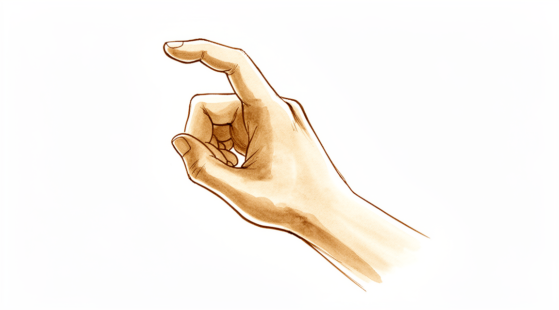
Ngón tay cò
Patients › Hand
Trigger finger causes painful catching or locking of a finger, often treated with splinting or steroid injection.
Những gì bạn đang cảm thấy
Bạn có thể nhận thấy đau ở lòng bàn tay, ngay tại vị trí ngón tay hoặc ngón cái gặp bàn tay. Vị trí này sẽ đau khi bạn ấn vào. Bạn cũng có thể cảm thấy một khối u nhỏ hoặc nút thắt tại đó. Đây thường là một vùng mô dày lên bao quanh gân của bạn, hoặc một sự sưng nhỏ trên chính gân đó.
Triệu chứng phổ biến nhất là hiện tượng kẹt hoặc khóa. Ngón tay hoặc ngón cái của bạn có thể bị kẹt ở tư thế cong. Bạn có thể nghe hoặc cảm thấy một tiếng bụp khi cố duỗi thẳng nó. Điều này thường xảy ra sau khi bạn nắm chặt thứ gì đó hoặc sử dụng bàn tay một cách mạnh mẽ. Vấn đề này thường trở nên nghiêm trọng hơn vào buổi sáng. Bạn có thể cảm thấy cứng khớp khi vừa thức dậy và sẽ linh hoạt hơn khi ngày trôi qua.
Trong các trường hợp nghiêm trọng hơn, ngón tay bị khóa chặt ở tư thế cong. Bạn có thể phải dùng tay kia để kéo nó thẳng ra. Nếu điều này xảy ra, ngón tay của bạn bị kẹt và bạn không thể tự di chuyển nó.
Những triệu chứng này có thể gây khó khăn cho các hoạt động hàng ngày. Các hành động đơn giản như với tay ra sau lưng để cài áo ngực, nhét áo vào quần, hoặc cầm vô lăng lái xe có thể trở nên đau đớn hoặc bất tiện. Bạn có thể gặp khó khăn khi ngủ nếu đặt bàn tay của mình ở tư thế cong lại.
Hội chứng ngón tay cò (trigger finger) phổ biến hơn ở phụ nữ và những người trên 45 tuổi. Nó ảnh hưởng đến ngón cái thường xuyên nhất, tiếp theo là ngón áp út, ngón giữa, ngón út và ngón trỏ. Nếu bạn mắc bệnh tiểu đường, bạn có nhiều khả năng phát triển tình trạng này. Việc bị hội chứng ngón tay cò ở nhiều ngón tay cùng lúc cũng có thể là dấu hiệu của bệnh tiểu đường.
Đôi khi, cơn đau ở khớp giữa của ngón tay bị nhầm lẫn với hội chứng ngón tay cò. Cơn đau này xuất phát từ tình trạng căng thẳng kéo dài lên khớp. Cơn đau có thể không biến mất hoàn toàn ngay cả sau khi bác sĩ phẫu thuật giải phóng vùng mô căng thẳng ở lòng bàn tay của bạn. Nếu bạn có các bệnh lý khác như viêm khớp dạng thấp, nhiều ngón tay có thể bị ảnh hưởng, thường là ngón giữa và ngón áp út.
Những gì thực sự đang xảy ra
Ngón tay bị kẹt là một vấn đề cơ học khiến ngón tay của bạn bị mắc kẹt. Điều này xảy ra do các gân uốn cong ngón tay cọ xát vào một dải mô chặt được gọi là dây chằng A1. Hãy tưởng tượng gân giống như một sợi dây và dây chằng giống như một ròng rọc. Khi sợi dây bị sưng hoặc ròng rọc bị thu hẹp, sợi dây sẽ bị mắc kẹt. Sự mắc kẹt này gây ra hiện tượng khóa hoặc kêu lách cách mà bạn cảm nhận được.
Sự sưng nề bắt nguồn từ những thay đổi trong chính mô. Dây chằng A1 dày lên và có thêm các mạch máu. Gân thường phát triển một cục nhỏ, hay còn gọi là nốt sần, khi đi qua dây chằng. Nốt sần này được cấu tạo từ các loại mô khác nhau không thể trượt mượt mà. Đây là lý do tại sao ngón tay của bạn có thể bị kẹt, đặc biệt là vào buổi sáng khi tình trạng cứng khớp cao hơn.
Bạn cũng có thể cảm thấy đau ở lòng bàn tay hoặc gần gốc ngón tay. Đôi khi cơn đau lan ra sau bàn tay. Tình trạng này ảnh hưởng đến hai đến ba phần trăm dân số. Nó phổ biến hơn ở phụ nữ và những người mắc bệnh tiểu đường. Thực tế, mười đến hai mươi phần trăm người mắc bệnh tiểu đường sẽ phát triển tình trạng này trong suốt cuộc đời. Nó cũng có thể xảy ra cùng với các vấn đề sức khỏe khác như rối loạn tuyến giáp hoặc viêm khớp dạng thấp.
Ngón cái, ngón áp út và ngón giữa thường bị ảnh hưởng nhiều nhất. Nếu bạn mắc bệnh Dupuytren, một tình trạng gây dày lên ở lòng bàn tay, nguy cơ của bạn sẽ cao hơn. Điều này là do mô dư thừa ở lòng bàn tay có thể gây kích ứng gân tại dây chằng. Vấn đề này hoàn toàn mang tính cơ học. Gân đơn giản là không thể trượt tự do qua đường đi bình thường của nó.
Những gì chúng tôi có thể làm về vấn đề này
Bạn có thể bắt đầu với các biện pháp tự chăm sóc đơn giản tại nhà. Kéo giãn nhẹ nhàng, nẹp vào ban đêm và luân phiên chườm nóng hoặc lạnh có thể giúp ích. Một loại nẹp đặc biệt chặn khớp ngón tay chính giúp đỡ 77% người dùng. Một loại nẹp cho khớp đầu ngón tay giúp đỡ khoảng một nửa số người dùng. Những phương pháp này mang lại sự giảm triệu chứng trong thời gian ngắn và cải thiện chức năng. Bạn có thể thử nẹp đơn độc như một bước đầu tiên có nguy cơ thấp. Nó mang lại sự giảm triệu chứng tương tự như tiêm steroid. Hãy dành cho việc điều trị bảo tồn một cơ hội công bằng trước khi chuyển sang các phương pháp điều trị mạnh hơn.
Nếu tự chăm sóc không đủ, bác sĩ phẫu thuật của bạn có thể khuyên dùng tiêm corticosteroid. Loại thuốc này làm giảm sưng trong bao hoạt dịch của gân. Đây là một phương pháp điều trị đầu tiên hiệu quả. Đối với bệnh nhân không mắc bệnh tiểu đường, một hoặc hai lần tiêm mang lại sự giảm triệu chứng trong 65% đến 90% các trường hợp. Khoảng 60% bệnh nhân đạt được thành công chỉ sau một lần tiêm. Sự giảm triệu chứng này thường kéo dài. Đối với những người mắc bệnh tiểu đường, kết quả khó dự đoán hơn và phụ thuộc vào việc kiểm soát đường huyết. Tiêm lặp lại mang lại sự giảm triệu chứng trong một năm hoặc hơn ở 50% bệnh nhân. Nẹp và tiêm mang lại kết quả tương đương. Bác sĩ phẫu thuật của bạn sẽ chọn lựa chọn tốt nhất cho tình huống cụ thể của bạn.
Phẫu thuật được xem xét khi tiêm và nẹp không hiệu quả. Thủ thuật này giải phóng dải mô dày bao quanh gân. Điều này cho phép ngón tay di chuyển tự do trở lại. Phẫu thuật mở là phương pháp chữa khỏi cho hầu hết bệnh nhân thất bại với điều trị bảo tồn. Hơn 90% bệnh nhân có kết quả thỏa mãn sau phẫu thuật. Khoảng 97% trải qua sự giải quyết hoàn toàn hiện tượng kẹt ngón. Phẫu thuật nói chung có nguy cơ thấp. Hầu hết bệnh nhân ngừng cần dùng thuốc giảm đau trong vòng sáu tuần. Bác sĩ phẫu thuật của bạn sẽ thảo luận xem việc giải phóng bằng phẫu thuật mở hay bằng kim là phù hợp với bạn.
Những điều cần biết
Ngón tay cái bập (trigger finger) thường bắt đầu với cảm giác bị kẹt, có thể xuất hiện và biến mất. Nếu không được điều trị, các triệu chứng có thể dai dẳng hoặc nặng hơn. Nhiều bệnh nhân nhận thấy rằng điều trị bảo tồn mang lại hiệu quả tốt. Một lần tiêm corticosteroid đơn lẻ có tỷ lệ thành công dài hạn là 45%. Thành công kéo dài hơn hai năm sau một lần tiêm đơn lẻ có khả năng dự báo sự giảm triệu chứng bền vững. Bệnh nhân nữ bị ngón tay cái bập lần đầu tiên có tỷ lệ thành công dài hạn cao nhất sau một lần tiêm. Nếu lần tiêm đầu tiên không hiệu quả, lần tiêm thứ hai hoặc thứ ba mang lại sự giảm triệu chứng dài hạn trong 39% các trường hợp. Nẹp cố định cũng là một lựa chọn hiệu quả trong ngắn hạn. Nó mang lại sự giảm triệu chứng và cải thiện chức năng tương đương với tiêm steroid.
Nếu bạn chọn phẫu thuật, phẫu thuật giải phóng ngón tay cái bập dạng mở thường là một thủ thuật có nguy cơ thấp. Nó mang lại kết quả dài hạn xuất sắc với không có tỷ lệ tái phát. Các biến chứng nghiêm trọng, chẳng hạn như tổn thương dây thần kinh hoặc hiện tượng dây chằng gân bị căng như dây cung (bowstringing), không xảy ra với phẫu thuật mở. Tuy nhiên, các vấn đề nhỏ vẫn có thể xảy ra. Khoảng 1 trong 20 ngón tay sẽ trải qua một biến cố bất lợi nhẹ, thoáng qua sau phẫu thuật giải phóng. Những vấn đề này có thể bao gồm đau tại vết sẹo hoặc cứng khớp tạm thời. Khoảng 1 trong 200 ngón tay cần phẫu thuật lần hai. Một số bệnh nhân vẫn có thể cảm thấy đau ở khớp ngón tay nếu các triệu chứng kéo dài trong thời gian dài trước khi phẫu thuật. Cơn đau này là do những thay đổi ở khớp và có thể không hoàn toàn biến mất sau khi giải phóng.
Trải nghiệm của bạn có thể khác nhau tùy thuộc vào tình trạng sức khỏe. Nếu bạn mắc bệnh tiểu đường, tiêm steroid có thể làm tăng mức đường huyết trong 5 ngày hoặc hơn. Bệnh nhân có bệnh tiểu đường không ổn định có thể được điều trị tốt hơn mà không cần tiêm. Nếu bạn mắc bệnh Dupuytren, kết quả điều trị khó dự đoán hơn so với ngón tay cái bập thông thường. Sở thích điều trị của bạn có thể thay đổi sau khi gặp bác sĩ phẫu thuật, nhưng những thay đổi này không ảnh hưởng đến sự hài lòng cuối cùng của bạn. Hầu hết bệnh nhân đều kiểm soát tốt tình trạng của họ bằng cách tiêm, nẹp cố định hoặc phẫu thuật. Mục tiêu là khôi phục chuyển động trơn tru và giảm đau.
Khi nào cần gặp bác sĩ
Hãy gặp bác sĩ đa khoa nếu bạn nhận thấy đau, kẹt hoặc một khối u ở lòng bàn tay. Viêm gân gấp ngón tay (trigger finger) ảnh hưởng đến 2% đến 3% dân số. Bệnh phổ biến hơn ở phụ nữ và những người mắc bệnh tiểu đường. Các triệu chứng thường trở nên nặng hơn vào buổi sáng. Bạn có thể cảm thấy đau ở gốc ngón tay. Ngón tay của bạn có thể bị kẹt hoặc khóa khi bạn uốn cong nó. Trong các trường hợp nghiêm trọng, bạn có thể cần dùng tay kia để duỗi thẳng nó. Nếu ngón tay của bạn bị khóa ở tư thế cong, hãy tìm kiếm sự giúp đỡ. Đau dai dẳng hoặc yếu đi ảnh hưởng đến giấc ngủ hoặc công việc cũng cần được đánh giá lại. Đánh giá sớm giúp quản lý hiệu quả các vấn đề cơ học này.
Evidence & references
Overview
- Female patients presenting with their first trigger finger have the highest rate of long-term treatment success after a single corticosteroid injection [1].
- Open trigger finger release is generally a low-risk procedure, although there is potential for complications, some requiring reoperation [2].
- Management of trigger finger with 2 steroid injections before surgery is the least costly treatment strategy [14].
- Percutaneous trigger finger release is confirmed to be efficacious and safe, with no clinical advantage shown for using pre- or postoperative ultrasonography [15].
- The use of ultrasound guidance in percutaneous trigger finger release raises questions regarding its safety and efficacy, although the clinical significance of these findings is unclear [19].
- Open surgery for trigger finger and trigger thumb is recommended due to excellent long-term results, no recurrence, and no serious complications such as nerve transection or bowstringing [28].
- Patients' preferences for trigger finger treatment often change after consulting with a hand surgeon and during treatment, but these choices do not affect treatment satisfaction [4].
- There are differences in treatment preferences and perceived advantages and disadvantages regarding idiopathic trigger finger between patients and hand surgeons, which might be addressed by a decision aid [35].
- There are no differences in patient-reported outcomes for pain or function or in reduction of trigger finger severity at 52 weeks among splint alone, steroid alone, and combination treatments [36].
- The trial comparing splint alone, steroid alone, and combination treatments recommends splinting alone as the least invasive option [36].
- Patients who undergo open trigger finger release surgery in the clinic have complication rates similar to reported complication rates of surgery performed in the operating room [37].
Anatomy & Pathophysiology
- Trigger finger (stenosing tenosynovitis) is characterized by mechanical impingement of the flexor tendons at the A1 pulley [10].
- Pathologic examination of affected pulleys demonstrates a proliferation of chondrocytes and increased type III collagen [10].
- The flexor digitorum profundus tendon often demonstrates a pathologic nodule, while the flexor digitorum superficialis is often unaffected [10].
- Fibrocartilaginous metaplasia occurs in the pulley and/or flexor digitorum superficialis tendon [7].
- Trigger finger occurs in 2% to 3% of the general population [10].
- Women are more commonly affected than men [10].
- Trigger finger is more common in patients with diabetes mellitus, with a 10% to 20% lifetime incidence in this population [10].
- Trigger finger is associated with hypothyroidism, sarcoidosis, rheumatoid arthritis, and septic tenosynovitis [10].
- The digits are affected in decreasing order of prevalence: thumb, ring, long, little, and index [10].
- Middle and ring finger involvement is most common in adults [7].
- Trigger finger is possibly associated with repetitive grasping activities [7].
- Triggering is often more pronounced in the morning than later in the day [9].
- Patients frequently note referred pain at the dorsal metacarpophalangeal or proximal interphalangeal area [7].
- Concomitant trigger finger and carpal tunnel syndrome occur in 40% to 60% of patients [7].
- In the thumb, a fourth pulley (variable annular pulley) is found in 75% of patients and may contribute to stenosis [7].
- Sonographic hallmarks of trigger finger include thickening and hypervascularization of the A1 pulley [43].
- In the thumb, the flexor tendon and A1 pulley thicken significantly only after patients exhibit triggering [77].
- Triggering in patients with rheumatoid arthritis is treated by tenosynovectomy and excision of one slip of the flexor digitorum superficialis rather than A1 pulley release to prevent ulnar drift [9].
- In patients with rheumatoid arthritis, the entire annular pulley system should be preserved to prevent further ulnar drift of the fingers [9].
- Concurrent Dupuytren’s disease is present in 16% of trigger finger cases, rising to 25% when considering only the middle and ring fingers [62].
- The incidence of concurrent trigger finger and Dupuytren’s disease increases with age, reaching 50% in patients aged 80 and older [70].
- Dupuytren’s disease may drive the development of trigger finger through mechanical irritation of the tendon at the A1 pulley by pretendinous fibers [71].
- In advanced Dupuytren’s contracture (Stages II or III), concomitant trigger finger is rarely seen, potentially due to reduced range of motion and less mechanical irritation at the A1 pulley [71].
- Patients with greater volar migration of the flexor tendons after carpal tunnel release are at a higher risk of developing trigger finger [73].
- Repetitive power grip and flexion, such as those involved in ocean rowing, increase the prevalence of trigger finger [75].
Classification
- The Green classification of trigger finger includes Grade I, defined as pain and tenderness at the A1 pulley [7].
- The Green classification of trigger finger includes Grade II, defined as catching of the finger [7].
- The Green classification of trigger finger includes Grade III, defined as locking of the finger that is passively correctable [7].
- The Green classification of trigger finger includes Grade IV, defined as a fixed, locked finger [7].
Clinical Presentation
- Trigger finger is the common term for stenosing tenosynovitis of the flexor tendons with mechanical impingement of the flexor tendons at the A1 pulley [10].
- Trigger finger occurs in 2% to 3% of the general population [10].
- Women are more commonly affected by trigger finger than men [10].
- Pathologic examination of affected pulleys demonstrates a proliferation of chondrocytes and increased type III collagen [10].
- The flexor digitorum profundus tendon often demonstrates a pathologic nodule, while the flexor digitorum superficialis is often unaffected [10].
- The digits are affected in the following order of decreasing prevalence: thumb, ring, long, little, and index [10].
- Trigger finger is more common in patients with systemic diseases such as diabetes mellitus (10% to 20% lifetime incidence), hypothyroidism, sarcoidosis, rheumatoid arthritis, and septic tenosynovitis [10].
- Gout can mimic infectious tenosynovitis with initial presentation of marked pain, erythema, swelling, and warmth due to monosodium urate precipitation eliciting a fulminant inflammatory reaction in the tenosynovium [10].
- Calcific tendinitis can resemble an infection and result in triggering due to calcium salt deposition in the tenosynovium, with males affected five times more frequently than females [10].
- Pseudogout involves calcium pyrophosphate dihydrate crystal deposition, often localized to the triangular fibrocartilage or within the carpal tunnel, with rhomboid-shaped crystals showing positive birefringence [10].
- Amyloidosis is characterized by the deposition of beta-2-microglobulin in thick, plaque-like accumulations along the flexor tendons and is most commonly seen in patients with renal failure undergoing peritoneal or hemodialysis [10].
- Physical examination findings may include tenderness to palpation of the flexor tendon at the level of the A1 pulley [10].
- Physical examination findings may include palpable triggering or pain with flexion and extension of the finger [10].
- Physical examination findings may include nodularity of the flexor tendon just proximal to the A1 pulley [10].
- Physical examination findings may include the presence of a volar retinacular ganglion cyst between the A1 and A2 pulleys [10].
- Physical examination findings may include the presence of a fixed flexion deformity of the proximal interphalangeal (PIP) joint [10].
- Flexor tenosynovitis is characterized by pain and tenderness in the palm at the proximal edge of the digital A1 pulley [9].
- Patients frequently note catching or triggering of the affected finger or thumb after forceful flexion [9].
- In more severe cases, the opposite hand must be used to force the finger or thumb passively into extension [9].
- In the most severe cases, the finger becomes locked in a flexed position [9].
- Triggering is often more pronounced in the morning than later in the day [9].
- Stenosing tenosynovitis is more common in diabetic patients than in nondiabetic patients [9].
- When multiple digits are involved, the possibility of diabetes should be considered [9].
- Green classification Grade I is defined as pain over the A1 pulley [10].
- Green classification Grade II is defined as mechanical catching of the digit without locking [10].
- Green classification Grade III is defined as mechanical locking of the digit which is passively correctable [10].
- Green classification Grade IV is defined as a fixed locked finger [10].
- Trigger thumb in adults is a distinctly separate entity from congenital trigger thumb [18].
- Stenosing tenosynovitis leading to inability to extend the flexed digit or flex the extended digit often produces a palpable triggering and is usually seen in individuals older than 45 years of age [18].
- When associated with a collagen disease, several fingers may be involved, most often the long and ring fingers [18].
- Patients may note a lump or knot in the palm, which may be the thickened area in the first annular pulley or a nodule or fusiform swelling of the flexor tendon just distal to it [18].
- The tendon nodule is usually just proximal to the anulus at the metacarpophalangeal joint level [18].
- In a rheumatoid patient, a nodule distal to the metacarpophalangeal joint may cause triggering [18].
- Patients may experience triggering after operative release because of catching of the tendon on the palmar aponeurosis transverse fibers, which usually resolves with time [18].
- Occasionally, a partially lacerated flexor tendon at this level heals with a nodule sufficiently large to cause triggering [18].
- Local tenderness may be present but is not a prominent complaint [18].
- Pressure accentuates the apparent snapping or triggering of the more distal joints [18].
- Patients frequently state that the problem is in the proximal interphalangeal joint with trigger finger or in the proximal interphalangeal joint with trigger thumb [18].
- Other conditions such as intraarticular disorders (e.g., loose bodies, degenerative joint disease, and fractures) and common extensor tendon subluxation can cause similar symptoms and must be considered to determine effective treatment for idiopathic trigger finger [18].
- Proximal interphalangeal joint pain in trigger finger patients results from long symptom duration and consequent joint pathology [20].
- Proximal interphalangeal joint pain is incompletely resolved after A1 pulley release, leading to worse surgical outcomes than expected [20].
- Pediatric trigger finger is a distinct ailment from adult trigger finger [31].
- A secondary cause must be sought whenever long fingers are affected in pediatric trigger finger cases [31].
Investigations
- Trigger finger is characterized by pain and tenderness in the palm at the proximal edge of the digital A1 pulley [9].
- Patients frequently note catching or triggering of the affected finger or thumb after forceful flexion [9].
- In more severe cases, the opposite hand must be used to force the finger or thumb passively into extension [9].
- In the most severe cases, the finger becomes locked in a flexed position [9].
- Triggering is often more pronounced in the morning than later in the day [9].
- Stenosing tenosynovitis is more common in diabetic patients than in nondiabetic patients [9].
- When multiple digits are involved, the possibility of diabetes should be considered [9].
- Physical examination may reveal tenderness to palpation of the flexor tendon at the level of the A1 pulley [10].
- Physical examination may reveal palpable triggering or pain with flexion and extension of the finger [10].
- Physical examination may reveal nodularity of the flexor tendon just proximal to the A1 pulley [10].
- Physical examination may reveal the presence of a volar retinacular ganglion cyst between the A1 and A2 pulleys [10].
- Physical examination may reveal the presence of a fixed flexion deformity of the proximal interphalangeal (PIP) joint [10].
- The Green classification grades trigger finger severity: Grade I is pain over the A1 pulley; Grade II is mechanical catching without locking; Grade III is mechanical locking which is passively correctable; Grade IV is a fixed locked finger [10].
- Thickening and hypervascularization of the A1 pulley are the hallmarks of trigger fingers on sonography [43].
- Ultrasound (US) can detect various lesions in clinical trigger fingers, and some US findings correlate with clinical findings [58].
- Observing the triggering of the digit guides the patient's treatment and assists in diagnosing the condition [21].
- Advanced imaging is critical for identifying bony prominences causing locking when common etiologies are absent, such as in cases of metacarpal head osteochondroma presenting as "trigger finger" [63].
- A volar wrist ganglion can present as trigger finger and may be treated with interventional radiological measures [60].
Treatment
Non-Operative Management
- Corticosteroid injection is an effective first-line intervention for the treatment of trigger finger [24].
- Corticosteroid injections are effective in 57% of patients with trigger finger [12].
- One or two local injections of 1 ml triamcinolone acetonide 10 mg/ml is an effective and safe method of treatment for trigger fingers compared to placebo injection [47].
- Corticosteroid injection is effective, with 60% of patients achieving success after one injection [18].
- Injection is “curative” in about 60% of patients initially [7].
- 65% to 90% of patients who do not have diabetes obtain relief of symptoms with one or two injections [10].
- Diabetic patients are generally less responsive to corticosteroid injection [7].
- In patients with diabetes, relief of symptoms after injection is less reliable and may depend on chronic glucose levels (hemoglobin A1c levels) [10].
- Patients with metabolic syndrome are at risk of poorer functional outcomes and treatment failure after a single corticosteroid injection than age- and sex-matched controls [22].
- Female patients presenting with their first trigger finger have the highest rate of long-term treatment success after a single corticosteroid injection [1].
- A single corticosteroid injection for trigger finger has a 45% long-term success rate [26].
- Success beyond two years likely predicts lasting symptom relief, though results vary by sex and number of affected digits [26].
- Thirty-nine percent of second and third corticosteroid injections for trigger finger yield long-term relief [27].
- Repeat injections provided symptomatic relief for a year or more in 50% of patients [18].
- There is no difference in effectiveness between soluble and insoluble steroid preparations [7].
- We recommend the use of an injection without lidocaine to treat trigger finger [46].
- Management of trigger finger with 2 steroid injections before surgery is the least costly treatment strategy [14].
- Splinting is an effective short-term conservative treatment for trigger finger, offering symptom relief and functional improvement comparable to corticosteroid injections [25].
- There are no differences in patient-reported outcomes for pain or function or in reduction of trigger finger severity at 52 weeks among splint alone, steroid alone, and combination treatments [36].
- The trial authors recommend splinting alone as the least invasive option given the lack of difference in outcomes [36].
- Initiating conservative treatment with the MCP joint blocking splint has value for patients with trigger finger and positive outcomes in 77% of subjects [48].
- Use of the DIP joint splint was effective in about half of subjects [48].
- Orthoses are effective for non-surgical management of pediatric and adult trigger finger using various orthotic options [50].
- Nonoperative methods include stretching, night splinting, and combinations of heat and ice [18].
Surgical Management
- Surgical release of the A1 pulley is curative in digits refractory to steroid injection [9].
- Surgical release of the A1 pulley provides satisfactory results in >90% of patients [10].
- Approximately 97% of patients have complete resolution after operative treatment [18].
- Open trigger finger release is generally a low-risk procedure, although there is potential for complications, some requiring reoperation [2].
- Minor complications (relatively high) include wound dehiscence, scar tenderness, and decreased ROM [7].
- Open release is an effective treatment for trigger finger with limited need for nonprescription drugs, since almost all patients reported full resolution of triggering within 6 weeks, and analgesic use was minimal [51].
- The safety and effectiveness of percutaneous trigger finger release using a needle or a push knife have literature support [18].
- The study confirms the efficacy and safety of percutaneous trigger finger release but shows no clinical advantage in using pre- or postoperative ultrasonography [15].
- Incomplete pulley release and damage to the flexor tendons and digital nerves, especially in the index finger and thumb, remain of some concern with limited exposure techniques [18].
- Trigger release should be done with a local block so that the cessation of triggering of a particular finger can be evaluated [18].
- Some adjacent finger triggering may become obvious only after a given finger is released; both can be released at the same surgical setting [18].
- Persistence of triggering is more common than recurrence [18].
- Procedure room-based treatment of trigger finger is less costly than release in the OR [55].
- Management of diabetic trigger finger with immediate surgical release in the clinic is the most cost-effective treatment strategy, assuming a corticosteroid injection failure rate of at least 34% [56].
- Database review found that preoperative hypoglycemia increased infection risk after both injection and release procedures [18].
Surgical Technique
- Local anesthetic infiltration in the palm proximal to the incision site is preferred for open release [6].
- The use of a pneumatic arm tourniquet may be helpful, although a high forearm Esmarch wrap usually is sufficient [6].
- A transverse incision about 2 cm long several millimeters distal to the distal palmar crease is used for middle, ring, and small trigger finger releases [6].
- A transverse incision several millimeters distal to the proximal palmar crease is used for index trigger finger releases [6].
- Trigger thumb releases can be done through incisions either distal or proximal to the metacarpophalangeal joint flexion crease [6].
- Alternative incisions for the fingers can be made obliquely or longitudinally between the metacarpophalangeal and distal palmar creases [6].
- Alternative incisions for the thumb can be made obliquely across the thumb metacarpophalangeal flexion crease [6].
- The digital nerves must be avoided, which on the thumb are more palmar and closer to the flexor sheath than might be anticipated [6].
- The thumb radial digital nerve is especially vulnerable [6].
- Subcutaneous tissues are spread away from the underlying annular pulley system to safely protect the digital nerves [6].
- Trigger thumbs require release of only the A1 pulley, whereas trigger digits require division of the A1 and A0, or proximal palmar pulley [6].
- Pulley division is usually accomplished with an initial opening of the pulley with a No. 15 knife blade and a pair of tenotomy scissors [6].
- For trigger thumb release, care must be taken to avoid cutting too far distally and disrupting the oblique pulley [6].
- The sheath is incised from proximal to distal, approximately 1 cm, and reassessed for triggering [6].
- The patient should actively flex and extend the digit; persistent triggering implies that either the A1 and palmar pulleys are incompletely released or an alternate site of triggering is present [6].
- When the distal A1 pulley edge is released, the divided pulley leaves are parallel rather than ending in a V-shaped pattern [6].
- After the tendon sheath has been released, the patient is encouraged to actively flex and extend the digit to ensure that the release is complete [6].
- The skin is closed and a small, dry compression dressing is applied [6].
- The compression dressing is removed after 48 hours for open release [6].
- Sutures are removed at 10 to 14 days for open release [6].
- Normal use of the finger or thumb is encouraged after open release [6].
- For percutaneous release, it is helpful to have the patient understand that the procedure might fail and that subsequent open release may be necessary [30].
- Local anesthetic is injected into the palmar skin and more deeply proximal to the intended release site [30].
- For middle, ring, and small fingers, the injection site is between the proximal and distal palmar creases; for the index finger, it is proximal to the proximal palmar crease [30].
- Orientation should be maintained along the flexor tendon sheaths in the midline of the digit being released [30].
- An 18- or 19-gauge needle may suffice for percutaneous release [30].
- The palm is turned up, resting the hand on a folded towel to permit slight hyperextension of the metacarpophalangeal joint [30].
- The needle is inserted onto the A1 pulley and oriented so that the bevel is longitudinally aligned parallel to the flexor tendons [30].
- The needle is moved proximally and distally along the A1 pulley, pressing firmly, to feel for a scraping or grating sensation as the sheath is incised [30].
- When the grating is eliminated, the needle is removed and triggering is checked as the patient flexes and extends the digit [30].
- Additional needle passes might be needed for percutaneous release [30].
- Injection of corticosteroid is optional during percutaneous release [30].
- The needle entry site is covered with an adhesive bandage or light nonrestrictive dressing after percutaneous release [30].
- Active hand and finger use is encouraged with stretching exercises after percutaneous release [30].
- Normal use of the finger or thumb is encouraged after percutaneous release [30].
Special Populations and Considerations
- In patients with rheumatoid arthritis (RA), preference is to excise a slip of the FDS tendon rather than to release the A1 pulley [7].
- In patients with RA, excision of an FDS slip is preferred because these patients are at risk for ulnar drift at the MCP joint, and release of the A1 pulley can exacerbate this drift [7].
- In patients with RA, the entire annular pulley system should be preserved to prevent further ulnar drift of the fingers [9].
- Triggering in RA patients is treated by tenosynovectomy and excision of one slip of the flexor digitorum superficialis [9].
- In case of trigger finger combined with Dupuytren’s disease, an isolated opening of the pulley without touching the Dupuytren tissue, followed by corticosteroid application in the area of the opened pulley, is a recommended strategy to avoid recurrence and faster recovery [64].
- In women with a trigger finger, the choice of treatment should take into account whether there are also one or more patient- or trigger-related factors that increase the risk of conversion to surgery [11].
- Patients' preferences for trigger finger treatment often change after consulting with a hand surgeon and during treatment, but these choices do not affect treatment satisfaction [4].
- Comparing patients and hand surgeons, there were some differences in treatment preferences and perceived advantages and disadvantages regarding idiopathic trigger finger [35].
Complications
Non-Operative Treatment Complications
- Corticosteroid injections may elevate serum glucose levels for 5 days or more in patients with diabetes mellitus [18].
- Patients with unstable diabetes may be better treated without corticosteroid injection due to the risk of hyperglycemia [18].
- Preoperative hypoglycemia increases infection risk after both trigger finger injection and release procedures [6, 18].
Operative Treatment Complications
- Open trigger finger release is generally a low-risk procedure, although there is potential for complications, some requiring reoperation [2].
- About 1 in 20 fingers will experience a mild, transient adverse event after surgical release of the A1 pulley for idiopathic trigger finger [42].
- About 1 in 200 patients require a second surgery after open A1 pulley release for idiopathic trigger finger [42].
- Minor complications after surgical release are relatively high and include wound dehiscence, scar tenderness, and decreased range of motion [7, 8].
- Persistence of triggering is more common than recurrence after surgical release [18].
- Incomplete pulley release remains a concern with limited exposure techniques [18].
- Damage to the flexor tendons and digital nerves remains a concern with limited exposure techniques, especially in the index finger and thumb [18].
- The radial digital nerve is at risk of iatrogenic injury during thumb trigger finger release due to its superficial location [7, 8].
- The thumb radial digital nerve is more palmar and closer to the flexor sheath than might be anticipated, making it especially vulnerable during release [6].
- Triggering may persist after operative release due to catching of the tendon on the palmar aponeurosis transverse fibers, although this usually resolves with time [18].
- Occasionally, a partially lacerated flexor tendon at the release level heals with a nodule sufficiently large to cause triggering [18].
- Release of the A1 pulley in patients with rheumatoid arthritis carries a chance of exacerbating ulnar drift at the metacarpophalangeal joint [7, 8].
- Patients with more lifetime trigger fingers and/or prior trigger finger releases for other fingers are more likely to need ulnar superficialis slip resection during surgery [41].
- The incidence and treatment outcome of cases in which trigger finger occurred in conjunction with Dupuytren's disease appeared less predictable than that of ordinary trigger finger [40].
- Proximal interphalangeal joint pain in trigger finger patients results from long symptom duration and consequent joint pathology, and is incompletely resolved after A1 pulley release, leading to worse surgical outcomes than expected [20].
Recovery
- Female patients presenting with their first trigger finger have the highest rate of long-term treatment success after a single corticosteroid injection [1].
- A single corticosteroid injection for trigger finger has a 45% long-term success rate [26].
- Success beyond two years after a single corticosteroid injection likely predicts lasting symptom relief [26].
- Long-term success rates for corticosteroid injections vary by sex and number of affected digits [26].
- Steroid injections are an effective first-line intervention for the treatment of trigger finger [24].
- Thirty-nine percent of second and third corticosteroid injections for trigger finger yield long-term relief [27].
- Splinting is an effective short-term conservative treatment for trigger finger, offering symptom relief and functional improvement comparable to corticosteroid injections [25].
- Patients' preferences for trigger finger treatment often change after consulting with a hand surgeon and during treatment, but these choices do not affect treatment satisfaction [4].
- Open trigger finger release is generally a low-risk procedure, although there is potential for complications, some requiring reoperation [2].
- About 1 in 20 fingers will experience a mild, transient adverse event after surgical release of the A1 pulley for idiopathic trigger finger [42].
- About 1 in 200 fingers have a second surgery after surgical release of the A1 pulley for idiopathic trigger finger [42].
- Open surgery for trigger finger and trigger thumb yields excellent long-term results with no recurrence and no serious complications such as nerve transection or bowstringing [28].
- Percutaneous trigger finger release is effective and safe, with no clinical advantage in using pre- or postoperative ultrasonography [15].
- Simultaneous steroid injection at the time of surgical release provides greater subjective improvement in the early period after percutaneous trigger finger release [74].
- There were no significant differences in scar quality or improvement in patient-reported disability with transverse or longitudinal incisions for trigger finger release [38].
- Proximal interphalangeal joint pain in trigger finger patients results from long symptom duration and consequent joint pathology, and is incompletely resolved after A1 pulley release, leading to worse surgical outcomes than expected [20].
- Physicians should consider the duration of preoperative symptoms and preoperative flexion contracture of the PIP joint when deciding timing of surgery for trigger finger patients [23].
- Patients with more lifetime trigger fingers and/or prior TFRs for other fingers are more likely to need ulnar superficialis slip resection (USSR) during trigger finger release [41].
- Patients requiring USSR may benefit from hand therapy [41].
- The incidence and treatment outcome of cases in which trigger finger occurred in conjunction with Dupuytren's disease appeared less predictable than that of ordinary trigger finger [40].
Key Evidence
- [L4] Female patients presenting with their first trigger finger have the highest rate of long-term treatment success after a single corticosteroid injection. [1] (10.2106/jbjs.n.00004)
- [L3] Open trigger finger release is generally a low-risk procedure, although there is potential for complications, some requiring reoperation. [2] (10.1007/s11552-014-9716-9)
- [Paper] Patients' preferences for trigger finger treatment often change after consulting with a hand surgeon and during treatment, but these choices do not affect treatment satisfaction. [4] (10.1007/s12593-015-0203-5)
- [L3] In women with a trigger finger, the choice of treatment should take into account whether there are also one or more patient- or trigger-related factors that increase the risk of conversion to surgery. [11] (10.1302/0301-620x.104b10.bjj-2022-0058.r3)
- [L1] Corticosteroid injections are effective in 57% of patients with trigger finger. [12] (10.5435/00124635-200703000-00006)
- [L2] Management of trigger finger with 2 steroid injections before surgery is the least costly treatment strategy. [14] (10.1016/j.jhsa.2009.02.029)
- [L4] The study confirms the efficacy and safety of percutaneous trigger finger release but shows no clinical advantage in using pre- or postoperative ultrasonography. [15] (10.1177/1753193413517992)
- [L5] While the clinical significance of these findings is unclear, it raises questions regarding the safety and efficacy of percutaneous trigger finger release, even when adding ultrasound guidance. [19] (10.1007/s11552-008-9137-8)
- [L4] Proximal interphalangeal joint pain in trigger finger patients results from long symptom duration and consequent joint pathology, and is incompletely resolved after A1 pulley release, leading to worse surgical outcomes than expected. [20] (10.1177/1753193418809771)
- [L4] Observing the triggering of the digit guides the patient's treatment, and the outlined technique assists in diagnosing and treating one of the most common hand conditions seen in practice. [21] (10.1016/j.jhsa.2011.12.014)
- [L3] Trigger finger patients with metabolic syndrome are at risk of poorer functional outcomes and treatment failure after a single corticosteroid injection than age- and sex-matched controls. [22] (10.1016/j.jhsa.2016.07.091)
- [L4] Physicians should consider the duration of preoperative symptoms and preoperative flexion contracture of the PIP joint when deciding timing of surgery for trigger finger patients. [23] (10.1016/j.jhsa.2018.06.023)
- [L4] Steroid injections were an effective first-line intervention for the treatment of trigger finger. [24] (10.1016/j.jhsa.2014.09.006)
- [L2] Splinting is an effective short-term conservative treatment for trigger finger, offering symptom relief and functional improvement comparable to corticosteroid injections. [25] (10.1016/j.jhsg.2025.100881)
- [L5] This commentary highlights that a single corticosteroid injection for trigger finger has a 45% long-term success rate, with success beyond two years likely predicting lasting symptom relief, though results vary by sex and number of affected digits. [26] (10.2106/jbjs.n.00832)
- [L4] Thirty-nine percent of second and third corticosteroid injections for trigger finger yield long-term relief. [27] (10.1016/j.jhsa.2017.02.001)
- [L4] The authors recommend open surgery for trigger finger and trigger thumb, citing excellent long-term results with no recurrence and no serious complications such as nerve transection or bowstringing. [28] (10.1007/s00402-008-0802-8)
- [L4] Pediatric trigger finger is a distinct ailment from adult trigger finger, and a secondary cause must be sought whenever long fingers are affected. [31] (10.1177/1558944715627634)
- [L4] Comparing patients and hand surgeons, there were some differences in treatment preferences and perceived advantages and disadvantages regarding idiopathic trigger finger—differences that might be addressed by a decision aid. [35] (10.1016/j.jhsa.2014.08.010)
- [L5] The author notes that the referenced trial found no differences in patient-reported outcomes for pain or function or in reduction of trigger finger severity at 52 weeks among splint alone, steroid alone, and combination treatments, leading the trial authors to recommend splinting alone as the least invasive option. [36] (10.1097/corr.0000000000002726)
- [L4] Patients who undergo open trigger finger release surgery in the clinic have complication rates similar to reported complication rates of surgery performed in the operating room. [37] (10.1016/j.jhsg.2022.01.008)
- [L2] There were no significant differences in scar quality or improvement in patient-reported disability with transverse or longitudinal incisions for trigger finger release. [38] (10.1177/1753193419859375)
- [L3] Patients with more lifetime trigger fingers and/or prior TFRs for other fingers are more likely to need USSR, and these patients may benefit from hand therapy. [41] (10.1016/j.jhsa.2024.08.013)
- [L4] This large retrospective series suggests that about 1 in 20 fingers will experience a mild, transient adverse event after surgical release of the A1 pulley for idiopathic trigger finger and that about 1 in 200 have a second surgery. [42] (10.1016/j.jhsa.2012.05.014)
- [L4] Thickening and hypervascularization of the A1 pulley are the hallmarks of trigger fingers on sonography. [43] (10.7863/jum.2008.27.10.1407)
- [L1] We recommend the use of an injection without lidocaine to treat trigger finger. [46] (10.1016/j.jhsa.2018.06.090)
- [L1] One or two local injections of 1 ml triamcinolonacetonide 10 mg/ml is an effective and safe method of treatment for trigger fingers compared to placebo injection. [47] (10.1136/ard.2007.073106)
- [L1] Initiating conservative treatment with the MCP joint blocking splint has value for patients with trigger finger and positive outcomes in 77% of subjects, whereas use of the DIP joint splint was effective in about half of subjects. [48] (10.1016/j.jhsa.2011.10.038)
- [L1] Orthoses are effective for non-surgical management of pediatric and adult trigger finger using various orthotic options. [50] (10.1016/j.jht.2023.05.016)
- [L2] Open release is an effective treatment for trigger finger with limited need for nonprescription drugs, since almost all patients reported full resolution of triggering within 6 weeks, and analgesic use was minimal. [51] (10.1016/j.jhsa.2025.12.006)
- [L4] The study demonstrates that procedure room-based treatment of trigger finger is less costly than release in the OR. [55] (10.1097/gox.0000000000002509)
- [L3] Management of diabetic trigger finger with immediate surgical release in the clinic is the most cost-effective treatment strategy, assuming a corticosteroid injection failure rate of at least 34%. [56] (10.1016/j.jhsa.2016.08.007)
- [L4] US can detect various lesions in clinical trigger fingers, and some US findings correlated with clinical findings. [58] (10.1007/s00296-009-1165-3)
- [L4] This is the first reported case of triggering pathology at the wrist to be treated with interventional radiological measures rather than open surgery and demonstrates the efficacy of the technique. [60] (10.1177/1753193412453699)
- [L4] This case supports the inclusion of metacarpal head osteochondroma in the differential diagnosis of a 'trigger finger.' Advanced imaging is critical for identifying bony prominences causing locking when common etiologies are absent. [63] (10.1016/j.jhsg.2023.03.010)
- [L3] Patients with greater volar migration of the flexor tendons after CTR are at a higher risk of developing trigger finger. [73] (10.1177/1753193413479506)
- [L1] Simultaneous steroid injection at the time of surgical release provides greater subjective improvement in the early period after percutaneous trigger finger release. [74] (10.1177/1753193418813771)
- [L3] This study provides new conclusive evidence that the repetitive power grip and flexion involved in rowing increase the prevalence of trigger finger. [75] (10.1177/1558944720918321)
- [L4] In the thumb, the flexor tendon and A1 pulley thickened significantly only after patients exhibited triggering. [77] (10.1016/j.jhsa.2012.06.027)
References
[1] Long-Term Outcomes Following a Single Corticosteroid Injection for Trigger Finger. Journal of Bone and Joint Surgery. 2014. DOI: 10.2106/jbjs.n.00004 [2] Risk Factors for Complications of Open Trigger Finger Release. HAND. 2014. DOI: 10.1007/s11552-014-9716-9 [4] Early Patient Satisfaction with Different Treatment Pathways for Trigger Finger and Thumb. Journal of Hand and Microsurgery. 2015. DOI: 10.1007/s12593-015-0203-5 [6] Campbell S Operative Orthopaedics 4 Volume Set. EXCISION OF NECROTIC MUSCLES COMBINED WITH NEUROLYSIS OF MEDIAN AND ULNAR NERVES FOR SEVERE CONTRACTURE > TRIGGER FINGER AND THUMB > SURGICAL RELEASE OF TRIGGER FINGER. [7] Miller S Review Of Orthopaedics. SECTION 16 PATELLAR TRACKING IN TOTAL KNEE ARTHROPLASTY > 2. Flexor tendon injury > 3. Stenosing tenosynovitis (trigger finger). [8] Miller S Review Of Orthopaedics. 2. Flexor tendon injury > 3. Stenosing tenosynovitis (trigger finger). [9] A Lange Medical Book Current Diagnosis Treatment In Orthopedics Fifth Edition. 9Hand Surgery > 2. Flexor Tenosynovitis (Trigger Finger and Trigger Thumb). [10] Aaos Comprehensive Orthopaedic Review 3. Tendinopathy of the Hand and Wrist* > II. Trigger Finger. [11] Factors associated with conversion to surgical release after a steroid injection in patients with a trigger finger. The Bone & Joint Journal. 2022. DOI: 10.1302/0301-620x.104b10.bjj-2022-0058.r3 [12] Corticosteroid Injections in the Treatment of Trigger Finger: A Level I and II Systematic Review. Journal of the American Academy of Orthopaedic Surgeons. 2007. DOI: 10.5435/00124635-200703000-00006 [14] Using Evidence to Minimize the Cost of Trigger Finger Care. The Journal of Hand Surgery. 2009. DOI: 10.1016/j.jhsa.2009.02.029 [15] A study of 60 patients with percutaneous trigger finger releases: clinical and ultrasonographic findings. Journal of Hand Surgery (European Volume). 2014. DOI: 10.1177/1753193413517992 [18] Campbell S Operative Orthopaedics 4 Volume Set. EXCISION OF NECROTIC MUSCLES COMBINED WITH NEUROLYSIS OF MEDIAN AND ULNAR NERVES FOR SEVERE CONTRACTURE > TRIGGER FINGER AND THUMB. [19] Ultrasound-Assisted Percutaneous Trigger Finger Release: Is it Safe?. HAND. 2008. DOI: 10.1007/s11552-008-9137-8 [20] Clinical significance of proximal interphalangeal joint pain in patients with trigger fingers. Journal of Hand Surgery (European Volume). 2018. DOI: 10.1177/1753193418809771 [21] An Observation on Trigger Fingers. The Journal of Hand Surgery. 2012. DOI: 10.1016/j.jhsa.2011.12.014 [22] Effect of Metabolic Syndrome on the Outcome of Corticosteroid Injection for Trigger Finger: Matched Case-Control Study. The Journal of Hand Surgery. 2016. DOI: 10.1016/j.jhsa.2016.07.091 [23] Factors Causing Prolonged Postoperative Symptoms Despite Absence of Complications After A1 Pulley Release for Trigger Finger. The Journal of Hand Surgery. 2019. DOI: 10.1016/j.jhsa.2018.06.023 [24] Long-Term Effectiveness of Corticosteroid Injections for Trigger Finger and Thumb. The Journal of Hand Surgery. 2015. DOI: 10.1016/j.jhsa.2014.09.006 [25] Efficacy of Splinting in Managing Adult Trigger Finger: A Systematic Review of Short-Term Outcomes. Journal of Hand Surgery Global Online. 2026. DOI: 10.1016/j.jhsg.2025.100881 [26] Doc, Will This Injection Make My Trigger Finger Go Away?. Journal of Bone and Joint Surgery. 2014. DOI: 10.2106/jbjs.n.00832 [27] Long-Term Effectiveness of Repeat Corticosteroid Injections for Trigger Finger. The Journal of Hand Surgery. 2017. DOI: 10.1016/j.jhsa.2017.02.001 [28] Long-term results of surgical release of trigger finger and trigger thumb in adults. Archives of Orthopaedic and Trauma Surgery. 2009. DOI: 10.1007/s00402-008-0802-8 [30] Campbell S Operative Orthopaedics 4 Volume Set. EXCISION OF NECROTIC MUSCLES COMBINED WITH NEUROLYSIS OF MEDIAN AND ULNAR NERVES FOR SEVERE CONTRACTURE > PERCUTANEOUS RELEASE OF TRIGGER FINGER. [31] Pediatric Trigger Finger due to Osteochondroma. HAND. 2016. DOI: 10.1177/1558944715627634 [35] Trigger Finger: Assessment of Surgeon and Patient Preferences and Priorities for Decision Making. The Journal of Hand Surgery. 2014. DOI: 10.1016/j.jhsa.2014.08.010 [36] CORR Insights®: Are There Differences in Pain Reduction and Functional Improvement Among Splint Alone, Steroid Alone, and Combination for the Treatment of Adults With Trigger Finger?. Clinical Orthopaedics & Related Research. 2023. DOI: 10.1097/corr.0000000000002726 [37] Office-Based Open Trigger Finger Release Has a Low Complication Rate. Journal of Hand Surgery Global Online. 2022. DOI: 10.1016/j.jhsg.2022.01.008 [38] A prospective, randomized clinical trial of transverse versus longitudinal incisions for trigger finger release. Journal of Hand Surgery (European Volume). 2019. DOI: 10.1177/1753193419859375 [40] 31. The Influence of Dupuytren’s Disease on Trigger Fingers and Vice Versa. n.d.. [41] Risk Factors for Requiring Ulnar Superficialis Slip Resection During Trigger Finger Release. The Journal of Hand Surgery. 2025. DOI: 10.1016/j.jhsa.2024.08.013 [42] Adverse Events of Open A1 Pulley Release for Idiopathic Trigger Finger. The Journal of Hand Surgery. 2012. DOI: 10.1016/j.jhsa.2012.05.014 [43] Sonographic Appearance of Trigger Fingers. Journal of Ultrasound in Medicine. 2008. DOI: 10.7863/jum.2008.27.10.1407 [46] Trigger Finger Corticosteroid Injection With and Without Lidocaine—A Randomized, Double-Blind Controlled Trial. The Journal of Hand Surgery. 2018. DOI: 10.1016/j.jhsa.2018.06.090 [47] Corticosteroid injections effective for trigger finger in adults in general practice: a double-blinded randomised placebo controlled trial. Annals of the Rheumatic Diseases. 2008. DOI: 10.1136/ard.2007.073106 [48] Trigger Finger Treatment: A Comparison of 2 Splint Designs. The Journal of Hand Surgery. 2012. DOI: 10.1016/j.jhsa.2011.10.038 [50] Orthotic intervention options to non-surgically manage adult and pediatric trigger finger: A systematic review. Journal of Hand Therapy. 2023. DOI: 10.1016/j.jht.2023.05.016 [51] Patient-Reported Symptom Relief and Use of Analgesics After Open Release of Trigger Finger: A Prospective, Multicenter Cohort Study With 6 Months Follow-Up. The Journal of Hand Surgery. 2026. DOI: 10.1016/j.jhsa.2025.12.006 [55] A Cost and Efficiency Analysis of the WALANT Technique for the Management of Trigger Finger in a Procedure Room of a Major City Hospital. Plastic and Reconstructive Surgery - Global Open. 2019. DOI: 10.1097/gox.0000000000002509 [56] Cost of Immediate Surgery Versus Non-operative Treatment for Trigger Finger in Diabetic Patients. The Journal of Hand Surgery. 2016. DOI: 10.1016/j.jhsa.2016.08.007 [58] Ultrasonographic assessment of clinically diagnosed trigger fingers. Rheumatology International. 2009. DOI: 10.1007/s00296-009-1165-3 [60] Volar wrist ganglion presenting as trigger finger. Journal of Hand Surgery (European Volume). 2012. DOI: 10.1177/1753193412453699 [62] Dupuytren S Disease And Related Hyperproliferative Disorders. 31. The Influence of Dupuytren’s Disease on Trigger Fingers and Vice Versa > 31.3 Patients Characteristics and Incidence of Concurrent Disease. [63] Metacarpal Neck Osteochondroma: An Atypical Cause of “Trigger Finger”. Journal of Hand Surgery Global Online. 2023. DOI: 10.1016/j.jhsg.2023.03.010 [64] Dupuytren S Disease And Related Hyperproliferative Disorders. 31. The Influence of Dupuytren’s Disease on Trigger Fingers and Vice Versa > 31.5 Discussion > 31.5.3 Surgical Strategies and Recommendations. [70] Dupuytren S Disease And Related Hyperproliferative Disorders. 31. The Influence of Dupuytren’s Disease on Trigger Fingers and Vice Versa > 31.5 Discussion. [71] Dupuytren S Disease And Related Hyperproliferative Disorders. 31. The Influence of Dupuytren’s Disease on Trigger Fingers and Vice Versa > 31.5 Discussion > 31.5.1 Why Is DD Concurring with TF?. [73] The relationship of trigger finger and flexor tendon volar migration after carpal tunnel release. Journal of Hand Surgery (European Volume). 2013. DOI: 10.1177/1753193413479506 [74] Effects of simultaneous steroid injection after percutaneous trigger finger release: a randomized controlled trial. Journal of Hand Surgery (European Volume). 2018. DOI: 10.1177/1753193418813771 [75] Trigger Finger From Ocean Rowing: An Observational Study. HAND. 2020. DOI: 10.1177/1558944720918321 [77] Sonographic Appearance of the Flexor Tendon, Volar Plate, and A1 Pulley With Respect to the Severity of Trigger Finger. The Journal of Hand Surgery. 2012. DOI: 10.1016/j.jhsa.2012.06.027


