Gãy xương đòn
Patients › Shoulder
Clavicle fractures — when conservative management is fine and when fixation is indicated.
Những gì bạn đang cảm thấy
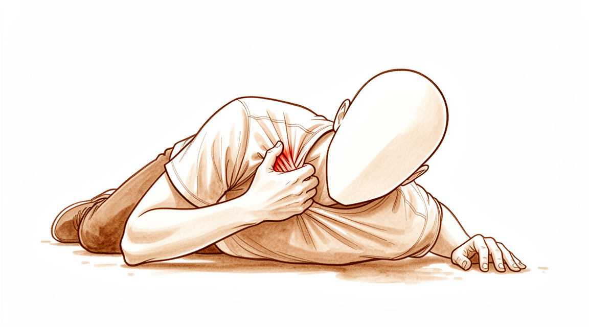
Bạn có thể cảm thấy đau nhói ở phía trước vai hoặc dọc theo xương đòn. Chấn thương này thường xảy ra sau khi ngã xuống vai hoặc bị tác động trực tiếp. Tình trạng này phổ biến ở nam giới, đặc biệt là trong các môn thể thao như bóng đá. Bạn có thể nhận thấy sưng và bầm tím trên xương. Vùng này sẽ nhạy cảm khi chạm vào.
Các hoạt động đơn giản hàng ngày trở nên khó khăn vì việc di chuyển cánh tay kéo căng xương bị tổn thương. Với tay ra sau lưng để cài áo ngực hoặc nhét áo vào quần có thể gây đau. Nhấc các vật, kể cả những vật nhẹ, có thể cảm thấy nặng nề hoặc không an toàn. Bạn có thể thấy khó giữ cánh tay xa khỏi cơ thể. Bác sĩ phẫu thuật của bạn sẽ khuyên bạn giữ cánh tay sát bên hông để giảm căng thẳng.
Cơn đau thường bùng phát vào ban đêm, khiến bạn khó tìm được tư thế ngủ thoải mái. Nằm nghiêng về phía bị thương thường là không thể. Thức dậy với cảm giác cứng khớp là điều phổ biến, đặc biệt nếu bạn di chuyển trong lúc ngủ. Sau khi vận động, cơn đau âm ỉ có thể tăng lên. Nghỉ ngơi thường giúp làm dịu cơn đau, nhưng bạn phải tránh mọi cử động gây ra những cơn đau nhói dữ dội.
Hầu hết các gãy xương đòn đều lành tốt với điều trị không phẫu thuật. Tuy nhiên, bạn cần theo dõi sát sao để đảm bảo xương không bị lệch vị trí. Các biến chứng phổ biến nhất là gãy xương không liền (non-unions), nơi xương không lành lại, và gãy xương liền lệch (malunions), nơi xương lành ở một vị trí hơi khác. Những điều này có thể gây đau kéo dài hoặc một khối u nhìn thấy được. Nếu bạn bị gãy xương đòn bên trong (gần ngực hơn), bản thân xương thường lành tốt nếu bạn vượt qua được giai đoạn chấn thương ban đầu. Nhưng hãy lưu ý rằng một tỷ lệ cao bệnh nhân bị loại gãy xương cụ thể này có thể đối mặt với các rủi ro sức khỏe nghiêm trọng trong vòng ba năm sau chấn thương.
Đối với các gãy xương bên ngoài bị lệch (gần khớp vai), điều trị không phẫu thuật dẫn đến tỷ lệ gãy xương không liền cao hơn nhưng vẫn mang lại chức năng vai tuyệt vời và nguy cơ thấp đối với các biến chứng khác. Thanh thiếu niên bị gãy xương hoàn toàn bị lệch thường thấy kết quả tuyệt vời sau năm năm nếu được điều trị không phẫu thuật. Bác sĩ phẫu thuật của bạn sẽ điều chỉnh việc chăm sóc dựa trên kiểu gãy xương cụ thể và kỳ vọng của bạn.
Những gì thực sự đang xảy ra
Xương đòn của bạn nối xương ức với xương bả vai. Nó đóng vai trò như một thanh chống giúp duy trì vai rộng và ổn định. Hầu hết các trường hợp gãy xương xảy ra khi bạn ngã trực tiếp xuống vai hoặc bị tác động mạnh vào vùng này. Những chấn thương này phổ biến ở nam giới và thường liên quan đến tai nạn xe hơi, té ngã hoặc hoạt động thể thao.
Khi xương bị gãy, sự căn chỉnh bình thường bị mất đi. Điều này có thể gây đau và hạn chế khả năng cử động cánh tay của bạn. Tin tốt là cơ thể bạn rất giỏi trong việc chữa lành xương này. Hầu hết các trường hợp gãy xương, đặc biệt là ở phần giữa, đều lành tốt mà không cần phẫu thuật. Ngay cả khi bạn chọn không phẫu thuật, bác sĩ phẫu thuật của bạn vẫn sẽ theo dõi bạn chặt chẽ. Điều này là do các mảnh xương có thể dịch chuyển nhẹ trong những ngày ngay sau khi chấn thương.
Đối với một số bệnh nhân, phẫu thuật là lựa chọn tốt hơn. Điều này thường đúng nếu vết gãy bị lệch hoàn toàn hoặc nếu xương bị ngắn lại từ 2 cm trở lên. Phẫu thuật giúp cố định xương tại chỗ để nó lành đúng cách. Đây là một thủ thuật an toàn và hiệu quả cho cả người lớn và thanh thiếu niên. Trên thực tế, hầu hết các biến chứng từ gãy xương đòn—dù được điều trị có phẫu thuật hay không—đều là các vấn đề liên quan đến cách xương lành, chẳng hạn như không liền lại hoặc lành ở vị trí hơi sai lệch.
Tỷ lệ biến chứng chung đối với chăm sóc phẫu thuật trung bình là 8,1%. Đối với thanh thiếu niên, điều trị không phẫu thuật thường dẫn đến ít biến chứng hơn và mức độ hài lòng tương tự so với phẫu thuật. Tuy nhiên, trong một số trường hợp chọn lọc khi vết gãy nghiêm trọng, phẫu thuật có thể mang lại kết quả tốt hơn. Bác sĩ phẫu thuật của bạn sẽ xem xét hình dạng cụ thể của vết gãy và mục tiêu cá nhân của bạn để quyết định hướng đi tốt nhất. Mục tiêu luôn là khôi phục chức năng và sự thoải mái cho vai của bạn một cách an toàn.
Những gì chúng tôi có thể làm về vấn đề này
Hầu hết các gãy xương đòn là do ngã xuống vai hoặc bị tác động trực tiếp. Phần lớn các trường hợp xảy ra ở nam giới và liên quan đến chấn thương giao thông, té ngã hoặc chấn thương thể thao. Bác sĩ phẫu thuật của bạn sẽ khám kỹ lưỡng để tìm các chấn thương khác ở vùng vai, đặc biệt nếu chấn thương liên quan đến lực nén. Hầu hết bệnh nhân bị gãy xương đòn đều có kết quả xuất sắc khi được điều trị bảo tồn. Điều này có nghĩa là bạn có thể mong đợi kết quả lâm sàng và chức năng tốt, bất kể bạn có chọn phẫu thuật hay không.
Bạn có thể bắt đầu với việc tự chăm sóc và vật lý trị liệu. Việc điều trị không phẫu thuật ban đầu là hợp lý vì bệnh nhân có kết quả chức năng tương tự ngay cả khi phẫu thuật bị trì hoãn. Cần theo dõi chặt chẽ do nguy cơ di lệch tiến triển trong giai đoạn quanh thời điểm chấn thương. Bác sĩ phẫu thuật của bạn có thể khuyến nghị trì hoãn đánh giá sau 6 tuần đối với gãy thân xương đòn di lệch. Thời điểm này cho phép dự đoán chính xác những bệnh nhân có khả năng liền xương với điều trị không phẫu thuật. Vật lý trị liệu nhằm khôi phục tầm vận động và sức mạnh khi xương lành. Sau khi gãy xương đòn đã lành, việc chụp hình ảnh X-quang thêm không cung cấp thông tin đáng kể nào.
Điều trị nội khoa tập trung vào giảm đau. Bác sĩ phẫu thuật của bạn có thể kê đơn thuốc giảm đau hoặc thuốc chống viêm để giúp bạn kiểm soát khó chịu trong quá trình lành xương. Đối với các trường hợp cụ thể như không liền xương, tiêm tủy xương là một phương pháp điều trị đầy hứa hẹn với tỷ lệ biến chứng thấp và thành công ban đầu. Tuy nhiên, phim X-quang đơn giản một mặt tiêu chuẩn của xương đòn là không đủ để xác định một cách đáng tin cậy mức độ ngắn lại của gãy xương đòn và nhu cầu phẫu thuật giữa các bác sĩ chỉnh hình được đào tạo chuyên khoa về y học vai/chấn thương thể thao. Bác sĩ phẫu thuật của bạn sẽ sử dụng phán đoán lâm sàng kết hợp với hình ảnh học để hướng dẫn việc chăm sóc cho bạn.
Phẫu thuật được xem xét khi điều trị bảo tồn đã đạt đến giới hạn. Bằng chứng chất lượng cao cho thấy điều trị phẫu thuật các gãy xương đòn di lệch ở người trưởng thành dẫn đến tỷ lệ liền xương cao hơn và kết quả báo cáo bởi bệnh nhân sớm tốt hơn so với điều trị không phẫu thuật, mặc dù kết quả dài hạn tương tự. Điều trị phẫu thuật các gãy thân xương đòn di lệch ở người trưởng thành có liên quan đến tỷ lệ liền xương cao hơn và kết quả báo cáo bởi bệnh nhân sớm tốt hơn so với điều trị không phẫu thuật. Một nhóm bệnh nhân chọn lọc với các gãy xương hoàn toàn di lệch, ngắn lại 2 cm trở lên, hoặc các chỉ định cụ thể được hưởng lợi từ cố định phẫu thuật, điều đã được chứng minh là dẫn đến kết quả cải thiện so với các biện pháp không phẫu thuật. Bác sĩ phẫu thuật của bạn sẽ thảo luận xem lựa chọn này có phù hợp với bạn hay không dựa trên đặc điểm gãy xương và kỳ vọng của bạn.
Những điều cần biết
Tiên lượng của bạn phụ thuộc phần lớn vào độ tuổi và vị trí của vết gãy. Đối với hầu hết người lớn bị gãy ở giữa xương đòn, phẫu thuật giúp xương liền nhanh hơn và chức năng vai sớm tốt hơn so với điều trị không phẫu thuật. Tuy nhiên, kết quả dài hạn tương tự nhau bất kể bạn có chọn phẫu thuật hay không. Nếu bạn là thiếu niên, điều trị không phẫu thuật thường được ưu tiên. Phương pháp này có tỷ lệ biến chứng thấp hơn và vẫn mang lại chức năng dài hạn xuất sắc, ngay cả khi xương liền ở vị trí hơi lệch.
Quá trình liền xương cần có thời gian. Với phẫu thuật, xương thường liền nhanh hơn so với không phẫu thuật. Trong một số trường hợp đòi hỏi vận động mạnh, chẳng hạn như vận động viên chuyên nghiệp, xương có thể liền sau khoảng 8,8 tuần. Không phẫu thuật, bác sĩ phẫu thuật có thể chờ đợi sáu tuần để xem xương có tự liền hay không trước khi đưa ra các quyết định tiếp theo. Hầu hết các trường hợp gãy xương là do té ngã hoặc va đập trực tiếp. Các vấn đề phổ biến nhất là gãy không liền (xương không liền) hoặc gãy liền lệch (xương liền ở vị trí không thẳng hàng). Những vấn đề này có thể xảy ra với cả hai phương pháp điều trị.
Biến chứng tương đối hiếm gặp. Tỷ lệ biến chứng trung bình đối với điều trị phẫu thuật là 8,1%. Đối với thanh thiếu niên, điều trị không phẫu thuật thậm chí còn ít biến chứng hơn. Nếu bạn bị gãy xương đòn trong (medial clavicle), tiên lượng của bạn thường tốt sau khi vượt qua giai đoạn chấn thương ban đầu, mặc dù cần lưu ý rằng một tỷ lệ lớn bệnh nhân bị loại gãy xương cụ thể này có thể phải đối mặt với các nguy cơ sức khỏe nghiêm trọng trong vòng ba năm. Đối với hầu hết các trường hợp gãy xương đòn khác, nguy cơ đau vai dài hạn hoặc hội chứng đau dưới mỏm quạ (subacromial pain syndrome - đau dưới xương bả vai) không tăng lên do chính chấn thương đó.
Nếu bạn chọn điều trị không phẫu thuật, việc theo dõi sát là rất quan trọng. Có nguy cơ các mảnh xương có thể tách xa nhau hơn trong những tuần đầu. Nếu bạn không phẫu thuật, bên vai của bạn có thể có nhiều triệu chứng hơn bên kia từ 10 đến 30 năm sau chấn thương. Nếu bạn có phẫu thuật, bạn có thể gặp phải nhiều biến chứng ngắn hạn hơn một chút, nhưng bạn sẽ có khả năng phục hồi chức năng sớm hơn. Một khi vết gãy đã liền hoàn toàn, việc chụp X-quang thêm thường không cung cấp thông tin mới. Bác sĩ phẫu thuật của bạn sẽ điều chỉnh kế hoạch điều trị dựa trên kiểu gãy xương cụ thể và mục tiêu cá nhân của bạn.
Khi nào cần gặp bác sĩ
Hãy gặp bác sĩ đa khoa nếu bạn có tình trạng đau dai dẳng không cải thiện khi nghỉ ngơi. Hãy yêu cầu được bác sĩ chuyên khoa khám lại nếu bạn nhận thấy tình trạng yếu hoặc mất ổn định ở khớp vai. Hãy tìm kiếm sự chăm sóc y tế nếu khớp vai của bạn bị khóa hoặc đột ngột mất sức chịu lực. Hãy liên hệ với bác sĩ phẫu thuật nếu các triệu chứng ảnh hưởng đến giấc ngủ hoặc công việc của bạn. Hãy tìm kiếm sự trợ giúp nếu tình trạng của bạn xấu đi đột ngột. Hầu hết các trường hợp gãy xương là do té ngã vào vai hoặc bị tác động trực tiếp. Phần lớn các trường hợp xảy ra ở nam giới và có liên quan đến chấn thương do phương tiện giao thông, té ngã hoặc chấn thương thể thao. Cần theo dõi chặt chẽ các trường hợp gãy xương đòn được điều trị không phẫu thuật để đảm bảo quá trình lành xương diễn ra đúng cách.
Evidence & references
Overview
- If patients with medial clavicle fractures survive the initial trauma, they can expect good clinical and functional outcomes regardless of whether surgical or nonsurgical management is chosen [1].
- Close follow-up of nonoperatively treated clavicle fractures is warranted due to potential displacement related to patient position and progressive displacement in the peri-injury period [2].
- The most common complications following clavicle fractures, whether treated operatively or non-operatively, are non-unions and malunions [7].
- Clavicle fixation is a safe and effective procedure in the pediatric population with a lack of serious complications [12].
- Specific treatment of clavicle fractures should be individualized based on fracture characteristics and patient expectations rather than broadly applied [14].
- Nonoperative treatment of adolescent clavicle fractures demonstrated lower complication rates and similar satisfaction and functional outcomes compared to operative treatment [17].
- Most mid-shaft clavicle fractures can be treated effectively by non-operative means, but a select group of patients with completely displaced fractures, shortening of 2 cm or more, or specific indications benefit from surgical fixation which has been shown to result in improved outcomes compared with non-operative measures [28].
- Although ORIF of displaced midshaft clavicle fractures remains controversial in the adolescent population, there may be additional circumstances beyond absolute indications for surgical intervention that warrant ORIF at initial presentation [30].
- Current evidence suggests that the majority of clavicular fractures in adolescents can and should be treated nonoperatively, although operative treatment with plate and screw application has consistently good outcomes with a low complication rate in selected cases [57].
- There is an increasing trend toward stabilization and fixation of markedly displaced midshaft clavicle fractures in adolescents due to concerns about symptomatic malunion and poor functional outcomes with nonsurgical management, though definitive indications for fixation in this population remain unclear [61].
- Patient selection for surgery may influence functional outcome after midshaft clavicle fracture [64].
Anatomy & Pathophysiology
- Clavicle fractures do not increase the occurrence of later subacromial pain syndrome [3].
- Protraction of the scapula is not suggested as a major risk factor for the development of subacromial pain syndrome [3].
- Evaluation of the extent of anatomic injury and understanding its mechanical consequences regarding shoulder and arm function is key in developing treatment protocols for acromioclavicular joint injuries [34].
- Hook plate and superolateral locking plate with coracoclavicular suture fixation constructs offer superior biomechanical stability for distal third clavicle fractures with coracoclavicular ligament disruption [39].
- These constructs potentially reduce complications associated with subacromial hardware [39].
- The position of the hook portion of a clavicle hook plate implant can predispose anatomic structures to post-operative complications of subacromial impingement and bony erosion [52].
- The clinical relevance of biomechanical studies on surgical fixation of midshaft clavicle fractures is arguable because none investigate the effect of tissue adaptation over time [54].
- Clavicle hook plate fixation changes scapular kinematics and scapulohumeral rhythm [56].
- Reliable bony union and improved shoulder function can be expected with thoughtful surgical planning, appropriate implant choice, and meticulous surgical technique for clavicle nonunion and malunion [58].
- In complex scapula and ipsilateral clavicle fractures, the question of stability is preoperatively less relevant than whether dislocated fragments lead to compromised shoulder function [63].
- Biomechanical analyses of four different repair techniques for lateral clavicle fracture with coracoclavicular ligament injury did not show any significance in load to failure or displacement after cyclic loading among the study groups [66].
- Plate fixation for displaced midshaft clavicular fractures does not improve shoulder function or general symptoms, and does not decrease limitations compared with nonoperative treatment in a sling [67].
- Suture stabilization of the acromioclavicular ligament plus clavicular hook plate fixation is conducive to restoring shoulder functions and has higher economic efficiency compared to total ligament repair with loop plates for acromioclavicular joint dislocation [70].
- The biphasic plate concept is aimed at improving the biomechanics of locked plating [71].
- Biomechanical evaluation showed effective fixation across all specimens at 500 cycles for unstable lateral clavicle fractures with coracoclavicular ligament disruption (Neer type IIB) [72].
- The specific design of the Locking Compression superior anterior clavicle plate provides higher strength and stiffness when compared to seven and ten hole reconstruction plates in midshaft clavicle fracture stabilisation [73].
- Clinical outcomes for treatment of unstable distal clavicle fractures with multiple Steinmann pins were evaluated using the Constant-Murley score, the University of California at Los Angeles (UCLA) Shoulder score, and the Disabilities of the Arm, Shoulder and Hand (DASH) score [74].
- Force concentration phenomena result from morphological mismatch, such as excessive inclination and improper occupation of the subacromial space, in acromion and hook plate fixation for acromioclavicular joint dislocation [76].
- Regardless of shape, subacromial erosion did not affect clinical outcomes nor cause rotator cuff lesions after plate removal in type 5 acromioclavicular joint dislocations [77].
- Inferior plates may be better equipped to resist in vivo loads experienced by the clavicle during early rehabilitation, particularly during shoulder flexion motions associated with eating, in comminuted midshaft clavicle fractures [78].
- Three patients (18%) experienced postoperative issues including plate prominence (2) and shoulder stiffness (1) in outcomes of internal fixation of clavicle and coracoclavicular stabilization for unstable distal clavicle fractures; none required reoperation [81].
Classification
- Clavicle fractures are the most commonly occurring fracture [5].
- The middle third is the most frequent site of clavicle fractures [5].
- The incidence of clavicle fractures is 1.23% [27].
- The most common complications following clavicle fractures, whether treated operatively or non-operatively, are non-unions and malunions [7].
- Clavicle malunion is a distinct clinical entity that can be treated successfully [9].
- Complication rates following surgical clavicle fracture care averaged 8.1% [20].
- The Utrecht Score for clavicle fractures is a compact yet complete tool developed to assess functional outcome specifically in patients with a clavicle fracture, consisting of patient-reported and objective measures [24].
- The Constant score was found to be reliable for assessing patients with clavicle fractures, especially at the group level [55].
- The presented classification system for lateral clavicle fractures, along with associated treatment algorithms, showed substantial inter- and intraobserver reliability [32].
- The modified Neer classification remains the predominantly cited classification system for distal clavicle fractures [33].
- The intra- and interobserver reliability of the modified Neer classification for distal clavicle fractures has been demonstrated to be inconsistent, which can lead to incorrect treatment choices and misclassifications in research [33].
- The interrater agreement of the modified Neer classification system for lateral clavicle fractures was fair [45].
- Additional 3D CT did not improve the overall level of interrater or intrarater agreement of the modified Neer classification system or associated treatment choice for lateral clavicle fractures [45].
- A new classification system for distal clavicle fractures demonstrated moderate interobserver and substantial intraobserver reliability, as well as reliability for the associated treatment choice [35].
Clinical Presentation
- Clavicle fractures are the most commonly occurring fracture [5].
- The middle third of the clavicle is the most frequent site of fracture [5].
- Adolescent clavicle fractures occur more commonly in male patients [46].
- Adolescent clavicle fractures occur during sports activities [46].
- Adolescent clavicle fractures are secondary to a direct blow to the shoulder [46].
- Adolescent clavicle fractures occur on the nondominant side [46].
- Clinicians must carefully examine patients with isolated clavicle fractures for concomitant injuries to the ipsilateral shoulder girdle, particularly in the context of compression mechanisms [18].
- Segmental fractures of the clavicle are easily missed [26].
- Distal fractures of the clavicle in children are rare [43].
- Most distal clavicle fractures in children can be treated conservatively [43].
- The most common complications following clavicle fractures, whether treated operatively or non-operatively, are non-unions and malunions [7].
- Clavicle malunion is a distinct clinical entity that can be treated successfully [9].
Investigations
- Clavicle fractures are the most commonly occurring fracture, with the middle third being the most frequent site [5].
- Segmental fractures of the clavicle are easily missed [26].
- Delayed diagnosis of subacromial, supracoracoid dislocation of the acromioclavicular joint with ipsilateral clavicle fracture is likely if careful examination of the patient's radiographs is not performed [85].
- Clinicians must carefully examine patients with isolated clavicle fractures for concomitant injuries to the ipsilateral shoulder girdle, particularly in the context of compression mechanisms [18].
- Preoperative MRI or diagnostic arthroscopy to evaluate glenohumeral associated injuries to distal clavicle fractures should be recommended [69].
- An upright chest radiograph should be obtained to evaluate midshaft clavicle fracture displacement, as it represents the physiologic stress across the fracture when considering nonoperative management [82].
- Close follow-up of nonoperatively treated clavicle fractures is warranted [2].
- Displacement of diaphyseal clavicle fractures is related to patient position and progressive displacement in the peri-injury period [2].
- Standard plain unilateral radiographs of the clavicle are insufficient to reliably determine the degree of shortening of clavicle fractures and the need for surgery among shoulder/sports medicine fellowship–trained orthopaedic surgeons [86].
- When clavicle shortening is considered in the decision to pursue operative management, the use of plain radiograph-based measurements is not recommended [83].
- Delayed assessment at 6 weeks following displaced midshaft clavicle fracture enables an accurate prediction of patients who are likely to have union with nonoperative management [31].
- Once clavicle fractures are healed, further radiographic imaging does not provide any notable information [4].
Treatment
- If patients with medial clavicle fractures survive the initial trauma, good clinical and functional outcomes are expected regardless of whether surgical or nonsurgical management is chosen [1].
- Close follow-up of nonoperatively treated clavicle fractures is warranted due to potential for progressive displacement in the peri-injury period [2].
- Initial nonsurgical management of clavicle fractures may be reasonable because patients had similar functional outcomes even when surgery was delayed [6].
- Operative treatment of displaced medial clavicle fractures provides an excellent long-term functional outcome [8].
- Nonoperative management of adolescent mid-shaft clavicle fractures results in excellent functional outcomes at long-term follow-up [10].
- Most patients with clavicle fractures have an excellent outcome using conservative management [11].
- Clavicle fixation is a safe and effective procedure in the pediatric population with a lack of serious complications [12].
- Specific treatment of clavicle fractures should not be broadly applied but rather should be individualized based on fracture characteristics and patient expectations [14].
- A more prolonged surveillance period is recommended in children with recurrent fractures of the clavicle [15].
- Nonoperative treatment of adolescent clavicle fractures demonstrated lower complication rates and similar satisfaction and functional outcomes compared to operative treatment [17].
- Functional outcome is excellent following the treatment of both acute and non-united clavicle fractures, but recovery occurs earlier following acute treatment [19].
- Most mid-shaft clavicle fractures can be treated effectively by non-operative means, but a select group of patients with completely displaced fractures, shortening of 2 cm or more, or specific indications benefit from surgical fixation which has been shown to result in improved outcomes compared with non-operative measures [28].
- Although ORIF of displaced midshaft clavicle fractures remains controversial in the adolescent population, there may be additional circumstances beyond absolute indications for surgical intervention that warrant ORIF at initial presentation [30].
- Bone marrow injection for the treatment of clavicle nonunion is promising, with low morbidity and preliminary success justifying further trials [36].
- Plate fixation of midshaft clavicle fractures for delayed union and non-union is a cost-effective intervention but functional deficits persist at long-term follow-up [37].
- Comparably excellent outcomes of severe clavicle fractures in adolescent athletes can be achieved with non-operative treatment [38].
- High-quality evidence shows that surgical treatment of displaced clavicle fractures in adults results in higher union rates and better early patient-reported outcomes compared with nonsurgical treatment, though long-term outcomes are similar [40].
- Superiorly applied plate fixation is an effective treatment for clavicular nonunion [41].
- Operative treatment of displaced midshaft clavicle fractures in adults is associated with higher union rates and better early patient-reported outcomes than non-operative treatment, though long-term outcomes are similar [42].
- Nonsurgical and surgical management provide similar results for distal clavicle fractures [44].
- Treatment of middle-third clavicle non-union after initial failure of conservative treatment with stable fixation and bone graft is a reliable, well-suited and effective treatment [49].
- A targeted approach to the management of mid-shaft clavicle fractures is needed, with simple fractures treated nonoperatively and complex displaced fractures considered for surgery to prevent non-union [50].
- Nondisplaced clavicle fractures continue to be treated conservatively with a simple sling until the fracture is healed according to radiographs and clinical assessment [51].
- In the studied unit, there is no clearly favoured method of internal fixation of lateral clavicle fractures [59].
- Ipsilateral os acromiale may be a relative contraindication to the clavicle hook plate [62].
Complications
- Non-unions and malunions are the most common complications following clavicle fractures, regardless of whether they are treated operatively or non-operatively [7].
- Complication rates following surgical clavicle fracture care average 8.1% [20].
- Limited incision plating of midshaft clavicle fractures achieves a low complication rate comparable to standard incision techniques [23].
- Clavicle pinning results in minimal complications [48].
- Surgical treatment of clavicle fractures in the pediatric population is associated with a lack of serious complications [12].
- Nonoperative management of displaced distal clavicle fractures is associated with higher nonunion rates [29].
- Nonoperative management of displaced distal clavicle fractures carries a low risk of complications and delayed surgery [29].
- The affected shoulder side is more symptomatic than the unaffected side 10 to 30 years after conservative treatment of midshaft clavicle fractures [65].
- Clavicle fractures do not increase the occurrence of later subacromial pain syndrome [3].
- Protraction of the scapula is not suggested as a major risk factor for the development of subacromial pain syndrome [3].
- Adolescent mid-shaft clavicle fracture displacement does not predict nonunion at long-term follow-up [10].
- Children with recurrent fractures of the clavicle require a more prolonged surveillance period [15].
Recovery
- Patients with medial clavicle fractures who survive the initial trauma can expect good clinical and functional outcomes regardless of whether surgical or nonsurgical management is chosen [1].
- Medial clavicle fractures have favorable functional outcomes and pain relief at minimum 1-year follow-up among patients who survive the trauma [13].
- A high proportion of patients with medial clavicle fractures die within 3 years of the injury [13].
- Conservative management of medial clavicle fractures results in excellent functional results [16].
- Operative treatment of displaced medial clavicle fractures provides an excellent long-term functional outcome [8].
- Nonoperative management of adolescent mid-shaft clavicle fractures results in excellent functional outcomes at long-term follow-up [10].
- Adolescent mid-shaft clavicular fracture displacement does not predict nonunion or inferior functional outcome at long-term follow-up [10].
- Initial nonsurgical management of clavicle fractures may be reasonable because patients had similar functional outcomes even when surgery was delayed [6].
- Nonoperative management of displaced distal clavicle fractures results in higher nonunion rates, but shoulder function remains excellent, and the risk of complications and delayed surgery are low [29].
- Patients had very good clinical outcomes following operative management of an extra-lateral distal clavicle fracture pattern [22].
- Functional outcome is excellent following the treatment of both acute and non-united clavicle fractures, but recovery occurs earlier following acute treatment [19].
- Patients reported a good quality of life and functional outcome after plating for midshaft clavicular fractures [53].
- Surgical treatment of midshaft clavicle fractures significantly reduces the nonunion rate and shortens the time to union as compared with the nonoperative approach [47].
- Surgical treatment of midshaft clavicle fractures leads to better shoulder functional scores at short- and long-term follow-up compared with nonoperative treatment [47].
- Surgical treatment of midshaft clavicle fractures has a slightly higher incidence of complications than nonoperative treatment [47].
- A limited incision approach for plating of acute midshaft clavicle fractures achieved good functional and radiographic outcomes with a low complication rate comparable to standard incision techniques [23].
- The prognosis for obtaining bony union after infected clavicle fractures is poor, with only two of six patients achieving union [75].
- Clavicle fractures do not increase the occurrence of later subacromial pain syndrome [3].
- The results do not suggest protraction of the scapula as a major risk factor for the development of subacromial pain syndrome [3].
Key Evidence
- [L5] If patients with medial clavicle fractures can survive the initial trauma, there is every reason to expect good clinical and functional outcomes, regardless of whether surgical or nonsurgical management is chosen. [1] (10.1097/corr.0000000000001916)
- [L2] Close follow-up of nonoperatively treated clavicle fractures is warranted. [2] (10.1016/j.jse.2018.01.004)
- [L4] The results do not suggest protraction of the scapula as a major risk factor for the development of SAPS. [3] (10.1016/j.xrrt.2024.01.008)
- [L3] Once clavicle fractures are healed, further radiographic imaging does not provide any notable information. [4] (10.5435/jaaos-d-17-00598)
- [L3] Initial nonsurgical management of clavicle fractures may be reasonable because patients had similar functional outcomes even when surgery was delayed. [6] (10.5435/jaaos-d-16-00130)
- [L4] Operative treatment of displaced medial clavicle fractures provides an excellent long-term functional outcome. [8] (10.1007/s00068-018-1024-6)
- [L4] Clavicle malunion is a distinct clinical entity that can be treated successfully. [9] (10.3109/17453674.2010.480939)
- [L3] Nonoperative management of adolescent mid-shaft clavicle fractures results in excellent functional outcomes at long-term follow-up. [10] (10.1302/0301-620x.103b5.bjj-2020-1929.r1)
- [L3] Most patients with clavicle fractures have an excellent outcome using conservative management. [11] (10.1016/j.jse.2019.06.022)
- [L4] Clavicle fixation is a safe and effective procedure in the pediatric population with a lack of serious complications. [12] (10.1177/2325967119s00056)
- [L4] Medial clavicle fractures have favorable functional outcomes and pain relief at minimum 1-year follow-up among those patients who survive the trauma, but a high proportion will die within 3 years of the injury. [13] (10.1097/corr.0000000000001839)
- [L5] Specific treatment of clavicle fractures should not be broadly applied but rather should be individualized based on fracture characteristics and patient expectations. [14] (10.1016/j.jse.2011.08.053)
- [L5] The authors recommend a more prolonged surveillance period in children with recurrent fractures of the clavicle. [15] (10.1097/bpb.0000000000000231)
- [Paper] Sixty eight patients with medial clavicle fractures were identified over a 5 year period, with excellent functional results seen following conservative management. [16] (10.1016/j.injury.2016.06.011)
- [L2] Nonoperative treatment of adolescent clavicle fractures demonstrated lower complication rates and similar satisfaction and functional outcomes compared to operative treatment. [17] (10.1177/2325967119s00428)
- [L4] Clinicians must carefully examine patients with isolated clavicle fractures for concomitant injuries to the ipsilateral shoulder girdle, particularly in the context of compression mechanisms. [18] (10.1177/03635465000280062301)
- [L3] Functional outcome is excellent following the treatment of both acute and non-united clavicle fractures, but recovery occurs earlier following acute treatment. [19] (10.1016/j.otsr.2017.03.021)
- [L3] Complication rates following surgical clavicle fracture care averaged 8.1%. [20] (10.1186/s12891-022-05075-5)
- [L4] The patients had very good clinical outcomes following operative management of an extra-lateral distal clavicle fracture pattern. [22] (10.1016/j.jse.2020.10.006)
- [L5] In this large cohort with long-term follow-up, a limited incision approach for plating of acute midshaft clavicle fractures achieved good functional and radiographic outcomes with a low complication rate comparable to the reported rate for standard incision techniques. [23] (10.1016/j.jse.2025.06.002)
- [L4] The Utrecht Score for clavicle fractures is a compact yet complete tool that was developed to assess functional outcome specifically in patients with a clavicle fracture, consisting of patient-reported and objective measures. [24] (10.1007/s00068-018-0979-7)
- [Case_report] The case highlights that segmental fractures of the clavicle are easily missed. [26] (10.1177/1758573214564496)
- [L4] The incidence of clavicle fractures was 1.23%. [27] (10.1016/j.injury.2011.04.008)
- [L4] Nonoperative management of displaced distal clavicle fractures results in higher nonunion rates, but shoulder function remains excellent, and risk of complications and delayed surgery are low. [29] (10.1016/j.jse.2023.12.006)
- [Case_report] Although ORIF of displaced midshaft clavicle fractures remains controversial in the adolescent population, there may be additional circumstances beyond absolute indications for surgical intervention that warrant ORIF at initial presentation. [30] (10.1016/j.xrrt.2023.03.004)
- [L1] Delayed assessment at 6 weeks following displaced midshaft clavicle fracture enables an accurate prediction of patients who are likely to have union with nonoperative management. [31] (10.2106/jbjs.19.00955)
- [L4] The presented classification system as well as associated treatment algorithms for lateral clavicle fractures showed substantial inter- and intraobserver reliability. [32] (10.1016/j.jse.2025.04.021)
- [L5] The modified Neer classification remains the predominantly cited classification system for distal clavicle fractures, yet its intra- and interobserver reliability has been demonstrated to be inconsistent, which can lead to incorrect treatment choices and misclassifications in research. [33] (10.1097/corr.0000000000001456)
- [L5] A comprehensive clinical approach emphasizing the evaluation of the extent of the anatomic injury and understanding its mechanical consequences regarding shoulder and arm function is a key in the development of guidelines for developing operative or non-operative treatment protocols and for establishing outcomes of the treatment protocols. [34] (10.1177/17585732221122335)
- [L3] The study demonstrated moderate interobserver and substantial intraobserver reliability of the new classification system and the associated treatment choice for distal clavicle fractures. [35] (10.1016/j.otsr.2018.05.015)
- [L4] Bone marrow injection for the treatment of clavicle nonunion is promising, with low morbidity and preliminary success justifying further trials. [36] (10.1016/j.jse.2006.05.001)
- [L3] Clavicle fixation for delayed and non-union is a cost-effective intervention but outcomes are worse compared to patients that unite with non-operative management. [37] (10.1177/1758573221990367)
- [L2] Comparably excellent outcomes of severe clavicle fractures in adolescent athletes can be achieved with non-operative treatment. [38] (10.1177/2325967121s00214)
- [L5] These constructs offer superior biomechanical stability in our model and potentially reduce complications associated with subacromial hardware. [39] (10.1016/j.xrrt.2025.100645)
- [L1] High-quality evidence shows that surgical treatment of displaced clavicle fractures in adults results in higher union rates and better early patient-reported outcomes compared with nonsurgical treatment, though long-term outcomes are similar. [40] (10.5435/jaaos-d-23-00472)
- [L4] Superiorly applied plate fixation is an effective treatment for clavicular nonunion. [41] (10.1016/j.jse.2008.05.046)
- [L4] Distal fractures of the clavicle in children are rare and most can be treated conservatively. [43] (10.1016/j.rboe.2015.12.006)
- [L4] Nonsurgical and surgical management provide similar results for distal clavicle fractures. [44] (10.5435/00124635-201107000-00002)
- [L3] The interrater agreement of the modified Neer classification system for lateral clavicle fractures was fair, and additional 3D CT did not improve the overall level of interrater or intrarater agreement of the classification system or associated treatment choice. [45] (10.1177/0363546515593949)
- [L4] Adolescent clavicle fractures occurred more commonly in male patients during sports, secondary to a direct blow to the shoulder, and on the nondominant side. [46] (10.1177/2325967120921344)
- [L1] Surgical treatment of midshaft clavicle fractures significantly reduces the nonunion rate and shortens the time to union as compared with the nonoperative approach and, despite a slightly higher incidence of complications, leads to better shoulder functional scores at short- and long-term follow-up. [47] (10.1177/0363546519826961)
- [L4] The technique of clavicle pinning resulted in minimal complications, short hospital stay and excellent functional outcomes. [48] (10.4103/0973-6042.57895)
- [L4] Treatment of middle-third clavicle non-union after initial failure of conservative treatment with stable fixation and bone graft is a reliable, well-suited and effective treatment. [49] (10.1016/j.otsr.2013.09.011)
- [L5] A targeted approach to the management of mid-shaft clavicle fractures is needed, with simple fractures treated nonoperatively and complex displaced fractures considered for surgery to prevent non-union. [50] (10.1016/j.injury.2020.11.066)
- [L4] Nondisplaced clavicle fractures continue to be treated conservatively with a simple sling until the fracture is healed according to radiographs and clinical assessment. [51] (10.3810/psm.2011.09.1930)
- [L5] The observed frequency of hook contact with surrounding subacromial structures in a static shoulder confirms that the position of the hook portion of the implant can predispose anatomic structures to the post-operative complications of subacromial impingement and bony erosion. [52] (10.1016/j.injury.2009.12.012)
- [L3] Patients reported a good quality of life and functional outcome after plating for midshaft clavicular fractures. [53] (10.1016/j.injury.2017.10.032)
- [L2] The clinical relevance of the biomechanical studies may be arguable since none investigate the effect of tissue adaptation over time. [54] (10.1016/j.injury.2018.02.017)
- [L4] The Constant score was found to be reliable for assessing patients with clavicle fractures, especially at the group level. [55] (10.1016/j.jse.2016.02.022)
- [L3] Clavicle hook plate fixation changes the scapular kinematics and scapulohumeral rhythm; thus, when clavicle hook plate fixation is complete, the implant should be promptly removed. [56] (10.1007/s00264-018-4003-y)
- [L4] Current evidence suggests that the majority of clavicular fractures in adolescents can and should be treated nonoperatively, although operative treatment with plate and screw application has consistently good outcomes with a low complication rate in selected cases. [57] (10.2106/jbjs.22.01036)
- [L5] Reliable bony union and improved shoulder function can be expected with thoughtful surgical planning, appropriate implant choice, and meticulous surgical technique. [58] (10.1016/j.jse.2013.01.022)
- [L4] In our unit there is no clearly favoured method of internal fixation of lateral clavicle fractures. [59] (10.1007/s00590-021-03173-z)
- [L5] There is an increasing trend toward stabilization and fixation of markedly displaced midshaft clavicle fractures in adolescents due to concerns about symptomatic malunion and poor functional outcomes with nonsurgical management, though definitive indications for fixation in this population remain unclear. [61] (10.5435/00124635-201301000-00002)
- [L4] Ipsilateral os acromiale may be a relative contraindication to the clavicle hook plate. [62] (10.1186/s12891-021-04841-1)
- [L4] The question of stability is preoperatively less relevant than the question of whether the dislocated fragments lead to compromised shoulder function. [63] (10.1007/s00068-018-0946-3)
- [L1] This review shows that patient selection for surgery may influence functional outcome after midshaft clavicle fracture. [64] (10.1177/1758573218777996)
- [L4] The affected shoulder side was more symptomatic than the unaffected side 10 to 30 years after the trauma when midshaft clavicle fractures were treated conservatively. [65] (10.1186/s13018-023-04450-9)
- [L5] The biomechanical analyses did not show any significance in load to failure or displacement after cyclic loading among the study groups. [66] (10.1007/s00167-017-4444-7)
- [L1] In addition, the procedure does not improve shoulder function or general symptoms, and it does not decrease limitations compared with nonoperative treatment in a sling. [67] (10.2106/jbjs.15.01394)
- [L1] Preoperative MRI or diagnostic arthroscopy to evaluate glenohumeral associated injuries to distal clavicle fractures should be recommended. [69] (10.1186/s13018-022-02919-7)
- [L3] This procedure was conducive to restoring shoulder functions and had higher economic efficiency. [70] (10.1186/s13018-025-06032-3)
- [L5] The biphasic plate concept is aimed at improving the biomechanics of locked plating. [71] (10.1016/j.injury.2020.04.032)
- [L5] Biomechanical evaluation showed effective fixation across all specimens at 500 cycles. [72] (10.1016/j.jse.2022.11.008)
- [L5] The specific design of the plate provides higher strength and stiffness when compared to reconstruction plates. [73] (10.1007/s00264-012-1671-x)
- [L4] Clinical outcomes were evaluated using the Constant-Murley score, the University of California at Los Angeles (UCLA) Shoulder score, and the Disabilities of the Arm, Shoulder and Hand (DASH) score. [74] (10.1097/bot.0000000000000850)
- [L4] The prognosis for obtaining bony union after infected clavicle fractures is poor, with only two of six patients achieving union. [75] (10.1097/01.blo.0000183088.60639.05)
- [L4] The force concentration phenomenon results from cases of morphological mismatch, such as excessive inclination and improper occupation of the subacromial space. [76] (10.1007/s00167-016-3987-3)
- [L3] Regardless of shape, subacromial erosion did not affect clinical outcomes nor cause rotator cuff lesions after plate removal. [77] (10.1186/s12891-021-04987-y)
- [L5] Inferior plates may be better equipped to resist the in vivo loads experienced by the clavicle during early rehabilitation after internal fixation, particularly during the shoulder flexion motions associated with eating. [78] (10.1016/j.clinbiomech.2010.12.007)
- [L4] Three patients (18%) experienced postoperative issues: plate prominence (2) and shoulder stiffness (1); none required reoperation. [81] (10.1177/17585732261456152)
- [L4] An upright chest radiograph should be obtained to evaluate midshaft clavicle fracture displacement, as it represents the physiologic stress across the fracture when considering nonoperative management. [82] (10.1097/bot.0000000000000727)
- [L4] When clavicle shortening is considered in the decision to pursue operative management, the use of plain radiograph-based measurements is not recommended. [83] (10.4055/cios.2016.8.4.367)
- [L5] Delayed diagnosis is likely if careful examination of the patient's radiographs is not performed. [85] (10.1177/2054270414527281)
- [L3] Standard plain unilateral radiographs of the clavicle are insufficient to reliably determine the degree of shortening of clavicle fractures and the need for surgery among shoulder/sports medicine fellowship–trained orthopaedic surgeons. [86] (10.1177/0363546514523926)
References
[1] CORR Insights®: What Are the Functional Outcomes and Pain Scores after Medial Clavicle Fracture Treatment?. Clinical Orthopaedics & Related Research. 2021. DOI: 10.1097/corr.0000000000001916 [2] Displacement of diaphyseal clavicle fractures related to patient position and progressive displacement in the peri-injury period. Journal of Shoulder and Elbow Surgery. 2018. DOI: 10.1016/j.jse.2018.01.004 [3] Clavicle fractures do not increase the occurrence of later subacromial pain syndrome. A registry-based case-control study with 15-25 years of follow-up of 131.838 persons from the Danish National Patient Register. JSES Reviews, Reports, and Techniques. 2024. DOI: 10.1016/j.xrrt.2024.01.008 [4] Potential Economic Benefits of Limited Clinical and Radiographic Follow-up After Plate Fixation of Midshaft Clavicle Fractures. Journal of the American Academy of Orthopaedic Surgeons. 2019. DOI: 10.5435/jaaos-d-17-00598 [5] 7. Clavicle Fractures: Epidemiology, Clinical Evaluation, Imaging, and Classification. n.d.. [6] Factors Affecting Functional Outcomes After Clavicle Fracture. Journal of the American Academy of Orthopaedic Surgeons. 2016. DOI: 10.5435/jaaos-d-16-00130 [7] 10. Complications of Clavicle Fractures—Diagnosis and Management. n.d.. [8] Displaced medial clavicle fractures: operative treatment with locking compression plate fixation. European Journal of Trauma and Emergency Surgery. 2018. DOI: 10.1007/s00068-018-1024-6 [9] Malunion after midshaft clavicle fractures in adults. Acta Orthopaedica. 2010. DOI: 10.3109/17453674.2010.480939 [10] Adolescent mid-shaft clavicular fracture displacement does not predict nonunion or inferior functional outcome at long-term follow-up. The Bone & Joint Journal. 2021. DOI: 10.1302/0301-620x.103b5.bjj-2020-1929.r1 [11] Plate fixation of clavicle fractures: comparison between early and delayed surgery. Journal of Shoulder and Elbow Surgery. 2020. DOI: 10.1016/j.jse.2019.06.022 [12] INCREASING VALUE: CAN MIDSHAFT CLAVICLE FRACTURES BE SURGICALLY TREATED AS AN OUTPATIENT?. Orthopaedic Journal of Sports Medicine. 2019. DOI: 10.1177/2325967119s00056 [13] What Are the Functional Outcomes and Pain Scores after Medial Clavicle Fracture Treatment?. Clinical Orthopaedics & Related Research. 2021. DOI: 10.1097/corr.0000000000001839 [14] Treatment of clavicle fractures: current concepts review. Journal of Shoulder and Elbow Surgery. 2012. DOI: 10.1016/j.jse.2011.08.053 [15] Post-traumatic nonunion of a clavicle fracture in a 9-year-old child. Journal of Pediatric Orthopaedics B. 2016. DOI: 10.1097/bpb.0000000000000231 [16] Natural history of medial clavicle fractures. Injury. 2016. DOI: 10.1016/j.injury.2016.06.011 [17] Two-year Functional Outcomes Of Operative Vs. Non-operative Treatment Of Completely Displaced Midshaft Clavicle Fractures In Adolescents: Results From A Prospective, Multicenter, Level 2 Study. Orthopaedic Journal of Sports Medicine. 2019. DOI: 10.1177/2325967119s00428 [18] Ipsilateral Clavicle Fracture, Sternoclavicular Joint Subluxation, and Long Thoracic Nerve Injury: An Unusual Constellation of Injuries Sustained during Wrestling. The American Journal of Sports Medicine. 2000. DOI: 10.1177/03635465000280062301 [19] Functional recovery following early mobilization after middle third clavicle osteosynthesis for acute fractures or nonunion: A case-control study. Orthopaedics & Traumatology: Surgery & Research. 2017. DOI: 10.1016/j.otsr.2017.03.021 [20] Surgical treatment, complications, reoperations, and healthcare costs among patients with clavicle fracture in England. BMC Musculoskeletal Disorders. 2022. DOI: 10.1186/s12891-022-05075-5 [22] Operative management of an extra-lateral distal clavicle fracture pattern: a study of 48 patients and a proposed update to the modified Neer classification. Journal of Shoulder and Elbow Surgery. 2021. DOI: 10.1016/j.jse.2020.10.006 [23] Limited incision plating of midshaft clavicle fractures: a case series of 1,038 patients. Journal of Shoulder and Elbow Surgery. 2026. DOI: 10.1016/j.jse.2025.06.002 [24] Development of the Utrecht Score for clavicle fractures: a short and complete clavicle score with patient-reported and objective measures. European Journal of Trauma and Emergency Surgery. 2018. DOI: 10.1007/s00068-018-0979-7 [26] Segmental clavicle fracture and acromio-clavicular joint disruption: an unusual case report. Shoulder & Elbow. 2014. DOI: 10.1177/1758573214564496 [27] A 2-year experience, management and outcome of 200 clavicle fractures. Injury. 2012. DOI: 10.1016/j.injury.2011.04.008 [28] 9. Operative Management of Clavicle Fractures: Indications, Techniques, and Outcomes. n.d.. [29] Are displaced distal clavicle fractures associated with inferior clinical outcomes following nonoperative management? A systematic review. Journal of Shoulder and Elbow Surgery. 2024. DOI: 10.1016/j.jse.2023.12.006 [30] Consequences of delayed surgical intervention of a displaced midshaft clavicle fracture: a case report. JSES Reviews, Reports, and Techniques. 2023. DOI: 10.1016/j.xrrt.2023.03.004 [31] Displaced Midshaft Clavicle Fracture Union Can Be Accurately Predicted with a Delayed Assessment at 6 Weeks Following Injury. Journal of Bone and Joint Surgery. 2020. DOI: 10.2106/jbjs.19.00955 [32] Differentiating and treating lateral clavicle fractures: a new simple classification system. Journal of Shoulder and Elbow Surgery. 2026. DOI: 10.1016/j.jse.2025.04.021 [33] Classifications in Brief: The Modified Neer Classification for Distal-third Clavicle Fractures. Clinical Orthopaedics & Related Research. 2020. DOI: 10.1097/corr.0000000000001456 [34] Acromioclavicular joint injuries revisited: Pathoanatomy, pathomechanics, and clinical presentation. Shoulder & Elbow. 2022. DOI: 10.1177/17585732221122335 [35] Distal clavicle fractures: A new classification system. Orthopaedics & Traumatology: Surgery & Research. 2018. DOI: 10.1016/j.otsr.2018.05.015 [36] Healing of a clavicle fracture nonunion with bone marrow injection. Journal of Shoulder and Elbow Surgery. 2007. DOI: 10.1016/j.jse.2006.05.001 [37] Plate fixation of midshaft clavicle fractures for delayed union and non-union is a cost-effective intervention but functional deficits persist at long-term follow-up. Shoulder & Elbow. 2021. DOI: 10.1177/1758573221990367 [38] Operative Versus Non-Operative Treatment of Severely Shortened or Comminuted Clavicle Fractures in Older Adolescent Athletes: Results from A Prospective, Multicenter, Level 2 Cohort Study. Orthopaedic Journal of Sports Medicine. 2021. DOI: 10.1177/2325967121s00214 [39] A biomechanical comparison of hook plate vs. superolateral locking plate with coracoclavicular suture fixation for distal third clavicle fractures with coracoclavicular ligament disruption. JSES Reviews, Reports, and Techniques. 2026. DOI: 10.1016/j.xrrt.2025.100645 [40] American Academy of Orthopaedic Surgeons Clinical Practice Guideline Summary on the Treatment of Clavicle Fractures. Journal of the American Academy of Orthopaedic Surgeons. 2023. DOI: 10.5435/jaaos-d-23-00472 [41] Nonunion of the clavicle treated with plate fixation: A review of forty-seven consecutive cases. Journal of Shoulder and Elbow Surgery. 2008. DOI: 10.1016/j.jse.2008.05.046 [42] AAOS CPG: Treatment of Clavicle Fractures. 2024. [43] Distal clavicle fractures in children. Revista Brasileira de Ortopedia (English Edition). 2016. DOI: 10.1016/j.rboe.2015.12.006 [44] Management of Distal Clavicle Fractures. Journal of the American Academy of Orthopaedic Surgeons. 2011. DOI: 10.5435/00124635-201107000-00002 [45] The Interrater and Intrarater Agreement of a Modified Neer Classification System and Associated Treatment Choice for Lateral Clavicle Fractures. The American Journal of Sports Medicine. 2015. DOI: 10.1177/0363546515593949 [46] Descriptive Epidemiology of Adolescent Clavicle Fractures: Results From the FACTS (Function after Adolescent Clavicle Trauma and Surgery) Prospective, Multicenter Cohort Study. Orthopaedic Journal of Sports Medicine. 2020. DOI: 10.1177/2325967120921344 [47] Midshaft Clavicle Fractures: Surgery Provides Better Results as Compared With Nonoperative Treatment: A Meta-analysis. The American Journal of Sports Medicine. 2019. DOI: 10.1177/0363546519826961 [48] Treatment of mid-shaft clavicle fractures: A comparative study. International Journal of Shoulder Surgery. 2009. DOI: 10.4103/0973-6042.57895 [49] Outcomes from surgical treatment of middle-third clavicle fractures non-union in adults: A series of 21 cases. Orthopaedics & Traumatology: Surgery & Research. 2014. DOI: 10.1016/j.otsr.2013.09.011 [50] Midshaft clavicle fracture – Nonoperative versus operative care. Injury. 2021. DOI: 10.1016/j.injury.2020.11.066 [51] Clavicle Fractures: A Review of the Literature and Update on Treatment. The Physician and Sportsmedicine. 2011. DOI: 10.3810/psm.2011.09.1930 [52] Subacromial morphometric assessment of the clavicle hook plate. Injury. 2010. DOI: 10.1016/j.injury.2009.12.012 [53] Plating for midshaft clavicular fractures: The impact on quality of life and functional outcome. Injury. 2017. DOI: 10.1016/j.injury.2017.10.032 [54] Surgical fixation of midshaft clavicle fractures: A systematic review of biomechanical studies. Injury. 2018. DOI: 10.1016/j.injury.2018.02.017 [55] High inter-rater reliability, agreement, and convergent validity of Constant score in patients with clavicle fractures. Journal of Shoulder and Elbow Surgery. 2016. DOI: 10.1016/j.jse.2016.02.022 [56] Evaluation of three-dimensional in vivo scapular kinematics and scapulohumeral rhythm between shoulders with a clavicle hook plate and contralateral healthy shoulders. International Orthopaedics. 2018. DOI: 10.1007/s00264-018-4003-y [57] Clavicular Fractures in the Adolescent. Journal of Bone and Joint Surgery. 2023. DOI: 10.2106/jbjs.22.01036 [58] Management of clavicle nonunion and malunion. Journal of Shoulder and Elbow Surgery. 2013. DOI: 10.1016/j.jse.2013.01.022 [59] Operative management of lateral third clavicle fractures: a comparison of internal fixation methods. European Journal of Orthopaedic Surgery & Traumatology. 2021. DOI: 10.1007/s00590-021-03173-z [61] Displaced Clavicle Fractures in Adolescents: Facts, Controversies, and Current Trends. Journal of the American Academy of Orthopaedic Surgeons. 2013. DOI: 10.5435/00124635-201301000-00002 [62] Os acromiale may be a contraindication of the clavicle hook plate: case reports and literature review. BMC Musculoskeletal Disorders. 2021. DOI: 10.1186/s12891-021-04841-1 [63] Decision-making for complex scapula and ipsilateral clavicle fractures: a review. European Journal of Trauma and Emergency Surgery. 2018. DOI: 10.1007/s00068-018-0946-3 [64] Does candidate for plate fixation selection improve the functional outcome after midshaft clavicle fracture? A systematic review of 1348 patients. Shoulder & Elbow. 2018. DOI: 10.1177/1758573218777996 [65] Long-term conservative treatment outcomes for midshaft clavicle fractures: a 10-to-30-year follow-up. Journal of Orthopaedic Surgery and Research. 2023. DOI: 10.1186/s13018-023-04450-9 [66] Lateral clavicle fracture with coracoclavicular ligament injury: a biomechanical study of 4 different repair techniques. Knee Surgery, Sports Traumatology, Arthroscopy. 2017. DOI: 10.1007/s00167-017-4444-7 [67] Plate Fixation Compared with Nonoperative Treatment for Displaced Midshaft Clavicular Fractures. Journal of Bone and Joint Surgery. 2017. DOI: 10.2106/jbjs.15.01394 [69] Concomitant glenohumeral injuries in patients with distal clavicle fractures undergoing arthroscopic-assisted surgery: a systematic review. Journal of Orthopaedic Surgery and Research. 2022. DOI: 10.1186/s13018-022-02919-7 [70] Suture stabilization of the acromioclavicular ligament plus clavicular hook plate fixation vs. total ligament repair with loop plates for acromioclavicular joint dislocation. Journal of Orthopaedic Surgery and Research. 2025. DOI: 10.1186/s13018-025-06032-3 [71] Biphasic Plating – In vivo study of a novel fixation concept to enhance mechanobiological fracture healing. Injury. 2020. DOI: 10.1016/j.injury.2020.04.032 [72] Biomechanical analysis of plating techniques for unstable lateral clavicle fractures with coracoclavicular ligament disruption (Neer type IIB). Journal of Shoulder and Elbow Surgery. 2023. DOI: 10.1016/j.jse.2022.11.008 [73] Biomechanical comparison of the Locking Compression superior anterior clavicle plate with seven and ten hole reconstruction plates in midshaft clavicle fracture stabilisation. International Orthopaedics. 2012. DOI: 10.1007/s00264-012-1671-x [74] Treatment of Unstable Distal Clavicle Fractures With Multiple Steinmann Pins—A Modification of Neer's Method: A Series of 56 Consecutive Cases. Journal of Orthopaedic Trauma. 2017. DOI: 10.1097/bot.0000000000000850 [75] Infection after Clavicle Fractures. Clinical Orthopaedics and Related Research. 2005. DOI: 10.1097/01.blo.0000183088.60639.05 [76] Morphological analysis of acromion and hook plate for the fixation of acromioclavicular joint dislocation. Knee Surgery, Sports Traumatology, Arthroscopy. 2016. DOI: 10.1007/s00167-016-3987-3 [77] Effect of subacromial erosion shape on rotator cuff and clinical outcomes after hook plate fixation in type 5 acromioclavicular joint dislocations: a retrospective cohort study. BMC Musculoskeletal Disorders. 2022. DOI: 10.1186/s12891-021-04987-y [78] The comminuted midshaft clavicle fracture: A biomechanical evaluation of plating methods. Clinical Biomechanics. 2011. DOI: 10.1016/j.clinbiomech.2010.12.007 [81] Unstable distal clavicle fractures: Outcomes of internal fixation of clavicle and coracoclavicular stabilization. Shoulder & Elbow. 2026. DOI: 10.1177/17585732261456152 [82] Positional Change in Displacement of Midshaft Clavicle Fractures: An Aid to Initial Evaluation. Journal of Orthopaedic Trauma. 2017. DOI: 10.1097/bot.0000000000000727 [83] Measurement of Clavicle Fracture Shortening Using Computed Tomography and Chest Radiography. Clinics in Orthopedic Surgery. 2016. DOI: 10.4055/cios.2016.8.4.367 [85] Subacromial, supracoracoid dislocation of the acromioclavicular joint with ipsilateral clavicle fracture: a case report with review of the literature and classification. JRSM Open. 2014. DOI: 10.1177/2054270414527281 [86] Intraobserver and Interobserver Agreement in the Classification and Treatment of Midshaft Clavicle Fractures. The American Journal of Sports Medicine. 2014. DOI: 10.1177/0363546514523926


