锁骨骨折
Patients › Shoulder
Clavicle fractures — when conservative management is fine and when fixation is indicated.
您的感受
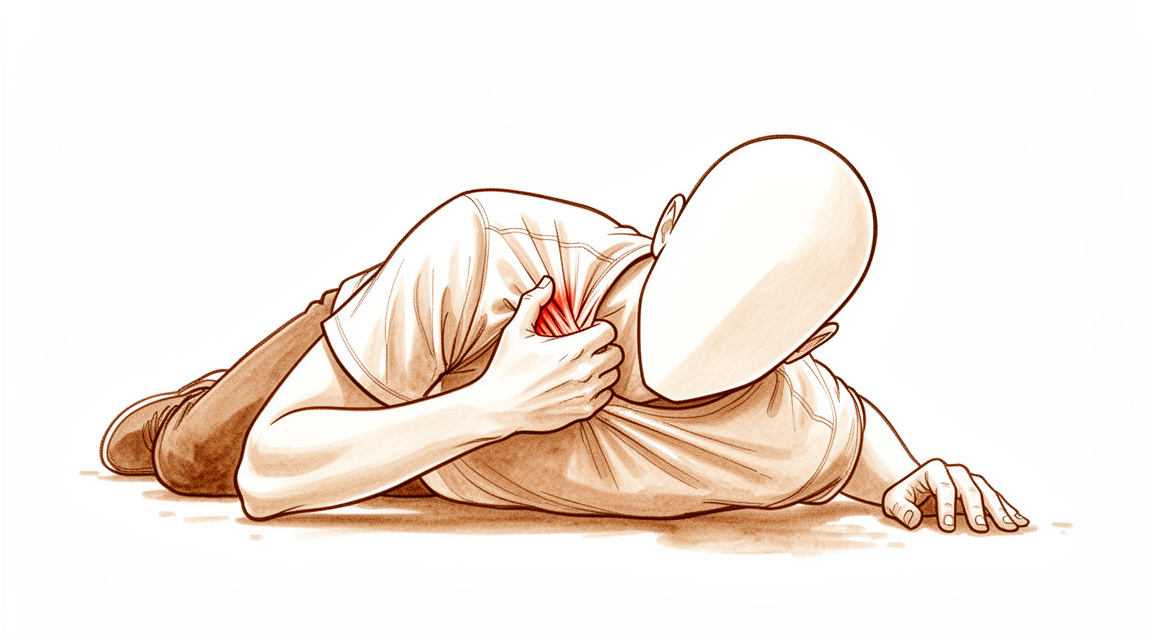
您可能会感到肩部前方或锁骨区域出现锐痛。这种损伤通常发生在肩部着地跌倒或直接遭受撞击之后。男性中较为常见,尤其是在橄榄球等运动中。您可能会注意到骨骼上方出现肿胀和瘀伤。该区域触压时会感到疼痛。
简单的日常活动变得困难,因为移动手臂会牵拉受伤的骨骼。将手伸到背后扣内衣或塞衬衫下摆可能会引起疼痛。提起物体,即使是轻物,也可能感觉沉重或不安全。您可能会发现难以将手臂保持在远离身体的位置。您的外科医生会建议您将手臂贴近身体侧方,以减少牵拉。
疼痛常在夜间加剧,使得找到舒适的睡眠姿势变得困难。通常无法侧卧于受伤的一侧。晨起时出现僵硬很常见,尤其是如果您在夜间移动过。活动后,酸痛感可能会增加。休息通常有助于缓解疼痛,但您必须避免任何会导致疼痛剧烈加剧的动作。
大多数锁骨骨折通过非手术治疗愈合良好。然而,您需要密切随访,以确保骨骼没有发生移位。最常见的并发症包括骨不连(骨骼未能愈合)和畸形愈合(骨骼以略有不同的位置愈合)。这些情况可能导致持续性疼痛或可见的隆起。如果您患有内侧锁骨骨折(靠近胸部),只要挺过初始创伤,骨骼本身通常能愈合良好。但请注意,患有这种特定类型骨折的患者中,有较高比例可能在受伤后三年内面临严重的健康风险。
对于移位的远端骨折(靠近肩关节),非手术治疗会导致较高的骨不连发生率,但仍能提供良好的肩关节功能和其他并发症的低风险。对于完全移位的骨折,青少年患者即使接受非手术治疗,五年后通常也能获得良好的结果。您的外科医生会根据您的具体骨折模式和期望制定个性化的治疗方案。
实际发生了什么
您的锁骨将胸骨与肩胛骨连接起来。它起到支撑杆的作用,使您的肩膀保持宽阔和稳定。大多数骨折发生在直接摔倒到肩膀上或该区域受到猛烈撞击时。这些损伤在男性中很常见,通常与车祸、跌倒或运动有关。
当骨头断裂时,正常的排列就会丧失。这会导致疼痛并限制您手臂的活动。好消息是您的身体非常擅长愈合这种骨头。大多数骨折,尤其是中间部分的骨折,无需手术即可良好愈合。即使您选择不进行手术,您的外科医生也会密切观察您。这是因为受伤后的最初几天,骨头碎片可能会发生轻微移位。
对于某些患者,手术是更好的选择。如果骨折完全移位或骨头缩短 2 厘米或更多,通常就是这种情况。手术有助于将骨头固定在原位,以便其正确愈合。这是一项对成人和青少年都安全有效的程序。事实上,锁骨骨折的绝大多数并发症——无论是否接受手术治疗——都是骨头愈合问题,例如未能愈合或愈合位置略有偏差。
外科治疗的整体并发症率平均为 8.1%。对于青少年,非手术治疗通常比手术导致更少的并发症,且满意度水平相似。然而,在骨折严重的特定情况下,手术可以带来更好的结果。您的外科医生会根据您骨折的具体形态和个人目标来决定最佳路径。目标始终是安全地恢复您的肩膀功能和舒适度。
我们能采取的措施
大多数锁骨骨折是由于肩部着地跌倒或直接撞击所致。此类骨折多见于男性,常与车辆交通事故、跌倒或运动损伤有关。您的外科医生会仔细检查您是否伴有肩部区域的其他损伤,尤其是当受伤机制涉及挤压时。大多数锁骨骨折患者通过保守治疗可获得极佳的治疗效果。这意味着,无论您是否选择手术,均可期待良好的临床和功能结果。
您可以从自我管理和物理治疗开始。初始非手术治疗是合理的,因为即使手术延迟,患者的功能结果也相似。由于在损伤周围期存在进行性移位的风险,因此需要进行密切随访。对于移位的中段锁骨骨折,您的外科医生可能会建议在伤后6周进行评估。这一时机有助于准确预测哪些患者可能通过非手术治疗实现骨愈合。物理治疗旨在随着骨骼愈合恢复关节活动度和肌力。一旦锁骨骨折愈合,进一步的影像学检查不再提供显著的临床信息。
药物治疗主要侧重于缓解疼痛。您的外科医生可能会开具止痛药或抗炎药,以帮助您在愈合过程中管理不适。对于特定情况,如骨不连,骨髓注射是一种前景良好的治疗方法,具有低发病率和初步的成功率。然而,对于经过肩关节/运动医学专科培训的骨科医生而言,标准的锁骨单侧X线平片不足以可靠地确定锁骨骨折的缩短程度以及手术需求。您的外科医生将结合临床判断和影像学检查来指导您的治疗。
当保守治疗达到极限时,会考虑手术治疗。高质量证据表明,与保守治疗相比,成人移位性锁骨骨折的手术治疗具有更高的骨愈合率和更好的早期患者报告结局,尽管长期结局相似。成人移位性中段锁骨骨折的手术治疗与非手术治疗相比,具有更高的骨愈合率和更好的早期患者报告结局。部分完全移位、缩短2厘米或以上或具有特定适应证的患者可从手术固定中获益,研究表明其结局优于非手术治疗措施。您的外科医生将根据您的骨折特征和期望值,与您讨论该选项是否适合您。
预期情况
您的预后主要取决于您的年龄以及骨折的具体位置。对于大多数锁骨中段骨折的成年人而言,与保守治疗相比,手术治疗能带来更快的愈合速度和更好的早期肩关节功能。然而,无论选择手术与否,长期结果是相似的。如果是青少年患者,通常更倾向于非手术治疗。非手术治疗并发症发生率较低,即使骨骼在轻微移位的位置愈合,仍能提供良好的长期功能。
愈合需要时间。接受手术后,骨骼通常比不手术时融合得更快。在一些高强度需求的情况下,例如职业运动员,愈合时间可能约为 8.8 周。如果不进行手术,外科医生可能会等待六周,观察骨骼是否自行愈合,然后再做出进一步的决定。大多数骨折由跌倒或直接撞击引起。最常见的问题是骨不连(骨骼未愈合)或畸形愈合(骨骼在错位的位置愈合)。这两种情况均可能发生在任何治疗路径中。
并发症相对少见。外科治疗的平均并发症率为 8.1%。对于青少年,非手术治疗引起的并发症更少。如果您患有锁骨内侧骨折,一旦度过急性损伤期,预后通常良好,但需要注意的是,患有这种特定类型骨折的患者中,有相当比例的人在三年内可能面临严重的健康风险。对于大多数其他锁骨骨折,损伤本身并不会增加长期肩痛或肩峰下疼痛综合征(肩胛骨下方的疼痛)的风险。
如果选择非手术治疗,密切随访至关重要。在最初几周内,骨折碎片可能会进一步分离。如果不进行手术,在创伤发生 10 到 30 年后,患侧肩膀的症状可能比健侧更明显。如果进行手术,您可能会经历稍多的短期并发症,但很可能更早恢复功能。一旦骨折完全愈合,进一步的 X 光检查通常不会提供新的信息。您的外科医生将根据您的具体骨折类型和个人目标制定治疗方案。
何时就诊
若疼痛持续且休息后无改善,请咨询全科医生。若发现肩部无力或不稳,请要求专科医生评估。若肩部出现卡住或脱位,请及时就医。若症状影响睡眠或工作,请联系您的外科医生。若病情突然加重,请立即寻求帮助。大多数骨折是由于跌倒时肩部着地或直接撞击所致。大多数患者为男性,且与交通事故、跌倒或运动损伤有关。对非手术治疗锁骨骨折的患者进行密切随访是必要的,以确保正确愈合。
Evidence & references
Overview
- If patients with medial clavicle fractures survive the initial trauma, they can expect good clinical and functional outcomes regardless of whether surgical or nonsurgical management is chosen [1].
- Close follow-up of nonoperatively treated clavicle fractures is warranted due to potential displacement related to patient position and progressive displacement in the peri-injury period [2].
- The most common complications following clavicle fractures, whether treated operatively or non-operatively, are non-unions and malunions [7].
- Clavicle fixation is a safe and effective procedure in the pediatric population with a lack of serious complications [12].
- Specific treatment of clavicle fractures should be individualized based on fracture characteristics and patient expectations rather than broadly applied [14].
- Nonoperative treatment of adolescent clavicle fractures demonstrated lower complication rates and similar satisfaction and functional outcomes compared to operative treatment [17].
- Most mid-shaft clavicle fractures can be treated effectively by non-operative means, but a select group of patients with completely displaced fractures, shortening of 2 cm or more, or specific indications benefit from surgical fixation which has been shown to result in improved outcomes compared with non-operative measures [28].
- Although ORIF of displaced midshaft clavicle fractures remains controversial in the adolescent population, there may be additional circumstances beyond absolute indications for surgical intervention that warrant ORIF at initial presentation [30].
- Current evidence suggests that the majority of clavicular fractures in adolescents can and should be treated nonoperatively, although operative treatment with plate and screw application has consistently good outcomes with a low complication rate in selected cases [57].
- There is an increasing trend toward stabilization and fixation of markedly displaced midshaft clavicle fractures in adolescents due to concerns about symptomatic malunion and poor functional outcomes with nonsurgical management, though definitive indications for fixation in this population remain unclear [61].
- Patient selection for surgery may influence functional outcome after midshaft clavicle fracture [64].
Anatomy & Pathophysiology
- Clavicle fractures do not increase the occurrence of later subacromial pain syndrome [3].
- Protraction of the scapula is not suggested as a major risk factor for the development of subacromial pain syndrome [3].
- Evaluation of the extent of anatomic injury and understanding its mechanical consequences regarding shoulder and arm function is key in developing treatment protocols for acromioclavicular joint injuries [34].
- Hook plate and superolateral locking plate with coracoclavicular suture fixation constructs offer superior biomechanical stability for distal third clavicle fractures with coracoclavicular ligament disruption [39].
- These constructs potentially reduce complications associated with subacromial hardware [39].
- The position of the hook portion of a clavicle hook plate implant can predispose anatomic structures to post-operative complications of subacromial impingement and bony erosion [52].
- The clinical relevance of biomechanical studies on surgical fixation of midshaft clavicle fractures is arguable because none investigate the effect of tissue adaptation over time [54].
- Clavicle hook plate fixation changes scapular kinematics and scapulohumeral rhythm [56].
- Reliable bony union and improved shoulder function can be expected with thoughtful surgical planning, appropriate implant choice, and meticulous surgical technique for clavicle nonunion and malunion [58].
- In complex scapula and ipsilateral clavicle fractures, the question of stability is preoperatively less relevant than whether dislocated fragments lead to compromised shoulder function [63].
- Biomechanical analyses of four different repair techniques for lateral clavicle fracture with coracoclavicular ligament injury did not show any significance in load to failure or displacement after cyclic loading among the study groups [66].
- Plate fixation for displaced midshaft clavicular fractures does not improve shoulder function or general symptoms, and does not decrease limitations compared with nonoperative treatment in a sling [67].
- Suture stabilization of the acromioclavicular ligament plus clavicular hook plate fixation is conducive to restoring shoulder functions and has higher economic efficiency compared to total ligament repair with loop plates for acromioclavicular joint dislocation [70].
- The biphasic plate concept is aimed at improving the biomechanics of locked plating [71].
- Biomechanical evaluation showed effective fixation across all specimens at 500 cycles for unstable lateral clavicle fractures with coracoclavicular ligament disruption (Neer type IIB) [72].
- The specific design of the Locking Compression superior anterior clavicle plate provides higher strength and stiffness when compared to seven and ten hole reconstruction plates in midshaft clavicle fracture stabilisation [73].
- Clinical outcomes for treatment of unstable distal clavicle fractures with multiple Steinmann pins were evaluated using the Constant-Murley score, the University of California at Los Angeles (UCLA) Shoulder score, and the Disabilities of the Arm, Shoulder and Hand (DASH) score [74].
- Force concentration phenomena result from morphological mismatch, such as excessive inclination and improper occupation of the subacromial space, in acromion and hook plate fixation for acromioclavicular joint dislocation [76].
- Regardless of shape, subacromial erosion did not affect clinical outcomes nor cause rotator cuff lesions after plate removal in type 5 acromioclavicular joint dislocations [77].
- Inferior plates may be better equipped to resist in vivo loads experienced by the clavicle during early rehabilitation, particularly during shoulder flexion motions associated with eating, in comminuted midshaft clavicle fractures [78].
- Three patients (18%) experienced postoperative issues including plate prominence (2) and shoulder stiffness (1) in outcomes of internal fixation of clavicle and coracoclavicular stabilization for unstable distal clavicle fractures; none required reoperation [81].
Classification
- Clavicle fractures are the most commonly occurring fracture [5].
- The middle third is the most frequent site of clavicle fractures [5].
- The incidence of clavicle fractures is 1.23% [27].
- The most common complications following clavicle fractures, whether treated operatively or non-operatively, are non-unions and malunions [7].
- Clavicle malunion is a distinct clinical entity that can be treated successfully [9].
- Complication rates following surgical clavicle fracture care averaged 8.1% [20].
- The Utrecht Score for clavicle fractures is a compact yet complete tool developed to assess functional outcome specifically in patients with a clavicle fracture, consisting of patient-reported and objective measures [24].
- The Constant score was found to be reliable for assessing patients with clavicle fractures, especially at the group level [55].
- The presented classification system for lateral clavicle fractures, along with associated treatment algorithms, showed substantial inter- and intraobserver reliability [32].
- The modified Neer classification remains the predominantly cited classification system for distal clavicle fractures [33].
- The intra- and interobserver reliability of the modified Neer classification for distal clavicle fractures has been demonstrated to be inconsistent, which can lead to incorrect treatment choices and misclassifications in research [33].
- The interrater agreement of the modified Neer classification system for lateral clavicle fractures was fair [45].
- Additional 3D CT did not improve the overall level of interrater or intrarater agreement of the modified Neer classification system or associated treatment choice for lateral clavicle fractures [45].
- A new classification system for distal clavicle fractures demonstrated moderate interobserver and substantial intraobserver reliability, as well as reliability for the associated treatment choice [35].
Clinical Presentation
- Clavicle fractures are the most commonly occurring fracture [5].
- The middle third of the clavicle is the most frequent site of fracture [5].
- Adolescent clavicle fractures occur more commonly in male patients [46].
- Adolescent clavicle fractures occur during sports activities [46].
- Adolescent clavicle fractures are secondary to a direct blow to the shoulder [46].
- Adolescent clavicle fractures occur on the nondominant side [46].
- Clinicians must carefully examine patients with isolated clavicle fractures for concomitant injuries to the ipsilateral shoulder girdle, particularly in the context of compression mechanisms [18].
- Segmental fractures of the clavicle are easily missed [26].
- Distal fractures of the clavicle in children are rare [43].
- Most distal clavicle fractures in children can be treated conservatively [43].
- The most common complications following clavicle fractures, whether treated operatively or non-operatively, are non-unions and malunions [7].
- Clavicle malunion is a distinct clinical entity that can be treated successfully [9].
Investigations
- Clavicle fractures are the most commonly occurring fracture, with the middle third being the most frequent site [5].
- Segmental fractures of the clavicle are easily missed [26].
- Delayed diagnosis of subacromial, supracoracoid dislocation of the acromioclavicular joint with ipsilateral clavicle fracture is likely if careful examination of the patient's radiographs is not performed [85].
- Clinicians must carefully examine patients with isolated clavicle fractures for concomitant injuries to the ipsilateral shoulder girdle, particularly in the context of compression mechanisms [18].
- Preoperative MRI or diagnostic arthroscopy to evaluate glenohumeral associated injuries to distal clavicle fractures should be recommended [69].
- An upright chest radiograph should be obtained to evaluate midshaft clavicle fracture displacement, as it represents the physiologic stress across the fracture when considering nonoperative management [82].
- Close follow-up of nonoperatively treated clavicle fractures is warranted [2].
- Displacement of diaphyseal clavicle fractures is related to patient position and progressive displacement in the peri-injury period [2].
- Standard plain unilateral radiographs of the clavicle are insufficient to reliably determine the degree of shortening of clavicle fractures and the need for surgery among shoulder/sports medicine fellowship–trained orthopaedic surgeons [86].
- When clavicle shortening is considered in the decision to pursue operative management, the use of plain radiograph-based measurements is not recommended [83].
- Delayed assessment at 6 weeks following displaced midshaft clavicle fracture enables an accurate prediction of patients who are likely to have union with nonoperative management [31].
- Once clavicle fractures are healed, further radiographic imaging does not provide any notable information [4].
Treatment
- If patients with medial clavicle fractures survive the initial trauma, good clinical and functional outcomes are expected regardless of whether surgical or nonsurgical management is chosen [1].
- Close follow-up of nonoperatively treated clavicle fractures is warranted due to potential for progressive displacement in the peri-injury period [2].
- Initial nonsurgical management of clavicle fractures may be reasonable because patients had similar functional outcomes even when surgery was delayed [6].
- Operative treatment of displaced medial clavicle fractures provides an excellent long-term functional outcome [8].
- Nonoperative management of adolescent mid-shaft clavicle fractures results in excellent functional outcomes at long-term follow-up [10].
- Most patients with clavicle fractures have an excellent outcome using conservative management [11].
- Clavicle fixation is a safe and effective procedure in the pediatric population with a lack of serious complications [12].
- Specific treatment of clavicle fractures should not be broadly applied but rather should be individualized based on fracture characteristics and patient expectations [14].
- A more prolonged surveillance period is recommended in children with recurrent fractures of the clavicle [15].
- Nonoperative treatment of adolescent clavicle fractures demonstrated lower complication rates and similar satisfaction and functional outcomes compared to operative treatment [17].
- Functional outcome is excellent following the treatment of both acute and non-united clavicle fractures, but recovery occurs earlier following acute treatment [19].
- Most mid-shaft clavicle fractures can be treated effectively by non-operative means, but a select group of patients with completely displaced fractures, shortening of 2 cm or more, or specific indications benefit from surgical fixation which has been shown to result in improved outcomes compared with non-operative measures [28].
- Although ORIF of displaced midshaft clavicle fractures remains controversial in the adolescent population, there may be additional circumstances beyond absolute indications for surgical intervention that warrant ORIF at initial presentation [30].
- Bone marrow injection for the treatment of clavicle nonunion is promising, with low morbidity and preliminary success justifying further trials [36].
- Plate fixation of midshaft clavicle fractures for delayed union and non-union is a cost-effective intervention but functional deficits persist at long-term follow-up [37].
- Comparably excellent outcomes of severe clavicle fractures in adolescent athletes can be achieved with non-operative treatment [38].
- High-quality evidence shows that surgical treatment of displaced clavicle fractures in adults results in higher union rates and better early patient-reported outcomes compared with nonsurgical treatment, though long-term outcomes are similar [40].
- Superiorly applied plate fixation is an effective treatment for clavicular nonunion [41].
- Operative treatment of displaced midshaft clavicle fractures in adults is associated with higher union rates and better early patient-reported outcomes than non-operative treatment, though long-term outcomes are similar [42].
- Nonsurgical and surgical management provide similar results for distal clavicle fractures [44].
- Treatment of middle-third clavicle non-union after initial failure of conservative treatment with stable fixation and bone graft is a reliable, well-suited and effective treatment [49].
- A targeted approach to the management of mid-shaft clavicle fractures is needed, with simple fractures treated nonoperatively and complex displaced fractures considered for surgery to prevent non-union [50].
- Nondisplaced clavicle fractures continue to be treated conservatively with a simple sling until the fracture is healed according to radiographs and clinical assessment [51].
- In the studied unit, there is no clearly favoured method of internal fixation of lateral clavicle fractures [59].
- Ipsilateral os acromiale may be a relative contraindication to the clavicle hook plate [62].
Complications
- Non-unions and malunions are the most common complications following clavicle fractures, regardless of whether they are treated operatively or non-operatively [7].
- Complication rates following surgical clavicle fracture care average 8.1% [20].
- Limited incision plating of midshaft clavicle fractures achieves a low complication rate comparable to standard incision techniques [23].
- Clavicle pinning results in minimal complications [48].
- Surgical treatment of clavicle fractures in the pediatric population is associated with a lack of serious complications [12].
- Nonoperative management of displaced distal clavicle fractures is associated with higher nonunion rates [29].
- Nonoperative management of displaced distal clavicle fractures carries a low risk of complications and delayed surgery [29].
- The affected shoulder side is more symptomatic than the unaffected side 10 to 30 years after conservative treatment of midshaft clavicle fractures [65].
- Clavicle fractures do not increase the occurrence of later subacromial pain syndrome [3].
- Protraction of the scapula is not suggested as a major risk factor for the development of subacromial pain syndrome [3].
- Adolescent mid-shaft clavicle fracture displacement does not predict nonunion at long-term follow-up [10].
- Children with recurrent fractures of the clavicle require a more prolonged surveillance period [15].
Recovery
- Patients with medial clavicle fractures who survive the initial trauma can expect good clinical and functional outcomes regardless of whether surgical or nonsurgical management is chosen [1].
- Medial clavicle fractures have favorable functional outcomes and pain relief at minimum 1-year follow-up among patients who survive the trauma [13].
- A high proportion of patients with medial clavicle fractures die within 3 years of the injury [13].
- Conservative management of medial clavicle fractures results in excellent functional results [16].
- Operative treatment of displaced medial clavicle fractures provides an excellent long-term functional outcome [8].
- Nonoperative management of adolescent mid-shaft clavicle fractures results in excellent functional outcomes at long-term follow-up [10].
- Adolescent mid-shaft clavicular fracture displacement does not predict nonunion or inferior functional outcome at long-term follow-up [10].
- Initial nonsurgical management of clavicle fractures may be reasonable because patients had similar functional outcomes even when surgery was delayed [6].
- Nonoperative management of displaced distal clavicle fractures results in higher nonunion rates, but shoulder function remains excellent, and the risk of complications and delayed surgery are low [29].
- Patients had very good clinical outcomes following operative management of an extra-lateral distal clavicle fracture pattern [22].
- Functional outcome is excellent following the treatment of both acute and non-united clavicle fractures, but recovery occurs earlier following acute treatment [19].
- Patients reported a good quality of life and functional outcome after plating for midshaft clavicular fractures [53].
- Surgical treatment of midshaft clavicle fractures significantly reduces the nonunion rate and shortens the time to union as compared with the nonoperative approach [47].
- Surgical treatment of midshaft clavicle fractures leads to better shoulder functional scores at short- and long-term follow-up compared with nonoperative treatment [47].
- Surgical treatment of midshaft clavicle fractures has a slightly higher incidence of complications than nonoperative treatment [47].
- A limited incision approach for plating of acute midshaft clavicle fractures achieved good functional and radiographic outcomes with a low complication rate comparable to standard incision techniques [23].
- The prognosis for obtaining bony union after infected clavicle fractures is poor, with only two of six patients achieving union [75].
- Clavicle fractures do not increase the occurrence of later subacromial pain syndrome [3].
- The results do not suggest protraction of the scapula as a major risk factor for the development of subacromial pain syndrome [3].
Key Evidence
- [L5] If patients with medial clavicle fractures can survive the initial trauma, there is every reason to expect good clinical and functional outcomes, regardless of whether surgical or nonsurgical management is chosen. [1] (10.1097/corr.0000000000001916)
- [L2] Close follow-up of nonoperatively treated clavicle fractures is warranted. [2] (10.1016/j.jse.2018.01.004)
- [L4] The results do not suggest protraction of the scapula as a major risk factor for the development of SAPS. [3] (10.1016/j.xrrt.2024.01.008)
- [L3] Once clavicle fractures are healed, further radiographic imaging does not provide any notable information. [4] (10.5435/jaaos-d-17-00598)
- [L3] Initial nonsurgical management of clavicle fractures may be reasonable because patients had similar functional outcomes even when surgery was delayed. [6] (10.5435/jaaos-d-16-00130)
- [L4] Operative treatment of displaced medial clavicle fractures provides an excellent long-term functional outcome. [8] (10.1007/s00068-018-1024-6)
- [L4] Clavicle malunion is a distinct clinical entity that can be treated successfully. [9] (10.3109/17453674.2010.480939)
- [L3] Nonoperative management of adolescent mid-shaft clavicle fractures results in excellent functional outcomes at long-term follow-up. [10] (10.1302/0301-620x.103b5.bjj-2020-1929.r1)
- [L3] Most patients with clavicle fractures have an excellent outcome using conservative management. [11] (10.1016/j.jse.2019.06.022)
- [L4] Clavicle fixation is a safe and effective procedure in the pediatric population with a lack of serious complications. [12] (10.1177/2325967119s00056)
- [L4] Medial clavicle fractures have favorable functional outcomes and pain relief at minimum 1-year follow-up among those patients who survive the trauma, but a high proportion will die within 3 years of the injury. [13] (10.1097/corr.0000000000001839)
- [L5] Specific treatment of clavicle fractures should not be broadly applied but rather should be individualized based on fracture characteristics and patient expectations. [14] (10.1016/j.jse.2011.08.053)
- [L5] The authors recommend a more prolonged surveillance period in children with recurrent fractures of the clavicle. [15] (10.1097/bpb.0000000000000231)
- [Paper] Sixty eight patients with medial clavicle fractures were identified over a 5 year period, with excellent functional results seen following conservative management. [16] (10.1016/j.injury.2016.06.011)
- [L2] Nonoperative treatment of adolescent clavicle fractures demonstrated lower complication rates and similar satisfaction and functional outcomes compared to operative treatment. [17] (10.1177/2325967119s00428)
- [L4] Clinicians must carefully examine patients with isolated clavicle fractures for concomitant injuries to the ipsilateral shoulder girdle, particularly in the context of compression mechanisms. [18] (10.1177/03635465000280062301)
- [L3] Functional outcome is excellent following the treatment of both acute and non-united clavicle fractures, but recovery occurs earlier following acute treatment. [19] (10.1016/j.otsr.2017.03.021)
- [L3] Complication rates following surgical clavicle fracture care averaged 8.1%. [20] (10.1186/s12891-022-05075-5)
- [L4] The patients had very good clinical outcomes following operative management of an extra-lateral distal clavicle fracture pattern. [22] (10.1016/j.jse.2020.10.006)
- [L5] In this large cohort with long-term follow-up, a limited incision approach for plating of acute midshaft clavicle fractures achieved good functional and radiographic outcomes with a low complication rate comparable to the reported rate for standard incision techniques. [23] (10.1016/j.jse.2025.06.002)
- [L4] The Utrecht Score for clavicle fractures is a compact yet complete tool that was developed to assess functional outcome specifically in patients with a clavicle fracture, consisting of patient-reported and objective measures. [24] (10.1007/s00068-018-0979-7)
- [Case_report] The case highlights that segmental fractures of the clavicle are easily missed. [26] (10.1177/1758573214564496)
- [L4] The incidence of clavicle fractures was 1.23%. [27] (10.1016/j.injury.2011.04.008)
- [L4] Nonoperative management of displaced distal clavicle fractures results in higher nonunion rates, but shoulder function remains excellent, and risk of complications and delayed surgery are low. [29] (10.1016/j.jse.2023.12.006)
- [Case_report] Although ORIF of displaced midshaft clavicle fractures remains controversial in the adolescent population, there may be additional circumstances beyond absolute indications for surgical intervention that warrant ORIF at initial presentation. [30] (10.1016/j.xrrt.2023.03.004)
- [L1] Delayed assessment at 6 weeks following displaced midshaft clavicle fracture enables an accurate prediction of patients who are likely to have union with nonoperative management. [31] (10.2106/jbjs.19.00955)
- [L4] The presented classification system as well as associated treatment algorithms for lateral clavicle fractures showed substantial inter- and intraobserver reliability. [32] (10.1016/j.jse.2025.04.021)
- [L5] The modified Neer classification remains the predominantly cited classification system for distal clavicle fractures, yet its intra- and interobserver reliability has been demonstrated to be inconsistent, which can lead to incorrect treatment choices and misclassifications in research. [33] (10.1097/corr.0000000000001456)
- [L5] A comprehensive clinical approach emphasizing the evaluation of the extent of the anatomic injury and understanding its mechanical consequences regarding shoulder and arm function is a key in the development of guidelines for developing operative or non-operative treatment protocols and for establishing outcomes of the treatment protocols. [34] (10.1177/17585732221122335)
- [L3] The study demonstrated moderate interobserver and substantial intraobserver reliability of the new classification system and the associated treatment choice for distal clavicle fractures. [35] (10.1016/j.otsr.2018.05.015)
- [L4] Bone marrow injection for the treatment of clavicle nonunion is promising, with low morbidity and preliminary success justifying further trials. [36] (10.1016/j.jse.2006.05.001)
- [L3] Clavicle fixation for delayed and non-union is a cost-effective intervention but outcomes are worse compared to patients that unite with non-operative management. [37] (10.1177/1758573221990367)
- [L2] Comparably excellent outcomes of severe clavicle fractures in adolescent athletes can be achieved with non-operative treatment. [38] (10.1177/2325967121s00214)
- [L5] These constructs offer superior biomechanical stability in our model and potentially reduce complications associated with subacromial hardware. [39] (10.1016/j.xrrt.2025.100645)
- [L1] High-quality evidence shows that surgical treatment of displaced clavicle fractures in adults results in higher union rates and better early patient-reported outcomes compared with nonsurgical treatment, though long-term outcomes are similar. [40] (10.5435/jaaos-d-23-00472)
- [L4] Superiorly applied plate fixation is an effective treatment for clavicular nonunion. [41] (10.1016/j.jse.2008.05.046)
- [L4] Distal fractures of the clavicle in children are rare and most can be treated conservatively. [43] (10.1016/j.rboe.2015.12.006)
- [L4] Nonsurgical and surgical management provide similar results for distal clavicle fractures. [44] (10.5435/00124635-201107000-00002)
- [L3] The interrater agreement of the modified Neer classification system for lateral clavicle fractures was fair, and additional 3D CT did not improve the overall level of interrater or intrarater agreement of the classification system or associated treatment choice. [45] (10.1177/0363546515593949)
- [L4] Adolescent clavicle fractures occurred more commonly in male patients during sports, secondary to a direct blow to the shoulder, and on the nondominant side. [46] (10.1177/2325967120921344)
- [L1] Surgical treatment of midshaft clavicle fractures significantly reduces the nonunion rate and shortens the time to union as compared with the nonoperative approach and, despite a slightly higher incidence of complications, leads to better shoulder functional scores at short- and long-term follow-up. [47] (10.1177/0363546519826961)
- [L4] The technique of clavicle pinning resulted in minimal complications, short hospital stay and excellent functional outcomes. [48] (10.4103/0973-6042.57895)
- [L4] Treatment of middle-third clavicle non-union after initial failure of conservative treatment with stable fixation and bone graft is a reliable, well-suited and effective treatment. [49] (10.1016/j.otsr.2013.09.011)
- [L5] A targeted approach to the management of mid-shaft clavicle fractures is needed, with simple fractures treated nonoperatively and complex displaced fractures considered for surgery to prevent non-union. [50] (10.1016/j.injury.2020.11.066)
- [L4] Nondisplaced clavicle fractures continue to be treated conservatively with a simple sling until the fracture is healed according to radiographs and clinical assessment. [51] (10.3810/psm.2011.09.1930)
- [L5] The observed frequency of hook contact with surrounding subacromial structures in a static shoulder confirms that the position of the hook portion of the implant can predispose anatomic structures to the post-operative complications of subacromial impingement and bony erosion. [52] (10.1016/j.injury.2009.12.012)
- [L3] Patients reported a good quality of life and functional outcome after plating for midshaft clavicular fractures. [53] (10.1016/j.injury.2017.10.032)
- [L2] The clinical relevance of the biomechanical studies may be arguable since none investigate the effect of tissue adaptation over time. [54] (10.1016/j.injury.2018.02.017)
- [L4] The Constant score was found to be reliable for assessing patients with clavicle fractures, especially at the group level. [55] (10.1016/j.jse.2016.02.022)
- [L3] Clavicle hook plate fixation changes the scapular kinematics and scapulohumeral rhythm; thus, when clavicle hook plate fixation is complete, the implant should be promptly removed. [56] (10.1007/s00264-018-4003-y)
- [L4] Current evidence suggests that the majority of clavicular fractures in adolescents can and should be treated nonoperatively, although operative treatment with plate and screw application has consistently good outcomes with a low complication rate in selected cases. [57] (10.2106/jbjs.22.01036)
- [L5] Reliable bony union and improved shoulder function can be expected with thoughtful surgical planning, appropriate implant choice, and meticulous surgical technique. [58] (10.1016/j.jse.2013.01.022)
- [L4] In our unit there is no clearly favoured method of internal fixation of lateral clavicle fractures. [59] (10.1007/s00590-021-03173-z)
- [L5] There is an increasing trend toward stabilization and fixation of markedly displaced midshaft clavicle fractures in adolescents due to concerns about symptomatic malunion and poor functional outcomes with nonsurgical management, though definitive indications for fixation in this population remain unclear. [61] (10.5435/00124635-201301000-00002)
- [L4] Ipsilateral os acromiale may be a relative contraindication to the clavicle hook plate. [62] (10.1186/s12891-021-04841-1)
- [L4] The question of stability is preoperatively less relevant than the question of whether the dislocated fragments lead to compromised shoulder function. [63] (10.1007/s00068-018-0946-3)
- [L1] This review shows that patient selection for surgery may influence functional outcome after midshaft clavicle fracture. [64] (10.1177/1758573218777996)
- [L4] The affected shoulder side was more symptomatic than the unaffected side 10 to 30 years after the trauma when midshaft clavicle fractures were treated conservatively. [65] (10.1186/s13018-023-04450-9)
- [L5] The biomechanical analyses did not show any significance in load to failure or displacement after cyclic loading among the study groups. [66] (10.1007/s00167-017-4444-7)
- [L1] In addition, the procedure does not improve shoulder function or general symptoms, and it does not decrease limitations compared with nonoperative treatment in a sling. [67] (10.2106/jbjs.15.01394)
- [L1] Preoperative MRI or diagnostic arthroscopy to evaluate glenohumeral associated injuries to distal clavicle fractures should be recommended. [69] (10.1186/s13018-022-02919-7)
- [L3] This procedure was conducive to restoring shoulder functions and had higher economic efficiency. [70] (10.1186/s13018-025-06032-3)
- [L5] The biphasic plate concept is aimed at improving the biomechanics of locked plating. [71] (10.1016/j.injury.2020.04.032)
- [L5] Biomechanical evaluation showed effective fixation across all specimens at 500 cycles. [72] (10.1016/j.jse.2022.11.008)
- [L5] The specific design of the plate provides higher strength and stiffness when compared to reconstruction plates. [73] (10.1007/s00264-012-1671-x)
- [L4] Clinical outcomes were evaluated using the Constant-Murley score, the University of California at Los Angeles (UCLA) Shoulder score, and the Disabilities of the Arm, Shoulder and Hand (DASH) score. [74] (10.1097/bot.0000000000000850)
- [L4] The prognosis for obtaining bony union after infected clavicle fractures is poor, with only two of six patients achieving union. [75] (10.1097/01.blo.0000183088.60639.05)
- [L4] The force concentration phenomenon results from cases of morphological mismatch, such as excessive inclination and improper occupation of the subacromial space. [76] (10.1007/s00167-016-3987-3)
- [L3] Regardless of shape, subacromial erosion did not affect clinical outcomes nor cause rotator cuff lesions after plate removal. [77] (10.1186/s12891-021-04987-y)
- [L5] Inferior plates may be better equipped to resist the in vivo loads experienced by the clavicle during early rehabilitation after internal fixation, particularly during the shoulder flexion motions associated with eating. [78] (10.1016/j.clinbiomech.2010.12.007)
- [L4] Three patients (18%) experienced postoperative issues: plate prominence (2) and shoulder stiffness (1); none required reoperation. [81] (10.1177/17585732261456152)
- [L4] An upright chest radiograph should be obtained to evaluate midshaft clavicle fracture displacement, as it represents the physiologic stress across the fracture when considering nonoperative management. [82] (10.1097/bot.0000000000000727)
- [L4] When clavicle shortening is considered in the decision to pursue operative management, the use of plain radiograph-based measurements is not recommended. [83] (10.4055/cios.2016.8.4.367)
- [L5] Delayed diagnosis is likely if careful examination of the patient's radiographs is not performed. [85] (10.1177/2054270414527281)
- [L3] Standard plain unilateral radiographs of the clavicle are insufficient to reliably determine the degree of shortening of clavicle fractures and the need for surgery among shoulder/sports medicine fellowship–trained orthopaedic surgeons. [86] (10.1177/0363546514523926)
References
[1] CORR Insights®: What Are the Functional Outcomes and Pain Scores after Medial Clavicle Fracture Treatment?. Clinical Orthopaedics & Related Research. 2021. DOI: 10.1097/corr.0000000000001916 [2] Displacement of diaphyseal clavicle fractures related to patient position and progressive displacement in the peri-injury period. Journal of Shoulder and Elbow Surgery. 2018. DOI: 10.1016/j.jse.2018.01.004 [3] Clavicle fractures do not increase the occurrence of later subacromial pain syndrome. A registry-based case-control study with 15-25 years of follow-up of 131.838 persons from the Danish National Patient Register. JSES Reviews, Reports, and Techniques. 2024. DOI: 10.1016/j.xrrt.2024.01.008 [4] Potential Economic Benefits of Limited Clinical and Radiographic Follow-up After Plate Fixation of Midshaft Clavicle Fractures. Journal of the American Academy of Orthopaedic Surgeons. 2019. DOI: 10.5435/jaaos-d-17-00598 [5] 7. Clavicle Fractures: Epidemiology, Clinical Evaluation, Imaging, and Classification. n.d.. [6] Factors Affecting Functional Outcomes After Clavicle Fracture. Journal of the American Academy of Orthopaedic Surgeons. 2016. DOI: 10.5435/jaaos-d-16-00130 [7] 10. Complications of Clavicle Fractures—Diagnosis and Management. n.d.. [8] Displaced medial clavicle fractures: operative treatment with locking compression plate fixation. European Journal of Trauma and Emergency Surgery. 2018. DOI: 10.1007/s00068-018-1024-6 [9] Malunion after midshaft clavicle fractures in adults. Acta Orthopaedica. 2010. DOI: 10.3109/17453674.2010.480939 [10] Adolescent mid-shaft clavicular fracture displacement does not predict nonunion or inferior functional outcome at long-term follow-up. The Bone & Joint Journal. 2021. DOI: 10.1302/0301-620x.103b5.bjj-2020-1929.r1 [11] Plate fixation of clavicle fractures: comparison between early and delayed surgery. Journal of Shoulder and Elbow Surgery. 2020. DOI: 10.1016/j.jse.2019.06.022 [12] INCREASING VALUE: CAN MIDSHAFT CLAVICLE FRACTURES BE SURGICALLY TREATED AS AN OUTPATIENT?. Orthopaedic Journal of Sports Medicine. 2019. DOI: 10.1177/2325967119s00056 [13] What Are the Functional Outcomes and Pain Scores after Medial Clavicle Fracture Treatment?. Clinical Orthopaedics & Related Research. 2021. DOI: 10.1097/corr.0000000000001839 [14] Treatment of clavicle fractures: current concepts review. Journal of Shoulder and Elbow Surgery. 2012. DOI: 10.1016/j.jse.2011.08.053 [15] Post-traumatic nonunion of a clavicle fracture in a 9-year-old child. Journal of Pediatric Orthopaedics B. 2016. DOI: 10.1097/bpb.0000000000000231 [16] Natural history of medial clavicle fractures. Injury. 2016. DOI: 10.1016/j.injury.2016.06.011 [17] Two-year Functional Outcomes Of Operative Vs. Non-operative Treatment Of Completely Displaced Midshaft Clavicle Fractures In Adolescents: Results From A Prospective, Multicenter, Level 2 Study. Orthopaedic Journal of Sports Medicine. 2019. DOI: 10.1177/2325967119s00428 [18] Ipsilateral Clavicle Fracture, Sternoclavicular Joint Subluxation, and Long Thoracic Nerve Injury: An Unusual Constellation of Injuries Sustained during Wrestling. The American Journal of Sports Medicine. 2000. DOI: 10.1177/03635465000280062301 [19] Functional recovery following early mobilization after middle third clavicle osteosynthesis for acute fractures or nonunion: A case-control study. Orthopaedics & Traumatology: Surgery & Research. 2017. DOI: 10.1016/j.otsr.2017.03.021 [20] Surgical treatment, complications, reoperations, and healthcare costs among patients with clavicle fracture in England. BMC Musculoskeletal Disorders. 2022. DOI: 10.1186/s12891-022-05075-5 [22] Operative management of an extra-lateral distal clavicle fracture pattern: a study of 48 patients and a proposed update to the modified Neer classification. Journal of Shoulder and Elbow Surgery. 2021. DOI: 10.1016/j.jse.2020.10.006 [23] Limited incision plating of midshaft clavicle fractures: a case series of 1,038 patients. Journal of Shoulder and Elbow Surgery. 2026. DOI: 10.1016/j.jse.2025.06.002 [24] Development of the Utrecht Score for clavicle fractures: a short and complete clavicle score with patient-reported and objective measures. European Journal of Trauma and Emergency Surgery. 2018. DOI: 10.1007/s00068-018-0979-7 [26] Segmental clavicle fracture and acromio-clavicular joint disruption: an unusual case report. Shoulder & Elbow. 2014. DOI: 10.1177/1758573214564496 [27] A 2-year experience, management and outcome of 200 clavicle fractures. Injury. 2012. DOI: 10.1016/j.injury.2011.04.008 [28] 9. Operative Management of Clavicle Fractures: Indications, Techniques, and Outcomes. n.d.. [29] Are displaced distal clavicle fractures associated with inferior clinical outcomes following nonoperative management? A systematic review. Journal of Shoulder and Elbow Surgery. 2024. DOI: 10.1016/j.jse.2023.12.006 [30] Consequences of delayed surgical intervention of a displaced midshaft clavicle fracture: a case report. JSES Reviews, Reports, and Techniques. 2023. DOI: 10.1016/j.xrrt.2023.03.004 [31] Displaced Midshaft Clavicle Fracture Union Can Be Accurately Predicted with a Delayed Assessment at 6 Weeks Following Injury. Journal of Bone and Joint Surgery. 2020. DOI: 10.2106/jbjs.19.00955 [32] Differentiating and treating lateral clavicle fractures: a new simple classification system. Journal of Shoulder and Elbow Surgery. 2026. DOI: 10.1016/j.jse.2025.04.021 [33] Classifications in Brief: The Modified Neer Classification for Distal-third Clavicle Fractures. Clinical Orthopaedics & Related Research. 2020. DOI: 10.1097/corr.0000000000001456 [34] Acromioclavicular joint injuries revisited: Pathoanatomy, pathomechanics, and clinical presentation. Shoulder & Elbow. 2022. DOI: 10.1177/17585732221122335 [35] Distal clavicle fractures: A new classification system. Orthopaedics & Traumatology: Surgery & Research. 2018. DOI: 10.1016/j.otsr.2018.05.015 [36] Healing of a clavicle fracture nonunion with bone marrow injection. Journal of Shoulder and Elbow Surgery. 2007. DOI: 10.1016/j.jse.2006.05.001 [37] Plate fixation of midshaft clavicle fractures for delayed union and non-union is a cost-effective intervention but functional deficits persist at long-term follow-up. Shoulder & Elbow. 2021. DOI: 10.1177/1758573221990367 [38] Operative Versus Non-Operative Treatment of Severely Shortened or Comminuted Clavicle Fractures in Older Adolescent Athletes: Results from A Prospective, Multicenter, Level 2 Cohort Study. Orthopaedic Journal of Sports Medicine. 2021. DOI: 10.1177/2325967121s00214 [39] A biomechanical comparison of hook plate vs. superolateral locking plate with coracoclavicular suture fixation for distal third clavicle fractures with coracoclavicular ligament disruption. JSES Reviews, Reports, and Techniques. 2026. DOI: 10.1016/j.xrrt.2025.100645 [40] American Academy of Orthopaedic Surgeons Clinical Practice Guideline Summary on the Treatment of Clavicle Fractures. Journal of the American Academy of Orthopaedic Surgeons. 2023. DOI: 10.5435/jaaos-d-23-00472 [41] Nonunion of the clavicle treated with plate fixation: A review of forty-seven consecutive cases. Journal of Shoulder and Elbow Surgery. 2008. DOI: 10.1016/j.jse.2008.05.046 [42] AAOS CPG: Treatment of Clavicle Fractures. 2024. [43] Distal clavicle fractures in children. Revista Brasileira de Ortopedia (English Edition). 2016. DOI: 10.1016/j.rboe.2015.12.006 [44] Management of Distal Clavicle Fractures. Journal of the American Academy of Orthopaedic Surgeons. 2011. DOI: 10.5435/00124635-201107000-00002 [45] The Interrater and Intrarater Agreement of a Modified Neer Classification System and Associated Treatment Choice for Lateral Clavicle Fractures. The American Journal of Sports Medicine. 2015. DOI: 10.1177/0363546515593949 [46] Descriptive Epidemiology of Adolescent Clavicle Fractures: Results From the FACTS (Function after Adolescent Clavicle Trauma and Surgery) Prospective, Multicenter Cohort Study. Orthopaedic Journal of Sports Medicine. 2020. DOI: 10.1177/2325967120921344 [47] Midshaft Clavicle Fractures: Surgery Provides Better Results as Compared With Nonoperative Treatment: A Meta-analysis. The American Journal of Sports Medicine. 2019. DOI: 10.1177/0363546519826961 [48] Treatment of mid-shaft clavicle fractures: A comparative study. International Journal of Shoulder Surgery. 2009. DOI: 10.4103/0973-6042.57895 [49] Outcomes from surgical treatment of middle-third clavicle fractures non-union in adults: A series of 21 cases. Orthopaedics & Traumatology: Surgery & Research. 2014. DOI: 10.1016/j.otsr.2013.09.011 [50] Midshaft clavicle fracture – Nonoperative versus operative care. Injury. 2021. DOI: 10.1016/j.injury.2020.11.066 [51] Clavicle Fractures: A Review of the Literature and Update on Treatment. The Physician and Sportsmedicine. 2011. DOI: 10.3810/psm.2011.09.1930 [52] Subacromial morphometric assessment of the clavicle hook plate. Injury. 2010. DOI: 10.1016/j.injury.2009.12.012 [53] Plating for midshaft clavicular fractures: The impact on quality of life and functional outcome. Injury. 2017. DOI: 10.1016/j.injury.2017.10.032 [54] Surgical fixation of midshaft clavicle fractures: A systematic review of biomechanical studies. Injury. 2018. DOI: 10.1016/j.injury.2018.02.017 [55] High inter-rater reliability, agreement, and convergent validity of Constant score in patients with clavicle fractures. Journal of Shoulder and Elbow Surgery. 2016. DOI: 10.1016/j.jse.2016.02.022 [56] Evaluation of three-dimensional in vivo scapular kinematics and scapulohumeral rhythm between shoulders with a clavicle hook plate and contralateral healthy shoulders. International Orthopaedics. 2018. DOI: 10.1007/s00264-018-4003-y [57] Clavicular Fractures in the Adolescent. Journal of Bone and Joint Surgery. 2023. DOI: 10.2106/jbjs.22.01036 [58] Management of clavicle nonunion and malunion. Journal of Shoulder and Elbow Surgery. 2013. DOI: 10.1016/j.jse.2013.01.022 [59] Operative management of lateral third clavicle fractures: a comparison of internal fixation methods. European Journal of Orthopaedic Surgery & Traumatology. 2021. DOI: 10.1007/s00590-021-03173-z [61] Displaced Clavicle Fractures in Adolescents: Facts, Controversies, and Current Trends. Journal of the American Academy of Orthopaedic Surgeons. 2013. DOI: 10.5435/00124635-201301000-00002 [62] Os acromiale may be a contraindication of the clavicle hook plate: case reports and literature review. BMC Musculoskeletal Disorders. 2021. DOI: 10.1186/s12891-021-04841-1 [63] Decision-making for complex scapula and ipsilateral clavicle fractures: a review. European Journal of Trauma and Emergency Surgery. 2018. DOI: 10.1007/s00068-018-0946-3 [64] Does candidate for plate fixation selection improve the functional outcome after midshaft clavicle fracture? A systematic review of 1348 patients. Shoulder & Elbow. 2018. DOI: 10.1177/1758573218777996 [65] Long-term conservative treatment outcomes for midshaft clavicle fractures: a 10-to-30-year follow-up. Journal of Orthopaedic Surgery and Research. 2023. DOI: 10.1186/s13018-023-04450-9 [66] Lateral clavicle fracture with coracoclavicular ligament injury: a biomechanical study of 4 different repair techniques. Knee Surgery, Sports Traumatology, Arthroscopy. 2017. DOI: 10.1007/s00167-017-4444-7 [67] Plate Fixation Compared with Nonoperative Treatment for Displaced Midshaft Clavicular Fractures. Journal of Bone and Joint Surgery. 2017. DOI: 10.2106/jbjs.15.01394 [69] Concomitant glenohumeral injuries in patients with distal clavicle fractures undergoing arthroscopic-assisted surgery: a systematic review. Journal of Orthopaedic Surgery and Research. 2022. DOI: 10.1186/s13018-022-02919-7 [70] Suture stabilization of the acromioclavicular ligament plus clavicular hook plate fixation vs. total ligament repair with loop plates for acromioclavicular joint dislocation. Journal of Orthopaedic Surgery and Research. 2025. DOI: 10.1186/s13018-025-06032-3 [71] Biphasic Plating – In vivo study of a novel fixation concept to enhance mechanobiological fracture healing. Injury. 2020. DOI: 10.1016/j.injury.2020.04.032 [72] Biomechanical analysis of plating techniques for unstable lateral clavicle fractures with coracoclavicular ligament disruption (Neer type IIB). Journal of Shoulder and Elbow Surgery. 2023. DOI: 10.1016/j.jse.2022.11.008 [73] Biomechanical comparison of the Locking Compression superior anterior clavicle plate with seven and ten hole reconstruction plates in midshaft clavicle fracture stabilisation. International Orthopaedics. 2012. DOI: 10.1007/s00264-012-1671-x [74] Treatment of Unstable Distal Clavicle Fractures With Multiple Steinmann Pins—A Modification of Neer's Method: A Series of 56 Consecutive Cases. Journal of Orthopaedic Trauma. 2017. DOI: 10.1097/bot.0000000000000850 [75] Infection after Clavicle Fractures. Clinical Orthopaedics and Related Research. 2005. DOI: 10.1097/01.blo.0000183088.60639.05 [76] Morphological analysis of acromion and hook plate for the fixation of acromioclavicular joint dislocation. Knee Surgery, Sports Traumatology, Arthroscopy. 2016. DOI: 10.1007/s00167-016-3987-3 [77] Effect of subacromial erosion shape on rotator cuff and clinical outcomes after hook plate fixation in type 5 acromioclavicular joint dislocations: a retrospective cohort study. BMC Musculoskeletal Disorders. 2022. DOI: 10.1186/s12891-021-04987-y [78] The comminuted midshaft clavicle fracture: A biomechanical evaluation of plating methods. Clinical Biomechanics. 2011. DOI: 10.1016/j.clinbiomech.2010.12.007 [81] Unstable distal clavicle fractures: Outcomes of internal fixation of clavicle and coracoclavicular stabilization. Shoulder & Elbow. 2026. DOI: 10.1177/17585732261456152 [82] Positional Change in Displacement of Midshaft Clavicle Fractures: An Aid to Initial Evaluation. Journal of Orthopaedic Trauma. 2017. DOI: 10.1097/bot.0000000000000727 [83] Measurement of Clavicle Fracture Shortening Using Computed Tomography and Chest Radiography. Clinics in Orthopedic Surgery. 2016. DOI: 10.4055/cios.2016.8.4.367 [85] Subacromial, supracoracoid dislocation of the acromioclavicular joint with ipsilateral clavicle fracture: a case report with review of the literature and classification. JRSM Open. 2014. DOI: 10.1177/2054270414527281 [86] Intraobserver and Interobserver Agreement in the Classification and Treatment of Midshaft Clavicle Fractures. The American Journal of Sports Medicine. 2014. DOI: 10.1177/0363546514523926


