Gãy xương cánh tay gần
Patients › Shoulder
Proximal humerus fractures — Neer classification, sling management, and surgical options.
Những gì bạn đang cảm nhận
Bạn có khả năng sẽ cảm thấy đau ở vùng cánh tay trên và vai. Cơn đau này thường bắt nguồn từ một vết gãy xương gần khớp vai. Nếu vết gãy của bạn có liên quan đến xương yếu, được gọi là loãng xương, cơn đau có thể là một phần của một mô hình lớn hơn các vết thương do giòn xương. Các vết gãy không lệch, nơi các mảnh xương vẫn giữ nguyên vị trí, là phổ biến trong nhóm này. Ngay cả khi xương không bị dịch chuyển, những vết gãy này vẫn có thể gây ra tàn tật đáng kể và làm giảm cảm giác khỏe mạnh tổng thể của bạn.
Cơn đau có xu hướng trở nên tồi tệ hơn khi vận động. Bạn có thể thấy khó nâng cánh tay hoặc với tay lên cao. Các nhiệm vụ hàng ngày đơn giản trở nên khó khăn. Bạn có thể gặp khó khăn khi nhét áo vào quần hoặc với tay ra sau lưng để cài khuy áo ngực. Việc nâng các vật, ngay cả những vật nhẹ, có thể kích hoạt sự khó chịu sắc nhọn. Vì vai không ổn định, bất kỳ nỗ lực nào để sử dụng cánh tay đều có thể làm trầm trọng thêm tổn thương.
Nghỉ ngơi cánh tay thường giúp giảm đau. Tuy nhiên, bạn vẫn có thể trải qua các đợt bùng phát vào ban đêm. Nhiều bệnh nhân báo cáo rằng nằm ngủ ở bên bị tổn thương là đau đớn hoặc không thể. Thức dậy với vai cứng hoặc đau nhức là điều phổ biến. Bác sĩ phẫu thuật của bạn sẽ hướng dẫn bạn cách định vị cơ thể để thoải mái trong khi bảo vệ xương đang lành.
Nếu bạn dưới 65 tuổi, bác sĩ phẫu thuật của bạn có thể thảo luận xem phẫu thuật có mang lại lợi ích rõ ràng so với điều trị không phẫu thuật hay không. Đối với nhiều người trưởng thành trong nhóm tuổi này, bằng chứng không cho thấy lợi thế mạnh mẽ cho việc phẫu thuật. Hầu hết các vết gãy một phần lành tốt mà không cần phẫu thuật. Người lớn tuổi cũng thường được điều trị không phẫu thuật. Bất kể tuổi tác của bạn, mục tiêu là kiểm soát cơn đau và bảo vệ xương trong khi nó lành. Các biến chứng có thể xảy ra ở các giai đoạn khác nhau, vì vậy nhóm của bạn sẽ theo dõi bạn chặt chẽ. Bạn không đơn độc trong quá trình này; nhóm chăm sóc của bạn ở đó để hỗ trợ quá trình phục hồi của bạn và giúp bạn lấy lại chức năng một cách an toàn.
Những gì thực sự đang xảy ra
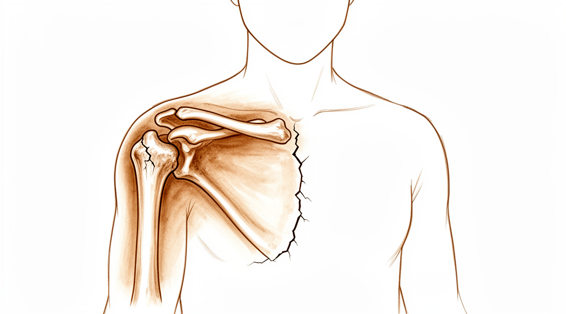
Vai của bạn là một khớp cầu và hốc, nơi xương cánh tay trên gặp xương bả vai. Đầu trên của xương cánh tay có hai gờ nhỏ được gọi là củ. Những gờ này đóng vai trò là điểm neo cho các gân xoay cuff. Các gân này là những bó sợi chắc khỏe giúp nâng và xoay cánh tay của bạn. Khi bạn gãy đầu trên của xương cánh tay, các điểm neo này có thể bị lệch khỏi vị trí.
Nếu các củ bị dịch chuyển xuống dưới, các gân sẽ mất độ căng đúng mức. Điều này làm sai lệch cơ học khớp. Ngay cả một sự dịch chuyển nhỏ 15 độ cũng có thể thay đổi cách khớp vận động và chịu lực. Sự sai lệch này gây đau và hạn chế vận động của bạn. Nó cũng khiến khớp khó lành hơn ở vị trí đúng.
Viêm nang khớp là lớp bao quanh vai. Nó giữ khớp lại với nhau và sản xuất dịch để duy trì độ trơn tru. Sau khi gãy xương, bao khớp này có thể trở nên cứng hoặc bị sẹo. Độ cứng này, cùng với bất kỳ tổn thương nào đối với các gân, làm giảm sức mạnh và tầm vận động của bạn. Bác sĩ phẫu thuật của bạn cần khôi phục lại giải phẫu để các mô này có thể hoạt động cùng nhau một lần nữa.
Đối với nhiều người, đặc biệt là người lớn tuổi, các mảnh xương quá vỡ để có thể sửa chữa bằng tấm và vít. Trong những trường hợp này, bác sĩ phẫu thuật của bạn có thể khuyên nên thay khớp. Thủ thuật này thay thế quả bóng bị hỏng bằng một cấy ghép bằng kim loại và nhựa. Lựa chọn này thường được chọn khi việc đưa cánh tay của bạn trở lại chuyển động là ưu tiên hàng đầu. Nó cung cấp sự hỗ trợ ổn định và cho phép bạn lấy lại chức năng ngay cả khi cấu trúc xương ban đầu bị tổn hại nghiêm trọng.
Những gì chúng tôi có thể làm về vấn đề này
Hầu hết các gãy xương một mảnh lành tốt mà không cần phẫu thuật. Trên thực tế, điều trị không phẫu thuật là tiêu chuẩn cho đa số các trường hợp. Bác sĩ phẫu thuật của bạn có thể sẽ khuyến nghị một giai đoạn bất động để cho xương liền lại. Bạn có thể mong đợi rằng thời gian nghỉ ngơi ngắn hạn và dài hạn đều mang lại kết quả tương tự, bất kể kiểu gãy xương. Phương pháp tiếp cận này đặc biệt phổ biến ở người lớn tuổi và trẻ em, những người có xương có tiềm năng tái tạo đáng kể.
Trong thời gian này, vật lý trị liệu đóng một vai trò quan trọng. Chuyên viên vật lý trị liệu của bạn sẽ hướng dẫn bạn thực hiện các chuyển động nhẹ nhàng để khôi phục tầm vận động. Mục tiêu là ngăn ngừa cứng khớp trong khi bảo vệ xương đang lành. Đối với các gãy xương hai mảnh có lệch ở bệnh nhân từ 60 tuổi trở lên, các nghiên cứu cho thấy không có sự khác biệt đáng kể về kết quả sau hai năm giữa phẫu thuật và điều trị không phẫu thuật. Do đó, bác sĩ phẫu thuật của bạn có thể khuyên bạn nên cho phương pháp điều trị bảo tồn một cơ hội công bằng trước khi xem xét các lựa chọn xâm lấn hơn.
Quản lý đau là cần thiết cho sự thoải mái của bạn. Bác sĩ phẫu thuật của bạn có thể kê đơn thuốc giảm đau hoặc thuốc chống viêm để kiểm soát sưng và khó chịu. Mặc dù bằng chứng nhấn mạnh sự thành công của việc điều trị không phẫu thuật, nhưng nó không đề cập chi tiết đến các phác đồ tiêm cụ thể như cortisone hoặc PRP cho loại gãy xương này. Thay vào đó, hãy tập trung vào việc tuân thủ lịch trình bất động và tham gia các buổi vật lý trị liệu. Effort nhất quán trong những tuần đầu tiên này đặt nền tảng cho quá trình hồi phục của bạn.
Phẫu thuật chỉ được xem xét khi điều trị bảo tồn đạt đến giới hạn hoặc khi kiểu gãy xương phức tạp. Điều này thường liên quan đến các gãy xương nghiêm trọng hơn, chẳng hạn như gãy xương ba hoặc bốn mảnh ở bệnh nhân lớn tuổi, nơi các mảnh xương bị lệch đáng kể. Trong những trường hợp này, bác sĩ phẫu thuật của bạn có thể khuyến nghị một thủ thuật để ổn định xương, chẳng hạn như sử dụng hệ thống đinh và tấm, hoặc trong một số trường hợp, thay khớp vai toàn phần ngược. Những lựa chọn này nhằm khôi phục chức năng và cung cấp độ bền lâu dài khi xương không thể tự lành đúng cách. Quyết định phụ thuộc vào tuổi tác, kiểu gãy xương cụ thể và sức khỏe tổng thể của bạn.
Những điều cần biết
Tiên lượng của bạn phụ thuộc phần lớn vào độ tuổi và số lượng mảnh xương liên quan. Hầu hết các trường hợp gãy xương một mảnh đều lành tốt mà không cần phẫu thuật. Ở người lớn tuổi, điều trị không phẫu thuật thường mang lại kết quả chức năng tốt. Tuy nhiên, nếu bạn trên sáu mươi tuổi, bác sĩ phẫu thuật có thể khuyến nghị phẫu thuật đối với các trường hợp gãy xương phức tạp ba hoặc bốn mảnh. Trong những trường hợp này, hệ thống đinh và tấm mới hoặc thay khớp vai ngược có thể mang lại chức năng dài hạn tốt hơn so với việc để gãy xương không được điều trị.
Quá trình hồi phục diễn ra dần dần. Đối với các trường hợp không phẫu thuật, thời gian bất động ngắn và dài đều cho kết quả tương tự. Bạn không cần lo lắng về việc độ dài chính xác của thời gian nghỉ ngơi có ảnh hưởng đến kết quả cuối cùng hay không. Nếu bạn được phẫu thuật, thời điểm thực hiện can thiệp sau năm ngày không ảnh hưởng đến kết quả cuối cùng. Điều này tạo sự linh hoạt cho đội ngũ chăm sóc sức khỏe trong việc lập kế hoạch an toàn. Hầu hết bệnh nhân có gãy xương phức tạp được điều trị bằng phẫu thuật đều đạt kết quả dài hạn tốt, mặc dù tỷ lệ biến chứng cao.
Hãy lưu ý rằng nguy cơ gặp các biến cố sức khỏe nghiêm trọng của bạn cao hơn sau chấn thương này. Tỷ lệ tử vong trong vòng một năm là 9,8%. Nguy cơ này tiếp tục tăng lên 28,2% sau năm năm. Nguy cơ tử vong tăng cao này tồn tại bất kể các yếu tố sức khỏe khác. Điều quan trọng là duy trì vận động và tuân thủ lời khuyên của bác sĩ phẫu thuật để giữ gìn sức khỏe tổng thể trong quá trình hồi phục.
Nếu bạn được thực hiện thay khớp vai ngược, chức năng của bạn có thể cải thiện đáng kể so với điều trị không phẫu thuật. Tuy nhiên, một số bệnh nhân nhận thấy sự giảm sút về chức năng và chất lượng cuộc sống theo thời gian. Sự thay đổi này xảy ra sau mốc hai năm nhưng nhìn chung không được coi là có ý nghĩa lâm sàng. Hầu hết bệnh nhân nhi hồi phục hoàn toàn với ít biến chứng. Đối với người lớn dưới sáu mươi lăm tuổi, phẫu thuật không phải lúc nào cũng mang lại lợi ích rõ rệt so với điều trị không phẫu thuật. Bác sĩ phẫu thuật sẽ cân nhắc các yếu tố này để lựa chọn phương pháp phù hợp nhất, hỗ trợ tốt nhất cho cuộc sống hàng ngày của bạn.
Khi nào cần gặp bác sĩ
Hãy gặp bác sĩ đa khoa nếu cơn đau không cải thiện khi nghỉ ngơi. Yêu cầu đánh giá bởi bác sĩ chuyên khoa nếu bạn cảm thấy yếu hoặc mất ổn định ở khớp vai. Liên hệ với bác sĩ phẫu thuật nếu cánh tay của bạn bị khóa hoặc đột ngột mất sức. Hãy tìm kiếm sự chăm sóc y tế nếu các triệu chứng ảnh hưởng đến giấc ngủ hoặc công việc của bạn. Tình trạng đau tăng lên đột ngột cần được chú ý ngay lập tức. Hầu hết các trường hợp gãy xương một mảnh lành tốt mà không cần phẫu thuật. Tuy nhiên, các biến chứng có thể xảy ra ở bất kỳ giai đoạn nào. Nguy cơ tử vong cao ở những người lớn tuổi bị gãy xương do loãng xương. Tỷ lệ tử vong sau một năm là 9,8%. Tỷ lệ tử vong sau năm năm tăng lên 28,2%. Nguy cơ không liền xương cao hơn so với suy nghĩ trước đây. Đừng bỏ qua các triệu chứng dai dẳng. Đánh giá sớm giúp bác sĩ phẫu thuật lựa chọn phương pháp điều trị phù hợp.
Evidence & references
Overview
- Non-operative management is associated with good outcomes in the majority of proximal humerus fractures in adults [1].
- Most older adults who sustain proximal humerus fractures continue to receive nonoperative treatment [4].
- Most one-part proximal humerus fractures are amenable to non-operative treatment with positive outcomes reported in the vast majority of cases [8].
- The available literature does not demonstrate a clear clinical benefit of operative treatment over nonoperative management of proximal humeral fractures in adult patients younger than 65 years [15].
- Both age and gender have an association with the definitive treatment patients received for proximal humerus fractures over the last decade [3].
- Most pediatric patients with proximal humerus fractures have favorable results, and complications are infrequent [12].
- Patients with pathologic humerus fractures had significantly higher complication rates compared with native humerus fractures after surgical treatment [29].
- Guidelines and treatment algorithms for native humerus fractures may not be generalizable for those of pathologic origin [29].
- The selection of reverse total shoulder arthroplasty (RTSA) over other surgical options is a current, reasonable, and safe option to treat proximal humerus fractures, particularly in those with higher Neer grades and/or in older patients [25].
- Patients with a proximal humerus fracture undergoing reverse total shoulder arthroplasty have significantly worse perioperative outcomes, including higher rates of complications, longer hospital stays, and higher costs, compared to patients with other indications [67].
- Prospective clinical trials with longer-term follow-up are required for definitive assessment of the ideal fixation construct for surgical management of two-part proximal humerus fractures [17].
- Besides age, most randomized controlled trials on surgical management of proximal humerus fractures do not include patient-specific variables within their inclusion and exclusion criteria [16].
Anatomy & Pathophysiology
- Inferior tuberosity displacement after prosthetic reconstruction of shoulder fractures is associated with diminished functional results [33].
- Inferior tuberosity positioning after hemiarthroplasty for proximal humerus fractures is associated with diminished function [40].
- Range of motion and strength thresholds can identify subjects with normal shoulder function [36].
- Shoulder flexion, extension, and abduction are only moderately correlated with patient-reported outcome measures (PROMs) [57].
- Holistic assessment of outcomes requires both subjective and objective outcomes [57].
- The changed position of the humeral head on the coronal plane does not affect final functional results in conservatively treated displaced proximal humerus fractures in the elderly [54].
- Bone quality significantly impacts implant anchorage in osteosynthesis for proximal humerus fractures [58].
- Positioning the arm in abduction and internal rotation may help mitigate deforming muscular forces in proximal humerus fractures [46].
- Rotator cuff tears are a detrimental factor and a major cause of painful shoulders in proximal humeral fractures with minimal displacement treated conservatively [63].
- The double plate strategy can increase the stability of the medial column of the proximal humerus and enhance the overall biomechanical property of the repaired proximal humerus [64].
- Reverse shoulder arthroplasty could be considered primary treatment for proximal humerus fractures, especially when optimal range of motion is of great importance to the patient [72].
- Glenoid loosening and severe scapular notching in reverse shoulder arthroplasty for proximal humerus fractures are related to poor positioning and/or incorrect orientation of the glenosphere [74].
Classification
- Proximal humerus fractures are osteoporotic injuries with increasing incidence due to aging populations [5].
- Accurate clinical evaluation, imaging, and classification are paramount for informed treatment decisions [5].
- Evaluation of classification systems for fractures of the proximal humerus with plain radiographs has yielded low interobserver reliability [32].
- The Mayo-FJD classification system for proximal humerus fractures allows high intraobserver and interobserver agreement using both radiographs and computed tomography [45].
- The use of artificial intelligence can accurately detect and classify proximal humerus fractures on plain shoulder AP radiographs [28].
- Morphologic classification of proximal humerus fractures as the sole basis for treatment algorithms and surgical success should be scrutinized [50].
- Current diagnosis coding practices (ICD-10) do not adequately capture the fracture complexity needed to conduct subgroup analysis for proximal humerus fractures [75].
- There is clear evidence of specific characteristics which differentiate proximal third humeral shaft fractures from those of midshaft and distal third [69].
Clinical Presentation
- Proximal humerus fractures are osteoporotic injuries with increasing incidence due to aging populations [5].
- Proximal humerus fractures are now typically osteoporotic fractures in women over 70, with prevalence increasing due to an aging population in poor general condition [19].
- There is a substantial mortality in patients with a proximal humerus fracture [6].
- Mortality at 1 year for fragility proximal humerus fractures is universally high regardless of risk factors [14].
- Surviving patients frequently have persistent symptoms that can be predicted as early as after 1 year [6].
- Complications associated with proximal humerus fractures are varied and can be categorized as occurring at the time of initial injury, during operative management, or as delayed sequelae [11].
- Most pediatric patients with proximal humerus fractures have favorable results, and complications are infrequent [12].
- Treatment algorithms and outcomes following proximal humerus fractures in patients less than or equal to 60 years of age are distinctly different from that of a more elderly population [13].
- Both age and gender have an association with the definitive treatment patients received for proximal humerus fractures over the last decade [3].
- Surgical treatment of proximal humerus fractures remains far from straightforward, with unpredictable outcomes where factors associated with poor results include being a woman, four-part fracture dislocation, and absence of metaphyseal head extension [37].
- Computed tomography improves the diagnostic accuracy but not the interobserver reliability of the Boileau classification of proximal humerus fracture sequelae [18].
- Computed tomography scan was more specific than radiographs in the assessment of proximal humerus fracture sequelae [18].
Investigations
- Proximal humerus fractures are osteoporotic injuries with increasing incidence due to aging populations [5].
- Accurate clinical evaluation, imaging, and classification are paramount for informed treatment decisions in proximal humerus fractures [5].
- Computed tomography improves the diagnostic accuracy of the Boileau classification of proximal humerus fracture sequelae [18].
- Computed tomography does not improve the interobserver reliability of the Boileau classification of proximal humerus fracture sequelae [18].
- Computed tomography scan is more specific than radiographs in the assessment of proximal humerus fracture sequelae [18].
- Artificial intelligence can accurately detect and classify proximal humerus fractures on plain shoulder AP radiographs [28].
- Convolutional neural networks proficiently rule out proximal humerus fractures on plain radiographs [76].
- The routine use of 3D-printed models may not be beneficial for classifying proximal humeral fracture patterns beyond the information gained from currently available imaging modalities [79].
- The routine use of 3D-printed models should be avoided as the sole determinant for recommending surgical intervention in proximal humeral fractures [79].
- In children with shoulder dislocation combined with proximal humerus fracture, bilateral anteroposterior shoulders x-ray is suggested routinely to confirm shoulder location in addition to palpation and anteroposterior and lateral humeral x-ray [83].
Treatment
Non-Operative Management
- Non-operative management is associated with good outcomes in the majority of proximal humerus fractures in adults [1].
- In the vast majority of cases, proximal humerus fractures may be treated nonoperatively [2].
- Over the past decade, most older adults who sustain proximal humerus fractures continue to receive nonoperative treatment [4].
- Most one-part proximal humerus fractures are amenable to non-operative treatment with positive outcomes reported in the vast majority of cases [8].
- Non-operative treatment of proximal humerus fractures seldom results in displacement that warrants operative intervention [24].
- There is little utility to the routine use of postoperative radiographs in follow-up of pediatric proximal humerus fractures [24].
- Proximal humerus fractures in children have tremendous potential for remodeling, making non-operative management the treatment of choice for most fractures [56].
- Most proximal humeral fractures in elderly patients can be treated nonoperatively with good functional outcomes [27].
- A majority of patients with proximal humeral fractures underwent non-operative treatment [41].
- Nonsurgical management of proximal humerus fractures decreased during the study period [35].
- Nonsurgical treatment should have a more prominent role in the treatment of proximal humeral fractures [48].
- Nonsurgical treatment provides better midterm outcomes compared to locking plate fixation for proximal humeral fractures [48].
- There is no significant difference in clinical outcomes at 2 years between surgery and non-operative treatment in patients 60 years of age or older with displaced 2-part fractures of the proximal humerus [39].
- The available literature does not demonstrate a clear clinical benefit of operative treatment over nonoperative management of proximal humeral fractures in adult patients younger than 65 years [15].
- Evidence-based recommendations to guide treatment of proximal humerus fractures are lacking, and no good evidence exists whether surgery is clearly superior to nonoperative treatment [65].
Operative Management
- Treatment algorithms and outcomes following proximal humerus fractures in patients less than or equal to 60 years of age are distinctly different from that of a more elderly population [13].
- Consensus when managing proximal humerus fractures is limited to specific scenarios, whereas lack of consensus still exists in others [7].
- Most RCTs on surgical management of proximal humerus fractures do not include patient-specific variables within their inclusion and exclusion criteria [16].
- Hemiarthroplasty and reverse prosthesis are indicated for complex proximal humerus fractures in patients no younger than 70 years of age [21].
- Reverse total shoulder replacement is a promising treatment for geriatrics with three- and four-part proximal humerus fractures aiming for a better long-term functional outcome [22].
- The selection of RTSA over other surgical options is a current, reasonable, and safe option to treat proximal humerus fractures, particularly in those with higher Neer grades and/or in older patients [25].
- Percutaneous treatment of selected proximal humeral fractures results in predictable union and good clinical results with a low rate of complications [26].
- No single fixation method is a panacea for proximal humeral fractures; choice of implant and method should be selected according to individual patient and fracture pattern characteristics based on clearly defined indications and contraindications [38].
- Minimally invasive plate osteosynthesis (MIPO) with PHILOS plate is a safe and effective option for the treatment of proximal humerus fractures, with good functional recovery and fewer complications, which are typically technique dependent [49].
- There are no significant differences in clinical outcomes or complication rates between standard components and fracture-specific components in reverse shoulder arthroplasty (RSA) for proximal humerus fractures [51].
Complications
- Proximal humerus fractures are associated with substantial mortality [6].
- Mortality at 1 year for fragility proximal humerus fractures is universally high regardless of risk factors [14].
- Surviving patients with proximal humerus fractures frequently have persistent symptoms that can be predicted as early as after 1 year [6].
- Complications associated with proximal humerus fractures are varied and can be categorized as occurring at the time of initial injury, during operative management, or as delayed sequelae [11].
- Low arthroplasty survival is observed after treatment for proximal humerus fracture sequelae [9].
- Patients with pathologic humerus fractures have significantly higher complication rates compared with native humerus fractures after surgical treatment [29].
- Guidelines and treatment algorithms for native humerus fractures may not be generalizable for those of pathologic origin [29].
- Predictive models using machine learning techniques demonstrate favorable discrimination and satisfactory-to-excellent performance in forecasting prolonged length of stay and serious adverse complications occurring within 30 days of surgical intervention for proximal humerus fracture [59].
- Most pediatric patients with proximal humerus fractures have favorable results, and complications are infrequent [12].
Recovery
- Both age and gender are associated with the definitive treatment received for proximal humerus fractures in patients older than fifty years [3].
- Most older adults who sustain proximal humerus fractures continue to receive nonoperative treatment [4].
- Treatment algorithms and outcomes for proximal humerus fractures in patients aged 60 years or younger are distinctly different from those in a more elderly population [13].
- Most proximal humeral fractures in elderly patients can be treated nonoperatively with good functional outcomes [27].
- Long-term treatment with reverse shoulder arthroplasty (RSA) for displaced 3- or 4-part proximal humerus fractures provides better functional outcomes compared to nonoperative treatment, a difference attributed to the deterioration of functional outcomes of the nonoperative treatment over time [44].
- There is substantial mortality in patients with a proximal humerus fracture, and surviving patients frequently have persistent symptoms that can be predicted as early as after 1 year [6].
- Mortality at 1 year for fragility proximal humerus fractures is universally high regardless of risk factors [14].
- Complications associated with proximal humerus fractures are varied and can be categorized as occurring at the time of initial injury, during operative management, or as delayed sequelae [11].
- Low arthroplasty survival is observed after treatment for proximal humerus fracture sequelae [9].
- Prospective clinical trials with longer-term follow-up are required for definitive assessment of the ideal fixation construct for surgical management of two-part proximal humerus fractures [17].
- After one year, long-term follow-up of fixed proximal humerus fractures may be unnecessary for those without symptoms [20].
- Reverse shoulder arthroplasty is used for the treatment of complex, displaced proximal humerus fractures in older individuals (≥ 65 years old) [30].
- It is a promising treatment for geriatrics with three- and four-part proximal humerus fractures aiming for a better long-term functional outcome [22].
- The locking plate provides satisfactory functional outcomes after a mid-term follow-up in patients with displaced proximal humerus fractures [23].
- ORIF of nonosteoporotic proximal humeral fractures with locking plates led to favorable functional and radiologic outcomes at a minimum of 10 years of follow-up [52].
- Percutaneous treatment of selected proximal humeral fractures results in predictable union and good clinical results with a low rate of complications [26].
- Minimally invasive treatment of displaced proximal humeral fractures in patients younger than 70 years using the Humerusblock yields good midterm clinical and radiological results [31].
- Timing of surgery does not impact outcomes of patients who underwent ORIF for proximal humerus fractures, with delays beyond 5 days not affecting outcome [84].
Key Evidence
- [L4] Non-operative management is associated with good outcomes in the majority of proximal humerus fractures in adults. [1] (10.5312/wjo.v5.i5.685)
- [L4] In the vast majority of cases, proximal humerus fractures may be treated nonoperatively. [2] (10.1155/2012/861598)
- [L3] Both age and gender have an association with the definitive treatment patients received for proximal humerus fractures over the last decade. [3] (10.1016/j.jseint.2021.11.007)
- [L4] Over the past decade, most older adults who sustain proximal humerus fractures continue to receive nonoperative treatment. [4] (10.1016/j.jseint.2021.08.006)
- [L3] Our results suggest that there is a substantial mortality in patients with a proximal humerus fracture, as we have previously reported, and that surviving patients frequently have persistent symptoms that can be predicted as early as after 1 year. [6] (10.1080/17453670510041295)
- [L5] Consensus when managing proximal humerus fractures is limited to specific scenarios, whereas lack of consensus still exists in others. [7] (10.1016/j.jse.2024.12.005)
- [L3] These results are pertinent when deciding on the treatment of proximal humerus fracture sequelae. [9] (10.1080/17453674.2020.1793548)
- [L5] Most pediatric patients with proximal humerus fractures have favorable results, and complications are infrequent. [12] (10.5435/jaaos-d-14-00033)
- [L4] Treatment algorithms and outcomes following proximal humerus fractures in patients less than or equal to 60 years of age are distinctly different from that of a more elderly population. [13] (10.1016/j.xrrt.2023.01.002)
- [L3] Mortality at 1 year for fragility proximal humerus fractures is universally high regardless of risk factors. [14] (10.1016/j.jse.2022.03.006)
- [L1] The available literature does not demonstrate a clear clinical benefit of operative treatment over nonoperative management of proximal humeral fractures in adult patients younger than 65 years. [15] (10.1016/j.xrrt.2021.04.014)
- [L2] Besides age, most RCTs on surgical management of proximal humerus fractures do not include patient-specific variables within their inclusion and exclusion criteria. [16] (10.1016/j.xrrt.2025.07.023)
- [L3] However, prospective clinical trials with longer-term follow-up are required for definitive assessment of the ideal fixation construct for surgical management of two-part proximal humerus fractures. [17] (10.1016/j.injury.2013.08.024)
- [L2] Computed tomography scan was more specific than radiographs in the assessment of proximal humerus fracture sequelae. [18] (10.1177/17585732221150785)
- [L2] Proximal humerus fractures are now typically osteoporotic fractures in women over 70, with prevalence increasing due to an aging population in poor general condition. [19] (10.1016/j.otsr.2012.05.013)
- [L3] After one-year, long-term follow-up of fixed proximal humerus fractures may be unnecessary for those without symptoms. [20] (10.1007/s00590-021-03099-6)
- [L4] They are indicated for complex proximal humerus fractures in patients no younger than 70 years of age. [21] (10.1016/j.otsr.2008.09.002)
- [L3] It is a promising treatment for geriatrics with three- and four-part proximal humerus fractures aiming for a better long-term functional outcome. [22] (10.1186/s12891-023-06669-3)
- [L4] The locking plate provides satisfactory functional outcomes after a mid-term follow-up in patients with displaced proximal humerus fractures. [23] (10.1007/s00590-010-0655-z)
- [Paper] Non-operative treatment of proximal humerus fractures seldom results in displacement that warrants operative intervention, and there is little utility to the routine use of postoperative radiographs in follow-up of these patients. [24] (10.1016/j.otsr.2016.09.022)
- [L5] The selection of RTSA over other surgical options is a current, reasonable, and safe option to treat proximal humerus fractures, particularly in those with higher Neer grades and/or in older patients. [25] (10.1097/corr.0000000000002430)
- [L4] Percutaneous treatment of selected proximal humeral fractures results in predictable union and good clinical results with a low rate of complications. [26] (10.1016/j.jse.2006.09.006)
- [L5] Most proximal humeral fractures in elderly patients can be treated nonoperatively with good functional outcomes. [27] (10.2106/jbjs.l.01293)
- [L4] The use of artificial intelligence can accurately detect and classify proximal humerus fractures on plain shoulder AP radiographs. [28] (10.1080/17453674.2018.1453714)
- [L3] After surgical treatment, patients with pathologic humerus fractures had significantly higher complication rates compared with native humerus fractures, suggesting that guidelines and treatment algorithms for native humerus fractures may not be generalizable for those of pathologic origin. [29] (10.1016/j.jse.2020.10.024)
- [L4] We report current and historical treatments, outcomes, and principles in reverse shoulder arthroplasty for treatment of complex, displaced proximal humerus fractures in older individuals ( ≥ 65 years old). [30] (10.1007/s12178-020-09597-0)
- [L4] Minimally invasive treatment of displaced proximal humeral fractures in patients younger than 70 years using the Humerusblock yields good midterm clinical and radiological results. [31] (10.1016/j.injury.2015.05.017)
- [L5] Evaluation of the classification systems for fractures of the proximal humerus with plain radiographs has yielded low interobserver reliability. [32] (10.1016/j.ocl.2008.05.002)
- [L5] These biomechanical observations may explain diminished functional results observed in patients treated with inferior tuberosity displacement after prosthetic reconstruction of shoulder fractures. [33] (10.1016/j.jse.2007.02.110)
- [L4] Nonsurgical management of proximal humerus fractures decreased during the study period. [35] (10.1016/j.jhsa.2020.03.022)
- [L3] Range of motion and strength thresholds can identify subjects with normal shoulder function. [36] (10.1016/j.jse.2010.06.005)
- [L5] Surgical treatment of proximal humerus fractures remains far from straightforward, with unpredictable outcomes where factors associated with poor results include being a woman, four-part fracture dislocation, and absence of metaphyseal head extension. [37] (10.1097/corr.0000000000002242)
- [L4] No single fixation method is a panacea for proximal humeral fractures; choice of implant and method should be selected according to individual patient and fracture pattern characteristics based on clearly defined indications and contraindications. [38] (10.1016/j.injury.2010.10.016)
- [L1] This trial found no significant difference in clinical outcomes at 2 years between surgery and non-operative treatment in patients 60 years of age or older with displaced 2-part fractures of the proximal humerus. [39] (10.1371/journal.pmed.1002855)
- [Abstract] These biomechanical changes may explain diminished function in patients with inferior tuberosity positioning after hemiarthroplasty for proximal humerus fractures. [40] (10.1016/j.jse.2007.02.027)
- [L3] A majority of patients with proximal humeral fractures underwent non-operative treatment. [41] (10.1186/s12891-019-2812-9)
- [L1] Long-term treatment with RSA for displaced 3- or 4-part proximal humerus fractures provides better functional outcomes compared to nonoperative treatment, a difference attributed to the deterioration of functional outcomes of the nonoperative treatment over time. [44] (10.1016/j.jse.2024.09.032)
- [L4] The Mayo-FJD classification system for proximal humerus fractures seems to allow high intraobserver and interobserver agreement using both radiographs and computed tomography. [45] (10.1016/j.jse.2023.02.035)
- [L5] These findings suggest that positioning the arm in abduction and internal rotation may help mitigate deforming muscular forces in proximal humerus fractures. [46] (10.5397/cise.2022.00885)
- [L3] Nonsurgical treatment should have a more prominent role in the treatment of proximal humeral fractures. [48] (10.1016/j.jse.2011.01.025)
- [L4] MIPO is a safe and effective option for the treatment of proximal humerus fractures, with good functional recovery and fewer complications, which are typically technique dependent. [49] (10.1016/j.aott.2016.10.003)
- [L2] Morphologic classification of proximal humerus fractures as the sole basis for treatment algorithms and surgical success should be scrutinized. [50] (10.1016/j.jseint.2022.02.006)
- [L1] This meta-analysis demonstrates no significant differences in clinical outcomes or complication rates between standard components and fracture-specific components in RSA, suggesting comparable performance in the treatment of proximal humerus fractures. [51] (10.1302/0301-620x.107b9.bjj-2024-1508.r2)
- [L3] ORIF of nonosteoporotic proximal humeral fractures with locking plates led to favorable functional and radiologic outcomes at a minimum of 10 years of follow-up. [52] (10.1097/corr.0000000000002895)
- [L2] However, the changed position of the humeral head on coronal plane does not affect the final functional results. [54] (10.4103/0973-6042.118911)
- [L3] Holistic assessment of outcomes with both subjective and objective outcomes are necessary, as shoulder flexion, extension, and abduction are only moderately correlated with PROMs. [57] (10.1016/j.jseint.2024.02.003)
- [L4] The paper reviews the biology and biomechanics of osteosynthesis for proximal humerus fractures, emphasizing that bone quality significantly impacts implant anchorage. [58] (10.1007/s00068-007-7089-2)
- [L3] Predictive models constructed using ML techniques demonstrated favorable discrimination and satisfactory-to-excellent performance in forecasting prolonged LOS and serious adverse complications occurring within 30 days of surgical intervention for proximal humerus fracture. [59] (10.1016/j.jseint.2024.02.005)
- [Paper] Rotator cuff tears are a detrimental factor and a major cause of painful shoulders. [63] (10.1007/s00264-004-0552-3)
- [L5] The double plate strategy can increase the stability of the medial column of the proximal humerus, and enhance the overall biomechanical property of the repaired proximal humerus. [64] (10.1186/s12891-024-08216-0)
- [L4] Evidence-based recommendations to guide treatment of proximal humerus fractures are lacking, and no good evidence exists whether surgery is clearly superior to nonoperative treatment. [65] (10.1016/j.ocl.2008.06.003)
- [Abstract] Patients with a proximal humerus fracture undergoing reverse total shoulder arthroplasty have significantly worse perioperative outcomes, including higher rates of complications, longer hospital stays, and higher costs, compared to patients with other indications. [67] (10.1016/j.jse.2015.05.005)
- [L4] There is clear evidence of specific characteristics which differentiate proximal third humeral shaft fractures from those of midshaft and distal third. [69] (10.1016/j.injury.2013.10.030)
- [L3] Therefore, reverse shoulder arthroplasty could be considered primary treatment, especially when optimal range of motion is of great importance to the patient. [72] (10.1177/17585732231190038)
- [L4] Glenoid loosening and severe scapular notching are related to poor positioning and/or incorrect orientation of the glenosphere. [74] (10.1016/j.otsr.2018.06.008)
- [L3] Current diagnosis coding practices do not adequately capture the fracture complexity needed to conduct subgroup analysis for proximal humerus fractures. [75] (10.1016/j.jse.2023.08.022)
- [L3] CNNs proficiently rule out proximal humerus fractures on plain radiographs. [76] (10.1302/0301-620x.106b11.bjj-2024-0264.r1)
- [L5] The routine use of 3D-printed models may not be beneficial for classifying proximal humeral fracture patterns beyond the information gained from currently available imaging modalities, and their use as the sole determinant for recommending surgical intervention should be avoided at this time. [79] (10.1097/corr.0000000000002017)
- [L5] In addition to palpation and anteroposterior and lateral humeral x-ray, we suggest adding bilateral anteroposterior shoulders xray routinely to confirm the shoulder location. [83] (10.1097/md.0000000000008977)
- [L3] Timing of surgery did not impact outcomes of patients who underwent ORIF for proximal humerus fractures. [84] (10.1016/j.jse.2025.02.019)
References
[1] Management of proximal humerus fractures in adults. World Journal of Orthopedics. 2014. DOI: 10.5312/wjo.v5.i5.685 [2] Evaluation and Management of Proximal Humerus Fractures. Advances in Orthopedics. 2012. DOI: 10.1155/2012/861598 [3] How age and gender influence proximal humerus fracture management in patients older than fifty years. JSES International. 2022. DOI: 10.1016/j.jseint.2021.11.007 [4] Trending a decade of proximal humerus fracture management in older adults. JSES International. 2022. DOI: 10.1016/j.jseint.2021.08.006 [5] 1. Clinical Evaluation, Imaging, and Classification of Proximal Humerus Fractures. n.d.. [6] Long-term outcome of a proximal humerus fracture predicted after 1 year. Acta Orthopaedica. 2005. DOI: 10.1080/17453670510041295 [7] Consensus statement on the treatment of proximal humerus fractures: a Delphi approach by the Neer Circle of the American Shoulder and Elbow Surgeons. Journal of Shoulder and Elbow Surgery. 2025. DOI: 10.1016/j.jse.2024.12.005 [8] 2. Non-operative Management of Proximal Humerus Fractures: Indications, Protocols, and Outcomes. n.d.. [9] Low arthroplasty survival after treatment for proximal humerus fracture sequelae: 3,245 shoulder replacements from the Nordic Arthroplasty Register Association. Acta Orthopaedica. 2020. DOI: 10.1080/17453674.2020.1793548 [11] 6. Complications of Proximal Humerus Fractures: Evaluation and Management. n.d.. [12] Evaluation and Management of Pediatric Proximal Humerus Fractures. Journal of the American Academy of Orthopaedic Surgeons. 2015. DOI: 10.5435/jaaos-d-14-00033 [13] Proximal humerus fracture management and outcomes are distinctly different for individuals 60 years of age or younger: a systematic review. JSES Reviews, Reports, and Techniques. 2023. DOI: 10.1016/j.xrrt.2023.01.002 [14] Morbidity and mortality of fragility proximal humerus fractures: a retrospective cohort study of patients presenting to a level one trauma center. Journal of Shoulder and Elbow Surgery. 2022. DOI: 10.1016/j.jse.2022.03.006 [15] Analyzing outcomes after proximal humerus fractures in patients <65 years: a systematic review and meta-analysis. JSES Reviews, Reports, and Techniques. 2021. DOI: 10.1016/j.xrrt.2021.04.014 [16] Randomized controlled trials investigating proximal humerus fractures lack consensus in inclusion criteria. JSES Reviews, Reports, and Techniques. 2025. DOI: 10.1016/j.xrrt.2025.07.023 [17] A comprehensive update on current fixation options for two-part proximal humerus fractures. Injury. 2014. DOI: 10.1016/j.injury.2013.08.024 [18] Computed tomography improves the diagnostic accuracy but not the interobserver reliability of the Boileau classification of proximal humerus fracture sequelae. Shoulder & Elbow. 2023. DOI: 10.1177/17585732221150785 [19] Epidemiology of proximal humerus fractures managed in a trauma center. Orthopaedics & Traumatology: Surgery & Research. 2012. DOI: 10.1016/j.otsr.2012.05.013 [20] No change in outcome ten years following locking plate repair of displaced proximal humerus fractures. European Journal of Orthopaedic Surgery & Traumatology. 2021. DOI: 10.1007/s00590-021-03099-6 [21] Three or four parts complex proximal humerus fractures: Hemiarthroplasty versus reverse prosthesis: A comparative study of 40 cases. Orthopaedics & Traumatology: Surgery & Research. 2009. DOI: 10.1016/j.otsr.2008.09.002 [22] Rehabilitation progress following reverse total shoulder replacement and internal fixation for geriatric three and four-part proximal humerus fractures – a propensity score matched comparison. BMC Musculoskeletal Disorders. 2023. DOI: 10.1186/s12891-023-06669-3 [23] Results of 131 consecutive operated patients with a displaced proximal humerus fracture: an analysis with more than two years follow-up. European Journal of Orthopaedic Surgery & Traumatology. 2010. DOI: 10.1007/s00590-010-0655-z [24] Reducing resource utilization during non-operative treatment of pediatric proximal humerus fractures. Orthopaedics & Traumatology: Surgery & Research. 2017. DOI: 10.1016/j.otsr.2016.09.022 [25] CORR Insights®: Short-term Complications for Proximal Humerus Fracture Surgery Have Decreased: An Analysis of the National Surgical Quality Improvement Program Database. Clinical Orthopaedics & Related Research. 2022. DOI: 10.1097/corr.0000000000002430 [26] Outcomes after percutaneous reduction and fixation of proximal humeral fractures. Journal of Shoulder and Elbow Surgery. 2007. DOI: 10.1016/j.jse.2006.09.006 [27] Proximal Humeral Fracture Treatment in Adults. Journal of Bone and Joint Surgery. 2014. DOI: 10.2106/jbjs.l.01293 [28] Automated detection and classification of the proximal humerus fracture by using deep learning algorithm. Acta Orthopaedica. 2018. DOI: 10.1080/17453674.2018.1453714 [29] Morbidity and mortality of surgically treated pathologic humerus fractures compared to native humerus fractures. Journal of Shoulder and Elbow Surgery. 2021. DOI: 10.1016/j.jse.2020.10.024 [30] Reverse Shoulder Arthroplasty for Proximal Humerus Fracture. Current Reviews in Musculoskeletal Medicine. 2020. DOI: 10.1007/s12178-020-09597-0 [31] Midterm outcome and complications after minimally invasive treatment of displaced proximal humeral fractures in patients younger than 70 years using the Humerusblock. Injury. 2015. DOI: 10.1016/j.injury.2015.05.017 [32] Classification and Imaging of Proximal Humerus Fractures. Orthopedic Clinics of North America. 2008. DOI: 10.1016/j.ocl.2008.05.002 [33] Biomechanical Assessment Of Inferior Tuberosity Placement During Hemiarthroplasty For 4-Part Proximal Humerus Fractures G.. Journal of Shoulder and Elbow Surgery. 2007. DOI: 10.1016/j.jse.2007.02.110 [35] Cost-Minimization Analysis and Treatment Trends of Surgical and Nonsurgical Treatment of Proximal Humerus Fractures. The Journal of Hand Surgery. 2020. DOI: 10.1016/j.jhsa.2020.03.022 [36] Does objective shoulder impairment explain patient-reported functional outcome? A study of proximal humerus fractures. Journal of Shoulder and Elbow Surgery. 2011. DOI: 10.1016/j.jse.2010.06.005 [37] CORR Insights®: What Factors Are Associated With Poor Shoulder Function and Serious Complications After Internal Fixation of Three-part and Four-part Proximal Humerus Fracture-dislocations?. Clinical Orthopaedics & Related Research. 2022. DOI: 10.1097/corr.0000000000002242 [38] New trends in fixation of proximal humeral fractures: A review. Injury. 2011. DOI: 10.1016/j.injury.2010.10.016 [39] Operative versus non-operative treatment for 2-part proximal humerus fracture: A multicenter randomized controlled trial. PLOS Medicine. 2019. DOI: 10.1371/journal.pmed.1002855 [40] Biomechanical Assessment Of Inferior Tuberosity Placement During Hemiarthroplasty For 4-Part Proximal Humerus Fractures. Journal of Shoulder and Elbow Surgery. 2007. DOI: 10.1016/j.jse.2007.02.027 [41] Readmissions, revisions, and mortality after treatment for proximal humeral fractures in three large states. BMC Musculoskeletal Disorders. 2019. DOI: 10.1186/s12891-019-2812-9 [44] Long-term outcomes of reverse shoulder arthroplasty versus nonoperative treatment for 3- or 4-part proximal humerus fractures in elderly patients: results from a prior randomized clinical trial. Journal of Shoulder and Elbow Surgery. 2025. DOI: 10.1016/j.jse.2024.09.032 [45] The Mayo-FJD Classification System For Proximal Humerus Fractures: Intra And Interobserver Agreement. Journal of Shoulder and Elbow Surgery. 2023. DOI: 10.1016/j.jse.2023.02.035 [46] Biomechanical investigation of arm position on deforming muscular forces in proximal humerus fractures. Clinics in Shoulder and Elbow. 2022. DOI: 10.5397/cise.2022.00885 [48] Locking plate versus nonsurgical treatment for proximal humeral fractures: better midterm outcome with nonsurgical treatment. Journal of Shoulder and Elbow Surgery. 2011. DOI: 10.1016/j.jse.2011.01.025 [49] Minimally invasive plate osteosynthesis with PHILOS plate for proximal humerus fractures. Acta Orthopaedica et Traumatologica Turcica. 2020. DOI: 10.1016/j.aott.2016.10.003 [50] The reliability of the Neer classification for proximal humerus fractures: a survey of orthopedic shoulder surgeons. JSES International. 2022. DOI: 10.1016/j.jseint.2022.02.006 [51] Standard compared with fracture-specific components in reverse shoulder arthroplasty for proximal humerus fractures. The Bone & Joint Journal. 2025. DOI: 10.1302/0301-620x.107b9.bjj-2024-1508.r2 [52] What Are the Long-term Outcomes of Locking Plates for Nonosteoporotic Three-part and Four-part Proximal Humeral Fractures With a Minimum 10-year Follow-up Period?. Clinical Orthopaedics & Related Research. 2023. DOI: 10.1097/corr.0000000000002895 [54] Relationship between the functional outcomes and radiological results of conservatively treated displaced proximal humerus fractures in the elderly: A prospective study. International Journal of Shoulder Surgery. 2013. DOI: 10.4103/0973-6042.118911 [56] 24. Proximal Humerus Fractures in the Adolescent Patient: Diagnosis, Management, and Complications. n.d.. [57] Achieving satisfactory functional outcomes in conservatively treated proximal humerus fractures: relationship between shoulder range of motion and patient-reported clinical outcome scores. JSES International. 2024. DOI: 10.1016/j.jseint.2024.02.003 [58] Biology and Biomechanics in Osteosynthesis of Proximal Humerus Fractures. European Journal of Trauma and Emergency Surgery. 2007. DOI: 10.1007/s00068-007-7089-2 [59] Preoperative factors predict prolonged length of stay, serious adverse complications, and readmission following operative intervention of proximal humerus fractures: a machine learning analysis of a national database. JSES International. 2024. DOI: 10.1016/j.jseint.2024.02.005 [63] Proximal humeral fractures with minimal displacement treated conservatively. International Orthopaedics. 2004. DOI: 10.1007/s00264-004-0552-3 [64] Biomechanical study of two different fixation methods for the treatment of Neer III proximal humerus fractures. BMC Musculoskeletal Disorders. 2024. DOI: 10.1186/s12891-024-08216-0 [65] Open Reduction and Internal Fixation of Proximal Humerus Fractures. Orthopedic Clinics of North America. 2008. DOI: 10.1016/j.ocl.2008.06.003 [67] Reverse Total Shoulder Arthroplasty Patients with a Proximal Humerus Fracture Have Significantly Worse Perioperative Outcomes than Other Indications: An Analysis of 5644 Cases. Journal of Shoulder and Elbow Surgery. 2015. DOI: 10.1016/j.jse.2015.05.005 [69] Proximal third humeral shaft fractures—A fracture entity not fully characterized by conventional AO classification. Injury. 2014. DOI: 10.1016/j.injury.2013.10.030 [72] Does primary treatment of proximal humerus fractures show favourable functional outcomes over secondary treatment with reverse shoulder arthroplasty?. Shoulder & Elbow. 2023. DOI: 10.1177/17585732231190038 [74] Reverse shoulder arthroplasty for proximal humerus fractures: Is the glenoid implant problematic?. Orthopaedics & Traumatology: Surgery & Research. 2018. DOI: 10.1016/j.otsr.2018.06.008 [75] ICD-10 diagnosis codes in electronic health records do not adequately capture fracture complexity for proximal humerus fractures. Journal of Shoulder and Elbow Surgery. 2024. DOI: 10.1016/j.jse.2023.08.022 [76] Detection, classification, and characterization of proximal humerus fractures on plain radiographs. The Bone & Joint Journal. 2024. DOI: 10.1302/0301-620x.106b11.bjj-2024-0264.r1 [79] CORR Insights®: 3D-printed Handheld Models Do Not Improve Recognition of Specific Characteristics and Patterns of Three-part and Four-part Proximal Humerus Fractures. Clinical Orthopaedics & Related Research. 2021. DOI: 10.1097/corr.0000000000002017 [83] Shoulder dislocation combined with proximal humerus fracture in children. Medicine. 2017. DOI: 10.1097/md.0000000000008977 [84] Delays beyond 5 days to surgery does not affect outcome following plate and screw fixation of proximal humerus fractures. Journal of Shoulder and Elbow Surgery. 2025. DOI: 10.1016/j.jse.2025.02.019


