锁骨固定术(切开复位内固定术,ORIF)
Patients › Rehabilitation
Rehabilitation after plate fixation of a clavicle fracture, gated on radiographic healing at review.
本方案旨在指导您在 Mater Private Hospital Rockhampton 由 Kieran Hirpara 医生进行锁骨(collarbone)骨折钢板固定术后的康复过程。以下每个阶段首先以通俗易懂的语言解释当前的情况及最重要的注意事项,随后附上供您的物理治疗师参考的结构化康复方案;请在首次物理治疗就诊时携带此页面或其 PDF 版本,以确保您的康复过程协调一致。您的物理治疗师可能会根据您的康复进展调整该计划。
如果您在术后对伤口有任何疑虑,请联系诊所。拍摄伤口照片并通过电子邮件发送以供审查通常很有帮助。
预期情况
手术使用钢板和螺钉将锁骨骨折断端固定于正确位置,以便骨骼愈合。钢板坚固,但仅相当于夹板,并非愈合骨组织的替代物:骨骼本身通常需要约6至12周才能愈合,此后数月内仍会持续强化(重塑)。康复进程围绕这一生物学过程分阶段进行:早期数周保护内固定,待骨折开始连接后恢复活动度,最后当骨骼能够承受负荷时,才进行负重和运动训练。
由于这是骨折,每个主要进展阶段(将手臂抬高超过肩高、加强力量、提举重物及重返运动)不仅取决于时间推移,还取决于X线片显示的骨折愈合情况,这一点将在您与Hirpara医生的复诊时得到确认。以下周数范围仅为典型情况,而非固定不变。
吸烟者和糖尿病患者骨骼愈合较慢,吸烟尤其可能延迟甚至阻止骨折愈合。如果您吸烟,骨折后的数周是戒烟的特别有利时机。
锁骨位于皮下浅表位置,因此肿胀消退后,常可触及(并看到)钢板。在最初几个月内,钢板可能在安全带或背包肩带压迫下产生压痛;随着该区域感觉减退,这种情况通常会缓解。如果骨折完全愈合后钢板仍引起不适,可在后续复诊时讨论取出钢板;这是一个独立的、从容做出的决定,通常在骨骼愈合后很久才进行。
整体进程概览:
- 第一阶段 — 保护期: 第0–3周
- 第二阶段 — 早期活动: 第3–6周
- 第三阶段 — 力量强化: 第6–12周
- 第四阶段 — 全面活动与运动重返: 第12周起
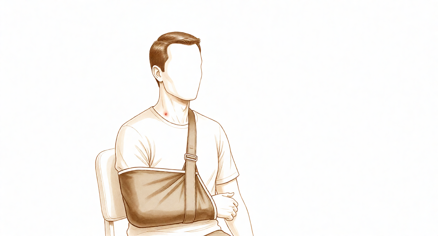
佩戴悬臂带
悬臂带可支撑手臂重量,缓解不适,并在早期保护正在愈合的骨骼。使用规则如下:
- 外出时尤其需要佩戴,以保护手臂并防止他人碰撞。睡觉时无需佩戴。
- 前三周内,大部分时间都应佩戴。之后可根据舒适度逐渐减少佩戴时间,大多数人到六周时即可停用。
- 洗澡、进行锻炼以及坐着且手臂有支撑的安静活动(如进食、书写、阅读)时,可取下悬臂带。
- 在家休息时,若注意保护,可取下悬臂带:坐着时手臂垫在枕头上,并保持手部低于肩高。
- 佩戴悬臂带期间禁止驾驶。 当您停用悬臂带,并能在驾驶时舒适且安全地操控车辆时,方可恢复驾驶,此情况将在您与 Hirpara 医生的复查中确认。
术后最初几天
如果手术中使用了神经阻滞,手臂在术后数小时内可能会感到麻木和沉重;在感觉恢复正常之前,请将其妥善置于吊带中保护。以下是早期阶段的一些实用建议:
- 在进行锻炼和物理治疗预约之前,先服用止痛药。
- 使用冰敷缓解疼痛和肿胀,每次约15–20分钟,用湿布包裹,切勿直接接触皮肤或伤口。
- 佩戴吊带时,放松肩部,让吊带承担手臂的重量。
- 注意姿势:保持耳朵、肩膀和髋部在一条直线上,避免肩膀向前耷拉;良好的姿势有助于保护骨折位置并防止僵硬。
- 从一开始就要保持手指、手腕、肘部和颈部的活动。
- 如果出现任何问题,请联系病房或告知您的物理治疗师。
第一阶段 — 保护期(第0–3周)
最初几周的重点是在骨折开始愈合时保护内固定。您将佩戴悬吊带,通过冰敷控制肿胀,并进行轻柔的练习以保持手臂其余部位的活动度:包括手、腕、肘和颈部,以及钟摆运动和低于肩高的轻柔辅助肩关节活动。最重要的规则是:肘部不要抬升至肩高以上,患侧手臂不要提举或搬运物品,不要通过手臂支撑身体起身,佩戴悬吊带期间禁止驾驶。
致物理治疗师:
目标
- 保护内固定及愈合中的骨骼和软组织
- 控制疼痛和肿胀
- 恢复肩关节低于90°抬高的被动活动范围
- 保持肘、腕、手及颈椎的全范围活动度
管理措施
- 根据需要应用冷疗及其他理疗手段;在练习和治疗前给予镇痛
- 检查悬吊带贴合度;教育患者正确使用悬吊带(保护性佩戴,尤其在外出时;根据本诊所惯例,夜间无需佩戴)及姿势
- 钟摆运动和桌面滑动练习
- 被动活动(PROM):在肩胛骨平面内进行外旋和内旋至舒适位;屈曲/侧平举/外展至最大90°
- 辅助主动活动(AAROM):使用棍棒在中立位进行外旋;仰卧位辅助屈曲至90°
- 主动活动(AROM):肘、腕、手及颈椎;握力练习(挤压球)
- 从第2周开始:抗阻腕屈曲/伸展及前臂旋转;轻柔的肩胛骨稳定及后缩练习
- 心血管训练:手臂佩戴悬吊带步行;手臂佩戴悬吊带骑固定自行车或卧式自行车
注意事项
- 禁止主动肩关节抬高
- 禁止肩关节屈曲或外展超过90°,包括被动活动
- 患侧手臂禁止提举或搬运物品;禁止手臂负重
- 佩戴悬吊带期间禁止驾驶
进阶标准
- 被动屈曲/侧平举至90°及外旋至约30°时无不适
- 静息时疼痛评分低于4/10
- 肘、腕和手主动活动范围完全恢复
- 伤口愈合,无并发症迹象
第二阶段 — 早期活动(第3–6周)
骨折开始愈合,但尚未完全愈合:此阶段旨在恢复活动度,而非力量。在舒适允许的情况下逐步停用吊带,被动及辅助活动度逐渐进展至全范围,并开始低于肩高时利用自身力量活动手臂,同时进行轻柔的肌肉激活(等长)练习。所提或所携带物品的重量不得超过一杯咖啡的重量,并避免任何强力拉伸。此阶段在约六周时以X线检查和Hirpara医生的复诊结束;该复诊(而非仅凭日历时间)是开启强化训练及肩高以上活动的关键。
致物理治疗师:
目标
- 逐步停用吊带(约在第6周时弃用)
- 在所有平面内将被动活动度进展至全范围
- 在低于90°时建立具有良好生物力学的主动活动度
- 开始轻柔的等长练习及肩周肌群训练
管理措施
- PROM(被动活动):在所有平面内进展至全范围,以耐受为度;禁止强力拉伸
- AAROM(辅助主动活动):仰卧位使用木棍进行屈曲,逐渐进展至直立位(躺椅式进展),墙壁及栏杆滑动,滑轮训练
- AROM(主动活动):抬举低于90°,无痛;仰卧位屈曲逐渐进展至站立位;坐位及侧卧位外旋
- 中立位下的肩袖等长练习;轻度的肩周肌群强化(肩胛骨后缩、低拉、中拉);轻度的肱二头肌和肱三头肌练习
- 监测代偿模式(耸肩、肩胛骨代偿)
- 心血管训练:步行;固定自行车
注意事项
- 禁止提起或携带超过约一杯咖啡重量的物品
- 在六周复诊确认骨折愈合前,禁止主动抬举超过90°
- 禁止对肩部进行强力拉伸,或采取引发疼痛的姿势
- 禁止通过手臂负重
- 佩戴吊带期间禁止驾驶
进阶标准
- 被动活动度达到全范围或接近全范围
- 主动抬举至90°,代偿极少且疼痛评分低于4/10
- X线显示骨折愈合满意,并经Hirpara医生复诊确认
第三阶段——强化训练(第6–12周)
一旦复查确认骨折愈合良好,肩关节活动将开始高于肩高,并在随后的几周内逐步建立完全主动活动范围。强化训练从轻柔开始:首先进行肌肉激活训练,然后使用弹力带,最后使用轻重量进行肩袖和肩胛骨肌肉的训练。在12周之前,提举重量保持较轻(不超过约2公斤),避免重体力或过头提举,此阶段不进行接触性运动。游泳和骑自行车通常在此阶段恢复,具体由您的物理治疗师指导。
致您的物理治疗师:
目标
- 在所有平面实现完全主动活动范围,且具有正常的生物力学机制
- 启动并推进肩袖及肩周肌群的强化训练
- 恢复正常日常活动
管理方案
- 在所有平面将主动活动范围(AROM)推进至90°以上,尽量减少代偿模式
- 根据需要拉伸:背阔肌、胸肌、后关节囊及睡眠者拉伸(sleeper stretches)
- 强化训练:等长肩袖训练,逐步过渡到使用弹力带进行抗阻外旋/内旋,初始阶段动作低于肩高;肩胛骨后缩及划船动作;肩胛平面抬举(scaption raises)、前锯肌训练及墙推(wall push-ups)在阶段后期进行
- 轻重量自由重量训练,根据耐受情况逐步推进:低负荷、高重复次数
- 有氧运动:固定自行车和步行;若复查获准,约8–10周开始游泳和跑步
注意事项
- 12周之前,提举重量不超过约2公斤
- 12周之前,避免过头或远离身体的重体力提举
- 不进行接触性运动;在阶段后期(约10–12周)之前,不进行增强式训练或冲击性负荷训练
- 强化训练应在舒适范围内进行,不应引发持续存在的疼痛
进阶标准
- 主动活动范围至少达到健侧的90%
- 肩袖及肩周肌群激活良好,抗阻训练时疼痛评分不超过3/10
- X光片显示骨折愈合进展良好,并经Hirpara医生复查确认
第四阶段 — 恢复全面活动与运动(第12周起)
最终阶段是逐步恢复较重的举重、手工劳动和运动。力量训练通过增加阻力、过头位以及(对于运动员)增强式、投掷和专项运动训练逐步推进。接触性和碰撞性运动(足球、橄榄球、骑马)需要X线显示骨折愈合,这将在您与Hirpara医生的复查中确认,最早通常在三个月到四个月左右,部分方案将碰撞性运动推迟至六个月。在骨骼未愈合前恢复运动有再骨折的风险,因此务必遵守这一限制。
物理治疗师须知:
目标
- 维持完全且无痛的活动范围
- 力量至少达到健侧的90%
- 逐步恢复手工劳动、娱乐和运动
管理
- 渐进性抗阻训练,包括离心负荷、过头位和功能模式(在耐受范围内)
- 节律性稳定训练和本体感觉训练;过头位运动员的增强式训练及间歇性投掷或球拍类运动计划
- 针对手工劳动者的工作特异性体能训练;在 unrestricted play(无限制比赛)前进行专项运动训练
- 回归运动的决策需个体化(接触性与非接触性、上肢需求),并与外科医生协调
进阶标准
- 完全且无痛的主动活动范围
- 测力计测试显示力量至少达到健侧的90%,且力量测试时无疼痛
- 完成分级回归运动计划,无疼痛或恐惧感
- 经与Hirpara医生复查确认X线骨折愈合后,方可进行接触性或碰撞性运动
术后康复方案
上述阶段改编自已发表的锁骨骨折固定术康复方案:包括马萨诸塞州总医院布里格姆运动医学科、猛犸骨科研究所、科罗拉多大学(Jonathan Bravman 医生)以及鲁什医疗中心中西部骨科(Brian Cole 医生),并结合了西萨福克郡和联合林肯郡 NHS 的物理治疗指南,以及基于运动员锁骨骨折系统综述的运动重返证据。周数范围为典型值而非固定值,您的康复进展由物理治疗师指导,并以 Dr Hirpara 复查时的骨折愈合情况为节点。本页面与诊所的一般康复建议配合使用;详见 术后疼痛管理 和 伤口护理。关于手术本身,请参阅 锁骨固定术。本方案背后的证据(手术与非手术试验数据、骨愈合率和运动重返率,以及已发表的外科医生方案)已汇总于证据部分,可从本页顶部下载 PDF 版本。
Evidence & references
Midshaft Clavicle Fracture — Operative vs Non-operative Management & Post-operative Rehabilitation (Plate ORIF)
Topic scope: (A) the decision between non-operative management and plate fixation for displaced midshaft clavicle fractures (the randomised-trial evidence on union, function and return to sport), and (B) post-operative rehabilitation after open reduction and internal fixation (ORIF) of the clavicle with a plate and screws. Distal-third and proximal-third fractures, which involve different fixation constructs, are noted only where they bear on the rehab principles.
Defining principle of the surgical rehab here: clavicle ORIF is a protect-the-fixation / protect-the-healing-fracture pathway, NOT an early-aggressive-motion pathway. The plate is a splint, not a substitute for healed bone — it neutralises load while the fracture itself unites over roughly 6–12 weeks and remodels for months afterwards. So the rehab is staged around fracture biology: a sling and below-shoulder-height-only motion early to protect the construct, range of motion progressed as the fracture knits (overhead motion deferred until the ~6-week x-ray), and strengthening / loading / collision sport withheld until radiographic union is confirmed. This is the opposite of a debridement or capsular-release pathway, where motion is the goal from day one and there is no fracture to protect. The single most important gate throughout is the x-ray, not the calendar — every major step up depends on how the fracture is healing.
A. THE OPERATIVE-vs-NON-OPERATIVE DECISION
Most clavicle fractures heal without surgery. The debate concerns completely displaced midshaft fractures (typically ≥100% displacement or ≥~2 cm shortening), where historic "all clavicles heal" teaching was overturned by randomised data.
The landmark trial — Canadian Orthopaedic Trauma Society (COTS) 2007
The COTS multicentre RCT randomised 132 patients with displaced midshaft clavicle fractures to plate ORIF vs non-operative sling treatment. Plate fixation produced a markedly lower nonunion rate (~2% vs ~23–24% non-operative), fewer symptomatic malunions, faster time to union, and better Constant and DASH scores at one year. This trial is the basis for offering surgery to active patients with completely displaced fractures — it did not establish that all such fractures require surgery. STRONG (RCT). [COTS 2007]
What later evidence tempered
- Meta-analyses of RCTs confirm operative fixation reduces nonunion and symptomatic malunion but show that much of the early functional advantage converges by 1 year, and comes at the cost of hardware-related reoperation. The decision is therefore shared and patient-specific (activity demands, displacement, comminution, smoking, occupation) rather than automatic. STRONG (SR/MA of RCTs). [Woltz-type meta-analysis; meta-regression, JSES 2020 — DOI 10.1016/j.jse.2020.02.011]
- A modern cohort comparison of dual mini-fragment plating vs non-operative care (mean 3.4-yr follow-up) found fewer union complications with fixation but similar patient-reported outcomes at final follow-up — echoing the "fixation buys reliable union, not necessarily a better long-term shoulder" theme. MODERATE (cohort). [DOI 10.1016/j.jse.2024.10.018]
- Heterogeneity between trials (how nonunion and displacement were defined, statistical handling of time-to-union) explains some of the apparent disagreement across studies — a caution against over-reading any single union statistic. MODERATE. [DOI 10.1016/j.jse.2012.03.015; meta-regression DOI 10.1016/j.jse.2020.02.011]
Construct choice (informs the rehab, not the patient's behaviour)
- Plate vs intramedullary fixation: an RCT comparing locked intramedullary nailing with plating found both achieve union; plates remain the workhorse for comminuted/displaced patterns. MODERATE (RCT). [DOI 10.1016/j.jse.2010.05.002]
- Plate position: superior plating is biomechanically strong but the plate lies directly under thin skin and is frequently symptomatic; anteroinferior plating lowers symptomatic hardware and removal rates. This is why patients commonly feel and see the plate, and why removal is a later, elective conversation. [Hardware-removal cohort, DOI 10.1016/j.jse.2017.03.011]
- Fixation reaches union even when delayed: immediate fixation vs delayed reconstruction of displaced midshaft fractures both restore objective strength and patient-oriented outcomes — reassuring that a fracture initially treated non-operatively can still be fixed successfully if it fails to unite. MODERATE (cohort). [DOI 10.1016/j.jse.2007.01.001]
B. POST-OPERATIVE REHABILITATION (plate ORIF)
The operation holds the fracture ends in position with a plate and screws so the bone can heal. Rehab is the same staged, fracture-protective sequence used across published surgeon and NHS protocols. Key facts that shape it:
- The plate neutralises load but the bone must unite biologically — typically 6–12 weeks to radiographic union, with remodelling for months after. Strengthening and loading that precede union risk implant loosening or re-fracture. Consensus / biomechanical.
- Bone healing is slower in smokers and in diabetics, and smoking can delay or prevent union — a modifiable risk worth addressing in the post-fracture window. Established.
- Overhead motion and strengthening are gated on the x-ray, not a fixed date — published protocols restrict elevation to ≤90° until early healing is confirmed (commonly the ~6-week review).
Consensus phased post-op timeline (plate ORIF)
| Phase | Window | Sling | ROM | Strengthening | Notes |
|---|---|---|---|---|---|
| I — Protection | Week 0–3 | Most of the time; off for showers/exercises/seated tasks; not required overnight | Passive/AAROM below 90° only — flexion/scaption/abduction capped at 90°, ER/IR in scapular plane to comfort; pendulums, table slides; full elbow/wrist/hand/cervical AROM | None at shoulder (grip + wrist only) | Protect fixation; settle pain/swelling; no driving while in sling; no lifting/carrying/weight-bearing through the arm |
| II — Early motion | Week 3–6 | Weaned as comfort allows; discarded by ~6 wk | Progress passive→full all planes (no forceful stretch); active motion below 90°; AAROM lawn-chair/pulley progression | Gentle isometrics + light periscapular work only | Recover movement, not strength. Lift ≤ a coffee cup. Phase ends with x-ray + review that gates overhead motion + strengthening |
| III — Strengthening | Week 6–12 | Off | AROM progresses above 90° once union confirmed; full active range built up | Cuff + scapular strengthening: isometric → bands → light weights; lift ≤ ~2 kg until 12 wk | Swimming/cycling typically return; no contact sport; no overhead/heavy lifting |
| IV — Return to activity & sport | Week 12 + | Off | Full, pain-free, maintained | Progressive heavy/eccentric/overhead loading; sport-specific + plyometric drills | Contact/collision sport needs radiographic union — typically ~3–4 months at the earliest, some protocols stage collision as late as 6 months |
The structure above matches the topic's patient protocol and is drawn from published surgeon ORIF protocols (Massachusetts General Brigham; Mammoth Orthopedic Institute; University of Colorado / Bravman; Midwest Orthopaedics at Rush / Cole) and NHS physiotherapy guidance (West Suffolk; United Lincolnshire). These protocols broadly agree on the sling ~3 weeks, ROM ≤90° early, overhead and strengthening after the ~6-week review, return to sport gated on union sequence; exact week boundaries vary by surgeon. WEAK / CONSENSUS — no rehab RCT defines the optimal regimen.
Return to sport — the evidence
- A systematic review of return to sport after clavicle fractures (Robertson & Wood, Br Med Bull 2016, 23 studies) found ~92% return to sport, at a mean of ~96 days (~3 months). MODERATE (SR of heterogeneous cohorts). [Robertson 2016]
- A more recent systematic review and meta-analysis reported mean return to play ~3.1 months operative vs ~3.9 months non-operative, with similar overall return rates but a higher rate of return to pre-injury level after operative treatment. MODERATE. [RTP SR-MA, JSES Rev 2024]
- In elite athletes specifically (e.g. NFL series), operative management has been used to achieve predictable, timely return — though selection bias makes these cohorts hard to generalise. WEAK (selected cohorts). [DOI 10.1177/0363546510372795]
The consistent signal: most athletes return by ~3 months, operative slightly faster and more reliably to pre-injury level — but union on x-ray, not the average timeline, governs clearance for collision sport.
C. KEY CONTROVERSIES / EVIDENCE QUALITY
- Who actually needs surgery. COTS established that fixation reduces nonunion/malunion in completely displaced midshaft fractures, but the early functional gap narrows by a year and fixation adds hardware reoperations. The modern position is shared decision-making for the active, completely-displaced patient — not routine fixation of all displaced fractures. Strong evidence, nuanced application.
- How big is the nonunion benefit, really? Reported nonunion rates vary with how "nonunion" and "displacement" are defined and how time-to-union is analysed; meta-regression shows this heterogeneity drives much of the between-study disagreement. Treat single headline figures with caution. Moderate.
- Hardware prominence and removal. Because the clavicle is subcutaneous, plates are often felt and sometimes symptomatic; removal rates depend heavily on plate position (anteroinferior < superior) and design (low-profile/dual). Removal is an elective, post-union decision. Moderate (cohorts).
- The rehab protocol itself is consensus. Phase timings come from surgeon patient-guidance documents and NHS leaflets, not a rehab RCT. The ≤90°-until-6-weeks and union-gated-sport principles are widely shared; precise week boundaries are not trial-derived. Weak/consensus.
D. EVIDENCE STRENGTH FLAGS (summary)
- STRONG (RCT / SR-MA of RCTs): plate fixation reduces nonunion and symptomatic malunion in displaced midshaft fractures (COTS 2007 RCT; meta-analyses), with early functional benefit that converges by ~1 year; plate vs IM nail both achieve union (RCT).
- MODERATE (cohorts / SR of cohorts): similar long-term PROs fixation vs non-op despite fewer union complications (dual-plate cohort 2024); return to sport ~92% at ~3 months, operative slightly faster/more reliable to pre-injury level (Robertson 2016 SR; RTP SR-MA 2024); hardware removal rate and its dependence on plate position; delayed fixation still succeeds.
- WEAK / CONSENSUS: the post-operative rehabilitation protocol itself (surgeon + NHS patient-guidance documents; no defining rehab RCT); elite-athlete operative series (selection bias).
CITATIONS
RAG corpus (180,000+ Orthopaedic articles) — clavicle-specific evidence
- Canadian Orthopaedic Trauma Society. Nonoperative treatment compared with plate fixation of displaced midshaft clavicular fractures: a multicenter, randomized clinical trial. J Bone Joint Surg Am. 2007;89(1):1–10. (also corpus-adjacent reanalysis: DOI 10.1016/j.jse.2012.03.015)
- Factors explaining heterogeneity in studies comparing surgical and nonsurgical treatment of midshaft clavicle fractures: a meta-regression analysis of RCTs and high-quality observational studies. J Shoulder Elbow Surg. 2020. DOI: 10.1016/j.jse.2020.02.011
- Dual mini-fragment plate fixation of midshaft clavicle fractures demonstrates fewer union complications but similar patient-reported outcomes compared to nonoperative management: a cohort study (mean 3.4-yr follow-up). J Shoulder Elbow Surg. 2024. DOI: 10.1016/j.jse.2024.10.018
- Locked intramedullary fixation vs plating for displaced and shortened mid-shaft clavicle fractures: a randomized clinical trial. J Shoulder Elbow Surg. 2010. DOI: 10.1016/j.jse.2010.05.002
- Does delay matter? Restoration of objectively measured shoulder strength and patient-oriented outcome after immediate fixation versus delayed reconstruction of displaced midshaft clavicle fractures. J Shoulder Elbow Surg. 2007. DOI: 10.1016/j.jse.2007.01.001
- Functional outcome of surgical treatment of symptomatic nonunion and malunion of midshaft clavicle fractures. J Shoulder Elbow Surg. 2007. DOI: 10.1016/j.jse.2006.12.002
- Plate fixation of midshaft clavicular fractures: patient-reported outcomes and hardware-related complications. J Shoulder Elbow Surg. 2015. DOI: 10.1016/j.jse.2015.09.029
- What is the hardware removal rate after anteroinferior plating of the clavicle? A retrospective cohort study. J Shoulder Elbow Surg. 2017. DOI: 10.1016/j.jse.2017.03.011
- A biomechanical and clinical comparison of midshaft clavicle plate fixation: are 2 screws as good as 3 on each side of the fracture? Orthop J Sports Med. 2017. DOI: 10.1177/2325967117725293
- Evolving management of middle-third clavicle fractures in the National Football League. Am J Sports Med. 2010. DOI: 10.1177/0363546510372795
- Effect of different statistical methods on union or time to union in a published study about clavicular fractures. J Shoulder Elbow Surg. 2012. DOI: 10.1016/j.jse.2012.03.015
- Treatment of clavicle fractures: current concepts review. J Shoulder Elbow Surg. 2011. DOI: 10.1016/j.jse.2011.08.053
Literature (URLs)
- Canadian Orthopaedic Trauma Society. Nonoperative treatment compared with plate fixation of displaced midshaft clavicular fractures: a multicenter RCT. JBJS 2007. https://journals.lww.com/jbjsjournal/fulltext/2007/01000/nonoperative_treatment_compared_with_plate.1.aspx
- Plate fixation versus nonoperative treatment for displaced midshaft clavicular fractures: a meta-analysis of RCTs. PubMed. https://pubmed.ncbi.nlm.nih.gov/28632595/
- Robertson GA, Wood AM. Return to sport following clavicle fractures: a systematic review. Br Med Bull. 2016;119(1):111–128. https://academic.oup.com/bmb/article-abstract/119/1/111/1744610
- Return to play following clavicular fracture — a systematic review and meta-analysis. JSES Rev Rep Tech. 2024. https://www.sciencedirect.com/science/article/pii/S2666639124001500
- Hardware removal after clavicle plating (rates, plate position): retrospective cohort. PubMed. https://pubmed.ncbi.nlm.nih.gov/28478898/
- Have new plate designs reduced hardware removal following midshaft clavicle fixation? J Clin Med. 2025. https://www.mdpi.com/2077-0383/14/18/6351
Published rehab protocols (patient-guidance — basis for the phase structure)
- Massachusetts General Brigham Sports Medicine. Rehabilitation Protocol for Clavicle ORIF. https://www.massgeneral.org/assets/MGH/pdf/orthopaedics/sports-medicine/physical-therapy/rehabilitation-protocol-for-clavicle-ORIF.pdf
- Crall T, Perumal J. Rehabilitation Guidelines for Clavicle Fracture S/P ORIF. Mammoth Orthopedic Institute. 2018. https://www.mammothortho.com/pdf/shoulder-clavicle-fx-orif-protocol.pdf
- Bravman JT. Clavicle ORIF Rehab Protocol. University of Colorado School of Medicine. https://www.sportsandshoulderdoc.com/pt-protocols/clavicle-orif.pdf
- Cole BJ. Clavicle Fracture ORIF Rehabilitation Protocol. Midwest Orthopaedics at Rush. https://www.briancolemd.com/wp-content/themes/ypo-theme/pdf/orif-clavicle-fracture-post-op-ver2.pdf
- West Suffolk NHS Foundation Trust. Clavicle ORIF — physiotherapy advice for patients after surgery. 2023. https://www.wsh.nhs.uk/CMS-Documents/Patient-leaflets/Physiotherapy/6857-1-Clavicle-open-reduction-internal-fixation-ORIF-physiotherapy-advice.pdf
- United Lincolnshire Teaching Hospitals NHS Trust. Clavicle Fracture ORIF — physiotherapy advice for patients after surgery. June 2025. https://www.ulh.nhs.uk/wp-content/uploads/2025/07/Clavicle-fracture.pdf


