

肘管松解术 资料
本方案指导您在 Mater Private Hospital Rockhampton 接受 Kieran Hirpara 医生进行的肘部尺神经松解术(肘管综合征松解术)后的康复过程。它说明了预期情况、早期数周的注意事项,以及有助于神经在组织稳定期间自由滑动的锻炼。请将此页面或其 PDF 文件带给您的物理治疗师或手部治疗师,以确保您的康复协调一致。您的治疗师可能会根据您康复的进展情况调整计划。
如果您对术后伤口有任何担忧,请联系诊所。通常,拍摄伤口照片并通过电子邮件发送以供审查会很有帮助。
两种不同的手术,两种略有不同的康复过程。 尺神经可以通过两种主要方式松解,您接受的具体手术类型会影响您早期的注意事项:
- 原位(简单)减压术: 在神经所在的原位进行松解,不移动神经。这是 Hirpara 医生进行的标准手术,康复速度较快:早期即可开始肘关节的轻柔全范围活动。不使用刚性支具。
- 前方(肌下)转位术(将神经提起并重新路由至肘部前方更受保护的位置):仅在神经在肘关节弯曲时相对于骨性突起(内上髁)发生半脱位或脱位的较少见情况下进行。这需要更谨慎的早期阶段,在神经和软组织在新位置稳定的最初几周内,避免肘关节屈曲和伸直达到极限范围。可佩戴简单吊带仅用于舒适。
请遵循以下针对您所接受手术的指导:大多数情况下为原位减压术,若您的神经不稳定则为转位术。
预期情况
关于伤口、肿胀及瘢痕管理,请参阅本诊所的伤口护理指南。
手术的目的是解除尺神经(支配小指和无名指感觉并驱动手部许多小肌肉的神经)所受的压迫。一旦压迫解除,神经便开始恢复,但神经愈合缓慢。
症状改善的速度很大程度上取决于术前神经受压的时间长短以及神经受刺激的程度。麻木感和针刺感通常最先缓解,有时在数天或数周内即可减轻。感觉丧失和手部力量恢复所需时间更长(通常为数月),且最终效果在术后一年内甚至更长时间仍可能持续改善。若术前神经长期受到严重刺激,部分感觉丧失或无力可能无法完全恢复;此时手术旨在阻止病情恶化,并为神经提供最佳的恢复机会。
比较这两种手术,对已发表试验的大型综述发现,原位简单减压术和前移术的总体效果相似,且简单减压术倾向于具有更少的伤口和软组织并发症 [1][2]。两者之间的选择由您的外科医生根据您的神经和肘部情况决定。
注意事项与限制
鼓励从术后初期开始,在舒适范围内进行手部轻度功能性活动,以完成日常任务,如自我护理、进食、穿衣、书写和打字。
早期的限制取决于您接受的具体手术:
- 原位(简单)减压术后(常规手术): 早期鼓励轻柔地活动肘部、前臂、手腕和手部全范围,以保持神经滑动。不使用支具。在前六周内,通过手臂进行的提举、抓握和负重应保持轻微,随后逐渐增加强度。
- 前方移位术后(仅当您的神经不稳定时): 前几周需保护肘部;避免强行将肘部完全弯曲或完全伸直,并避免长时间保持弯曲状态,以便神经在新的位置稳定下来。可佩戴简单吊带,仅用于舒适。神经滑动练习的开始时间比简单减压术后稍晚(通常在两到三周左右)。与原位减压术相同,在前六周内保持提举和阻力训练轻微,随后逐渐增加强度。
作为一般指导,提举和阻力强化训练在六周左右之前保持轻微,然后逐渐增加强度 [3][4]。
倚靠肘部(将肘部靠在硬表面上)会直接对神经施加压力,在恢复期间应避免。
伤口愈合后,疤痕按摩有助于保持神经上方皮肤和组织的柔软。伤口护理 页面提供了更多关于疤痕管理的信息。
这些是您手册中的练习,按照每张卡片上的说明进行。请按照 Dr Hirpara 和治疗师的指导开始:神经滑动练习的开始日期以及任何肘部活动范围限制,取决于您接受的具体手术。
您的锻炼
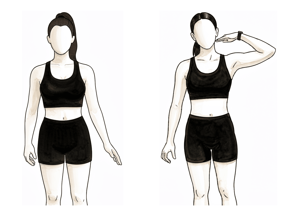
Kieran Hirpara 4.0
尺神经滑动
这些轻柔的动作可保持尺神经自由滑动,防止其粘连于愈合组织。在每个体位中仅以舒适为限平滑移动——轻微的牵拉感或刺痛感属正常现象,但应避免任何尖锐或强烈的针刺感。切勿强行拉伸。仅在外科医生或治疗师指示后开始(若曾接受转位手术,通常需延后开始——见下方注意事项)。
5次缓慢重复,每日2–3次
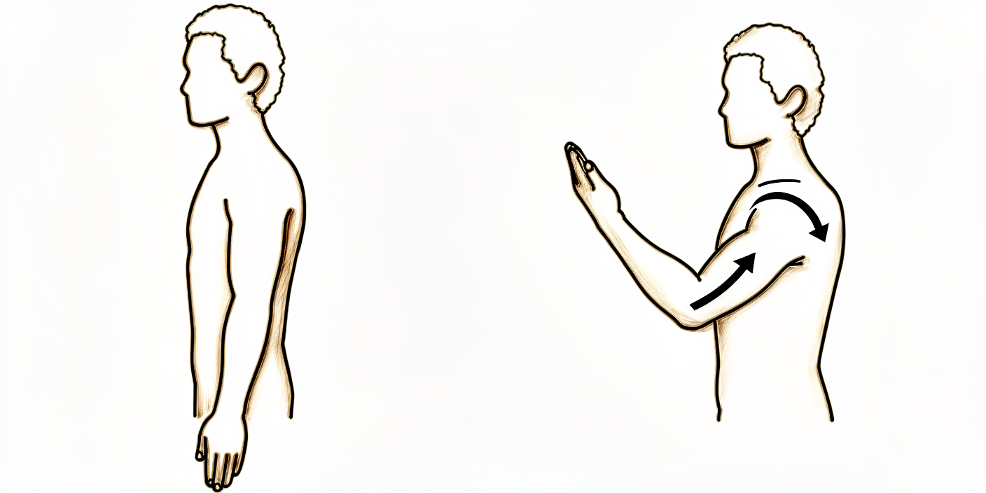
Kieran Hirpara 4.0
肘部弯曲(屈曲)
在上臂得到支撑的情况下,轻轻弯曲肘部,将手移向肩部,然后再放下。在舒适的范围内活动。如果您曾接受过移位手术,外科医生可能会要求您在最初几周内避免将肘部完全弯曲——请遵循您被给予的限制。
10次重复,3–4 次每日,在您的允许范围内
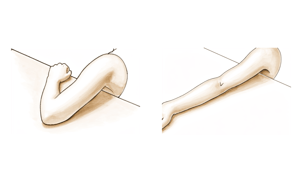
Kieran Hirpara 4.0
肘部伸直(伸展)
从弯曲位置开始,轻轻将肘部伸直至舒适的最大范围,然后恢复原位。如果您曾接受过转位手术,且在术后早期被要求避免将肘部完全伸直,则切勿强行将肘部完全伸直——只需达到您的外科医生设定的舒适活动范围即可。
10次重复,3–4 次每日,在您允许的范围内
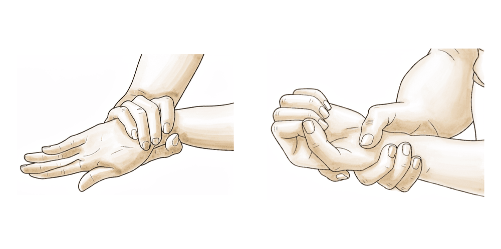
Kieran Hirpara 4.0
前臂旋转
将肘部紧贴身体一侧并弯曲成直角,缓慢地将手掌向上转向天花板,然后向下转向地面。在整个过程中,保持肘部紧贴身体。
每个方向10次,每日3–4次
Kieran Hirpara 4.0
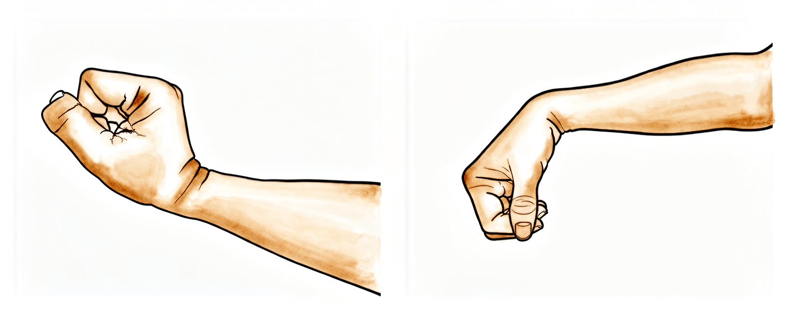
手腕和手指运动
轻轻上下屈伸手腕,并完全张开和握紧手指,先握成松拳,然后张开手指。这有助于保持整个肢体的活动,并在肘部恢复期间帮助减轻肿胀和僵硬。
每项10次,每日数次
请仅在Hirpara医生和您的手部治疗师的指导下开始下面这些锻炼动作,并严格保持在您被允许的范围内和限制内。尺神经滑动练习至关重要——它们能确保神经在新的位置保持滑动,而不是因瘢痕形成而固定,且需尽早开始。肘部屈曲、伸直和前臂旋转可防止肘部僵硬,而手腕和手指活动则维持其他部位的功能。避免长时间将肘部完全屈曲并保持静止,因为这正是最初刺激神经的原因。如果任何动作导致沿前臂延伸至小指和环指的尖锐或电击样疼痛,请立即停止。
恢复工作与活动
大多数人可在一至两周内恢复文职或轻体力工作,而较重、重复性或手工类工作通常需要约四至八周。当您的伤口能够耐受特定任务所涉及的接触和压力,且您能在上述注意事项范围内舒适地完成该任务时,即表示您已准备好从事该任务。如果您的工作较重、需要倚靠肘部或使用振动工具,请在术后复查时提出,以便制定计划(包括任何调整后的工作职责)。
驾驶通常在两至三周后恢复,前提是您已不再使用悬吊带,且能够控制车辆并在紧急情况下无痛地做出反应。恢复体育运动和过头活动通常需要六至十二周。
神经恢复遵循其自身较慢的时间线。麻木感通常最先消退,在数天至数周内缓解,而感觉减退和肌力则会在数月内持续改善,并可能在长达约一年的时间内继续好转。如果神经受压时间过长且程度严重,部分感觉减退或肌力可能无法完全恢复,此时手术的目标是阻止病情进一步进展。
方案之后
本方案与诊所的总体康复建议相辅相成:请参阅术后疼痛管理、伤口护理和手部治疗基础。上述分阶段计划与关于肘部尺神经减压术的已发表证据相符,您的持续康复将根据神经和肘部的进展情况,由您的物理治疗师或手部治疗师进行个体化指导。
参考文献
[1] Said J, Van Nest D, Foltz C, et al. Ulnar nerve in situ decompression versus transposition for idiopathic cubital tunnel syndrome: an updated meta-analysis. J Hand Microsurg. 2019;11(1):18–27. https://pmc.ncbi.nlm.nih.gov/articles/PMC6431285/ [2] Macadam SA, Gandhi R, Bezuhly M, Lefaivre KA. Simple decompression versus anterior subcutaneous and submuscular transposition of the ulnar nerve for cubital tunnel syndrome: a meta-analysis. J Hand Surg Am. 2008;33(8):1314.e1–12. https://pubmed.ncbi.nlm.nih.gov/18929194/ [3] Caliandro P, La Torre G, Padua R, Giannini F, Padua L. Treatment for ulnar neuropathy at the elbow. Cochrane Database Syst Rev. 2016;11:CD006839. https://www.cochranelibrary.com/cdsr/doi/10.1002/14651858.CD006839.pub4/full [4] Andrews K, Rowland A, Pranjal A, Ebraheim N. Cubital tunnel syndrome: anatomy, clinical presentation, and management. J Orthop. 2018;15(3):832–836. https://pmc.ncbi.nlm.nih.gov/articles/PMC6082832/
Evidence & references
This is the clinical evidence summary written for health professionals. It is technical, and it lists the research this page was built from. You do not need to read it to understand your treatment or to make a decision about it.
Topic scope: post-operative rehabilitation after surgical decompression of the ulnar nerve at the elbow. The single defining branch point is the operative technique: (A) in-situ (simple) decompression — an early-full-motion pathway; versus (B) anterior transposition (subcutaneous or submuscular) — a protected early phase that avoids end-range elbow flexion/extension for the first few weeks to protect the transposed nerve and its soft-tissue bed.
Defining principle of the rehab here: decompression relieves pressure on a nerve; it does not, by itself, create a load-bearing repair that needs months of protection. So the rehab is fundamentally an early-motion, nerve-glide pathway aimed at preventing perineural adhesion while the nerve recovers on its own (slow) biological timeline. The one variable that changes the early phase is whether the nerve was transposed — a transposed nerve sits in a new bed and end-range elbow excursion is restricted briefly to protect it, so nerve glides start later and elbow ROM is capped for a few weeks. Phase timings below are typical of published surgeon protocols and institutional consensus rather than trial-derived.
A. PROCEDURE CHOICE & OUTCOME EQUIVALENCE
- In-situ decompression and anterior transposition give equivalent clinical outcomes. Multiple meta-analyses of RCTs and comparative series find no significant difference in motor nerve conduction velocity or clinical outcome scores between simple decompression and transposition for idiopathic cubital tunnel syndrome. Strong (multiple SR/meta-analyses).
- Simple decompression carries a lower complication burden (wound, soft-tissue, devascularisation risk), and is often preferred where the nerve is stable and does not subluxate. Moderate–strong.
- Transposition is selected for nerve instability/subluxation, prior failed in-situ release, bony deformity, or a hostile cubital tunnel floor — surgeon's intra-operative judgement. Consensus.
- Endoscopic vs open in-situ decompression show comparable outcomes; choice does not change the rehab pathway (both early-motion). Moderate (SR).
B. POST-OPERATIVE REHABILITATION
Common principles (both pathways)
- Early digital, wrist and shoulder motion from day 1 to prevent stiffness and oedema.
- Ulnar nerve gliding to prevent perineural adhesion — timing differs by pathway (see below).
- No elbow leaning / direct pressure over the nerve during recovery.
- Wound: suture removal ~10–14 days; scar massage and desensitisation once healed.
- Nerve recovery is slow and graded: paraesthesia often improves first (days–weeks); numbness and intrinsic strength lag (months); final outcome continues to ~12 months. DASH, clinical findings and NCV improve postoperatively, with significant early gains by ~1 month in cohort data. Pre-operative severity/chronicity is the dominant predictor of incomplete recovery.
Phased timeline (typical of published surgeon protocols)
| Phase | In-situ (simple) decompression | Anterior transposition (SC / submuscular) |
|---|---|---|
| Week 0–2 | Soft dressing; early active full elbow ROM + digit/wrist/shoulder ROM; light ADLs | Splint/sling for comfort/protection (often elbow ~semi-flexed early); avoid end-range flexion AND extension, and avoid sustained/prolonged elbow flexion; digit/wrist/shoulder ROM |
| Week 2–6 | Progress to full unrestricted active ROM; scar massage + desensitisation once healed; nerve glides as tolerated | Suture out ~10–14d; gradually restore elbow ROM within set limits; scar/desensitisation; introduce nerve glides — typically deferred to this window |
| Week ~6+ | Strengthening / lifting built up as tolerated; return to full activity | Restrictions usually lifted ~6 wk; resistance strengthening from ~6 wk; build up gradually |
Dr Hirpara's practice parameters:
- Default operation = in-situ (simple) decompression; anterior submuscular transposition is reserved for a nerve that subluxates over the medial epicondyle. No rigid brace is used.
- Early elbow ROM: full active elbow motion from day 1 after in-situ decompression. After a transposition the elbow is protected from end-range flexion/extension for the first few weeks (a simple sling for comfort only — no rigid brace).
- Nerve glides: start early/as-tolerated after in-situ; start around 2–3 weeks after a transposition.
- Lifting: kept light (around ≤2 kg) for the first ~6 weeks, then resistance strengthening is built up gradually.
- Nerve recovery: paraesthesia settles first (days–weeks); numbness and intrinsic strength recover over months and can keep improving to ~12 months. Pre-operative severity/chronicity is the dominant predictor — long-standing severe compression may not fully recover, and surgery then aims to halt progression.
C. KEY CONTROVERSIES / EVIDENCE QUALITY
- Procedure equivalence is well supported (multiple meta-analyses); the complication-profile advantage of simple decompression drives the "in-situ first unless unstable" stance. Strong.
- The post-op rehab protocol itself is consensus/expert — drawn from surgeon patient-guidance protocols, not a rehab RCT. Phase timings are typical, not trial-derived. Weak/consensus.
- Nerve-glide evidence is stronger as a non-operative and adhesion-prevention measure than as a proven post-operative outcome-changer; biomechanical and clinical work supports gliding to reduce excursion-related symptoms. Moderate.
D. EVIDENCE STRENGTH FLAGS (summary)
- STRONG (SR / meta-analysis): clinical-outcome equivalence of in-situ decompression vs anterior transposition; lower complication rate with simple decompression.
- MODERATE (cohorts / SR): endoscopic vs open in-situ equivalence; post-op DASH/NCV improvement with early gains by ~1 month; nerve-gliding rationale.
- WEAK / CONSENSUS: the post-operative rehabilitation protocol (surgeon patient-guidance documents; no defining rehab RCT) — including the transposition early-ROM cap, nerve-glide start date, and the ~6-week lifting/strengthening threshold.
CITATIONS
RAG corpus (180,000+ Orthopaedic articles)
- Open vs retractor-endoscopic in-situ decompression of the ulnar nerve in cubital tunnel syndrome. Neurosurgery. DOI: 10.1227/neu.0b013e3182846dbd
- Randomized, prospective study comparing ulnar neurolysis in situ with submuscular transposition. Neurosurgery. DOI: 10.1227/01.neu.0000194847.04143.a1
- Open versus endoscopic in situ decompression in cubital tunnel syndrome: a systematic review. Int J Surg. 2016. DOI: 10.1016/j.ijsu.2016.09.012
- Simple decompression vs. subcutaneous anterior transposition of the ulnar nerve (2025). J Hand Surg Glob Online / XRRT. DOI: 10.1016/j.xrrt.2025.100630
- Cubital tunnel syndrome: current concepts. Curr Rev Musculoskelet Med. 2020. DOI: 10.1007/s12178-020-09650-y
- Predictors of surgical revision after in situ decompression of the ulnar nerve. J Shoulder Elbow Surg. 2015. DOI: 10.1016/j.jse.2014.12.015
- Clinical outcomes of ulnar nerve gliding exercise in the nonoperative treatment of cubital tunnel syndrome. JSES Int. 2025. DOI: 10.1016/j.jseint.2025.02.001
- Biomechanical analysis of ulnar nerve gliding and elongation. Clin Shoulder Elbow. 2024. DOI: 10.5397/cise.2024.00934
- Postoperative improvement in DASH score, clinical findings and nerve conduction velocity in cubital tunnel syndrome. Sci Rep. 2016. DOI: 10.1038/srep27497
Comparative-effectiveness literature (URLs)
- Said J, et al. Ulnar nerve in situ decompression versus transposition for idiopathic cubital tunnel syndrome: an updated meta-analysis. J Hand Microsurg. 2019. https://pmc.ncbi.nlm.nih.gov/articles/PMC6431285/
- Macadam SA, et al. Simple decompression versus anterior subcutaneous and submuscular transposition of the ulnar nerve: a meta-analysis. J Hand Surg Am. 2008. https://pubmed.ncbi.nlm.nih.gov/18929194/
- Caliandro P, et al. Treatment for ulnar neuropathy at the elbow. Cochrane Database Syst Rev. 2016;CD006839. https://www.cochranelibrary.com/cdsr/doi/10.1002/14651858.CD006839.pub4/full
- Andrews K, et al. Cubital tunnel syndrome: anatomy, clinical presentation, and management. J Orthop. 2018. https://pmc.ncbi.nlm.nih.gov/articles/PMC6082832/
Published rehab protocols (patient-guidance — basis for the phase structure)
- University of Virginia Orthopaedics — Cubital Tunnel Release, In-situ Rehabilitation Guidelines. https://med.virginia.edu/orthopaedic-surgery/wp-content/uploads/sites/242/2024/09/Cubital-tunnel-release-in-situ.pdf
- University of Virginia Orthopaedics — Cubital Tunnel Release, Anterior Subcutaneous Transposition. https://med.virginia.edu/orthopaedic-surgery/wp-content/uploads/sites/242/2024/09/Cubital-tunnel-release-anterior-subcutaneous-transposition.pdf
- AAOS OrthoInfo — Cubital Tunnel Release (patient recovery expectations). https://orthoinfo.aaos.org/en/treatment/cubital-tunnel-release/





