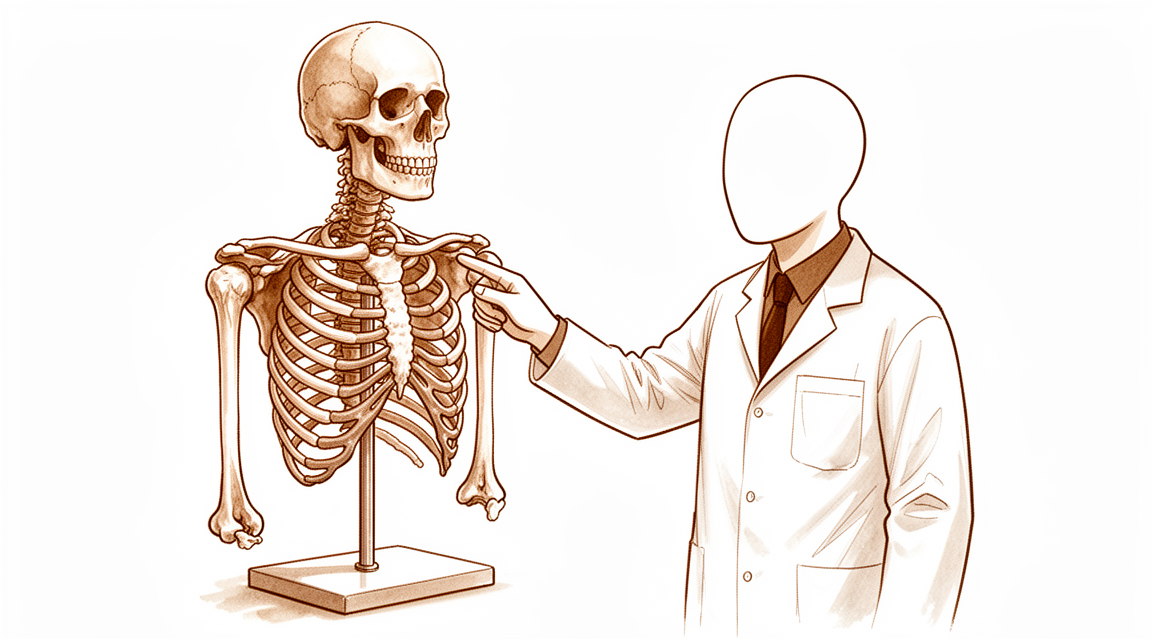
远端肱三头肌腱修复术
Patients › Rehabilitation
肘部远端肱三头肌腱修复后的保护性康复计划:初期将手臂置于90度角的简易吊带中休息,随后进行活动度恢复,接着进行主动伸直训练,最后分阶段谨慎进行力量训练。
本方案指导您在肱三头肌远端肌腱(将肱三头肌——负责伸直肘部的肌肉——锚定于肘尖的肌腱)手术修复后的康复过程,由基兰·希尔帕拉(Kieran Hirpara)医生在马特私人医院洛克汉普顿(Mater Private Hospital Rockhampton)负责。本方案从您的家庭锻炼计划开始,随后是专为您的物理治疗师或手部治疗师编写的结构化临床方案;请在首次治疗就诊时携带此页面或其PDF文件,以确保您的康复过程协调一致。您的治疗师可能会根据您的康复进展调整计划。
如果您对术后伤口有任何疑虑,请与诊室联系。拍摄伤口照片并发送电子邮件以供审查通常很有帮助。
预期情况
肱三头肌远端修复术将撕裂的肌腱重新附着于肘部鹰嘴处的骨面上。手术结束时,Hirpara 医生会在肘关节屈曲至直角(90°)时检查修复部位是否安全且完整。因此,您的肘关节将用简易悬吊带固定于约 90°,这是一个舒适的标准位置。无需使用铰链式支具,肘关节也不会保持接近伸直的位置。在进行锻炼和清洗时需取下悬吊带。
肌腱在两种情况下会承受负荷(受到牵拉),早期康复计划旨在同时防范这两种情况:
- 屈肘会牵拉修复部位。 因此,在最初约六周内,屈曲角度限制在 90°(直角)。相反,伸肘会使肌腱放松,因此在舒适范围内,完全伸直是被允许且鼓励的。
- 主动伸肘会锻炼肱三头肌,从而对修复部位产生牵拉力。 因此,在最初约六周内,您不应依靠自身力量主动伸肘;该方向的运动应轻柔地进行并借助外力辅助,而非由肱三头肌自身发力。
随后将分阶段逐步增加活动范围:约六周时增加屈曲幅度,约六周时开始主动伸肘,约十二周时开始抗阻(负重)力量训练。修复部位需数月时间才能逐渐成熟,因此较重负荷及重返运动是逐步恢复的,而非一次性进行。
注意事项与限制
- 在最初大约六周内,不要将肘关节弯曲超过直角(90°),因为弯曲会拉伸并牵拉修复部位。完全伸直是可以的,并且被鼓励进行。
- 在最初大约六周内,不要依靠肌肉力量主动伸直肘关节;应轻柔地借助外力帮助伸直,而不是通过肱三头肌发力。
- 在大约十二周内,不要进行任何抗阻伸直动作,也不要通过手臂进行推/压动作:禁止进行肱三头肌后踢、卧推或过头推举,也不要用患侧手臂从椅子或床上撑起身体。
- 按照指示佩戴呈90°角的吊带(不是支具,也不是保持完全伸直状态),在佩戴吊带期间或手臂无法安全控制方向盘时,不要驾驶车辆。
- 从一开始就要保持手、腕和肩部的活动,并在舒适范围内用手进行日常轻体力活动,前提是这些活动不涉及推、提重物或肘关节的强制弯曲。
关于伤口、肿胀和瘢痕管理,请参阅诊所的伤口护理指南。
您的锻炼
这些是您讲义中的锻炼项目。仅在 Hirpara 医生和您的物理治疗师指导下开始进行,并严格遵循您被允许的关节活动度和限制范围。早期锻炼旨在保持肘关节和前臂的活动,而不牵拉或锻炼修复部位:在受保护的 0–90° 活动范围内进行轻柔活动、辅助屈曲以及前臂旋转。肱三头肌等长收缩、主动伸直和弹力带训练属于后期阶段,在您获得明确许可之前不得开始。如果任何动作引起肘后部锐痛,请立即停止。
您的临床方案
本页面其余部分为肱三头肌远端腱修复术后的分期康复临床方案。本节内容将提供给您的物理治疗师或手治疗师,每个阶段均以通俗易懂的语言解释当前的康复重点。由于肘关节屈曲(会牵拉修复处)和主动或抗阻伸肘(会使肱三头肌收缩并压迫修复处)会对修复部位产生负荷,因此本方案在恢复关节活动度、主动伸肘及抗阻力量训练的过程中,对这两种负荷进行保护。
治疗前,请查阅患者的手术记录和既往病史,并与主刀医生沟通固定方式(经骨隧道 vs 缝线锚钉 footprint)、组织质量以及受保护的活动范围。Hirpara 医生的修复术式术中检查显示在屈曲 90° 时是安全的,术后使用简单吊带将肘关节固定于 90° 屈曲位(不使用铰链式支具,也不保持伸直位);受保护的活动范围为 0°–90°,伸肘活动以舒适为限,屈曲限制在 90°。
第一阶段 — 90°吊带保护下活动(第0至6周)
前六周旨在保护修复部位,同时防止肘关节僵硬。手臂置于简单的90°吊带中,仅在锻炼和卫生清洁时取下。肘关节仅在受保护的0–90°弧内活动,伸直至无痛舒适位,但屈曲不超过直角,且绝不在肱三头肌主动收缩下进行。
致物理治疗师:
教育与注意事项 - 使用90°简单吊带固定(不使用铰链式支具,不保持于近伸直位);仅在锻炼和清洗时取下 - 受保护的活动范围仅限0–90°:伸直可至完全/舒适位;屈曲不超过90° - 禁止主动或抗阻肘关节伸直(肱三头肌主动收缩会对修复部位产生负荷) - 患肢不可负重或推撑;在舒适范围内可进行轻量、无负荷的手部活动 - 早期轻柔进行肩关节被动活动(保护穿过肩部的肱二头肌长头)
管理 - 伤口:按医嘱处理手术敷料;监测感染迹象 - 水肿:抬高患肢,轻柔的手部泵血运动,必要时冰敷 - 锻炼:肘关节主动辅助活动(AAROM)/被动活动(PROM)在0–90°范围内(伸直至舒适位,屈曲限制在90°);腕关节、手部及握力的主动活动范围(ROM)锻炼;轻柔的肩关节活动;轻柔的前臂旋转;禁止主动伸直
进阶标准 - 伤口愈合;约在第6周时,0–90°活动范围舒适且可控
第二阶段——推进屈曲并启动主动伸展(第6至12周)
约6周后,解除屈曲限制,并将弯曲角度逐步推进至超过90°直至完全伸直。引入无阻力的主动伸直(伸展),并以等长收缩方式轻柔地重新激活肱三头肌。仍禁止进行抗阻伸展及负重。
供您物理治疗师参考:
评估 - 主动和被动活动范围(屈曲现逐步推进至超过90°,伸展);疼痛和肿胀;伤口/瘢痕复查
教育和注意事项 - 从约6周开始,逐步将屈曲角度推进至超过90°直至完全伸直 - 直至12周前,禁止进行抗阻伸展及通过手臂负重
管理 - 练习:第6–8周开始进行无阻力的主动向心伸展,在无痛范围内进行(用另一只手辅助降低/离心阶段);第8周进行轻量的次最大肱三头肌等长收缩;继续全弧度的活动度训练和前臂旋转;愈合后开始瘢痕管理
进阶标准 - 无痛的全范围活动度;良好的控制下完全主动伸展;疼痛评分≤3/10
第三阶段——强化与回归(第12周至16周及以后)
一旦恢复活动度并获准进行抗阻训练(约12周),即可开始强化训练,并逐步增加负荷:进行肱三头肌抗阻训练(先向心后离心),然后进行轻量的闭链负重训练,随后进行有限范围的推举动作。重返运动以达标为依据,最早在5至6个月时进行。
致您的物理治疗师:
评估 - 与对侧相比的肱三头肌力量;负荷下的疼痛/肿胀反应;根据需要进行功能性和运动/工作特异性测试
教育与注意事项 - 从约12周开始进行肱三头肌抗阻强化训练(向心→离心);逐步增加负荷 - 从约12周开始闭链负重训练(从轻量、小范围开始);从约14周开始进行轻量的推举动作(俯卧撑,有限范围)
管理 - 练习:渐进性抗阻肘关节伸展(弹力带→轻重量);分级闭链负荷;有限范围的推举;继续进行任何残留的活动度训练 - 一旦力量接近对称且功能恢复良好,可考虑出院 - 如果恢复出现平台期或结果不佳,考虑转诊回主治医生
重返运动标准 - 肱三头肌肌力5/5;无痛的高速度和运动特异性控制
重返工作与活动
从术后开始,鼓励在舒适范围内进行日常手部轻度活动(如进食、书写和轻度自理),前提是避免推、提或使肘关节屈曲超过其限制。由于在手臂佩戴悬吊带或无法安全控制方向盘期间不得驾驶,请在早期数周安排他人协助交通;待拆除悬吊带并经复查确认能够安全操控车辆后,方可恢复驾驶。
手臂的抗阻负荷与负重活动(推、压、提、拉)需等待至约12周,随后逐步增加。重返运动最早需5至6个月,其依据是恢复无痛的全范围关节活动度及充分、对称的肱三头肌力量,由Hirpara医生与您的物理治疗师评估判定,而非仅依据日历时间。较重的体力劳动遵循相同的基于标准的渐进原则。
术后方案
本方案与诊所的一般康复建议并行:请参阅术后疼痛管理、伤口护理和重返运动。上述分阶段计划反映了肱三头肌远端肌腱修复术后已发表的康复指南,您的持续康复将由Hirpara医生和您的物理治疗师根据肘关节的进展情况个体化指导。
Evidence & references
Distal Triceps Tendon Repair — Post-operative Rehabilitation (Evidence Brief)
Topic scope: post-operative rehabilitation after surgical reattachment of the avulsed distal triceps tendon to the olecranon (transosseous bone tunnels or suture-anchor footprint repair; best performed within ~3 weeks of injury). The extension mechanism is loaded by elbow flexion (passive stretch of the repair) and by active/resisted extension (triceps contraction), so the rehab cadence is built around protecting both, then restoring motion, then active extension, then resisted strength.
Defining principle: the repair is loaded in flexion and by triceps contraction, so early rehab limits flexion and blocks active/resisted extension while motion is restored, then releases active extension (~6 wk) and resisted extension (~12 wk) in steps, with return to sport at ~5–6 months. Dr Hirpara's stance: the repair is checked intra-operatively to be safe at 90° of flexion, so the elbow is rested in a simple sling at 90° (a standard, comfortable position — no hinged brace, and NOT held near extension) with a protected 0–90° arc (extension free to comfort, flexion capped at 90°) for ~6 weeks. This is deliberately less restrictive on early flexion than the published near-extension / 20°-flexion-lock guidelines, while keeping the key loading rules identical (no active extension to 6 wk, no resisted extension to 12 wk).
Evidence base and corpus note
No RCT and no large prospective cohort defines the rehab cadence for distal triceps repair. The phased timeline rests on a published institutional clinical-care guideline (Ohio State Sports Medicine, 2021), which itself cites the core review literature, corroborated by several surgeon and physiotherapy phased protocols. The local RAG corpus is thin on triceps-specific phased rehab (rotator-cuff and biceps content dominates), but it does contain the key biomechanical repair-strength papers, which inform how early and how aggressively one can mobilise. The week-by-week timeline is therefore carried by the published clinical-care guideline, with the corpus supplying the repair-strength evidence that justifies the cadence.
Key principles and controversies
- Early motion vs prolonged immobilisation. Classic teaching favours protective immobilisation (splint 2–6 wk, flexion-limited brace) because the triceps insertion is loaded in flexion. A counter-trend pushes accelerated early ROM where fixation is strong — a cadaveric study comparing dynamic-tape with standard suture fixation under an intense early-rehab protocol found the novel construct biomechanically superior, i.e. fixation strength is the rate-limiter for how early one can mobilise.
- Suture-anchor vs transosseous (bone-tunnel) repair strength. Carpenter et al. (JSES 2018) found no difference in tendon displacement between transosseous cruciate tunnels and suture-anchor repair when the number of sutures is equalised; the technique by Sarokhan & Leung (Arthrosc Tech 2019) cites Clark et al. (2014) finding anatomic (knotless) footprint repair superior to transosseous cruciate repair. Stronger anatomic footprint fixation is the lever that justifies earlier/more aggressive flexion and earlier resisted extension.
- Flexion-limit progression. No consensus on the exact ramp — the OSU guideline locks at 20° then advances ~15°/5 days; others use ~10°/week or an open 0–60° arc. All converge on full passive flexion by ~6 weeks, with active extension deferred to ~6 weeks and resisted extension to ~12 weeks. KH's variant keeps the elbow at 90° in a simple sling with a free 0–90° arc — less restrictive on early flexion, same loading deferrals.
- Strength athletes / high demand. Retrospective series in strength athletes report satisfactory return to sport but underline that resisted extension and pressing loads are the highest-risk re-rupture activities, supporting the firm 12-week resisted-extension / pressing block.
Phased timeline
| Phase | Window | Sling / ROM ceiling | Exercises | Criteria to progress |
|---|---|---|---|---|
| I — Protected motion | Weeks 0–6 | Simple sling at 90° (no hinged brace, not near extension), off for exercises. Protected arc 0–90°: extension free to comfort, flexion capped at 90°. No active extension. | AAROM/PROM elbow within 0–90°; wrist/hand/grip AROM; gentle shoulder ROM; forearm rotation | Wound healed; comfortable, controlled 0–90° arc at ~6 wk |
| II — Advance flexion + active extension | Weeks 6–12 | Release flexion cap; progress flexion past 90° toward full. No resisted extension / weight-bearing. | Wk 6–8 active concentric extension no resistance (assist eccentric with other arm); wk 8 light submaximal triceps isometrics | Full painless ROM; full active extension with good control; pain ≤3/10 |
| III — Strengthening & return | Weeks 12–16+ | Resisted triceps strengthening (concentric → eccentric) from ~12 wk; CKC weight-bearing from ~12 wk (light, small range); limited-range pressing ~wk 14 | Progressive resisted extension; graded loading; sport-/work-specific progression | 5/5 triceps strength; pain-free high-velocity / sport-specific control |
| Return to sport | ~5–6 months | Criterion-based, at the earliest | — | Full pain-free ROM + symmetrical triceps strength |
Evidence strength flags
- MODERATE (protocol cadence): the phased timeline (no active extension to ~6 wk, resisted extension to ~12 wk, return to sport ~5–6 mo) — anchored to the OSU Sports Medicine clinical-care guideline and corroborating surgeon/PT protocols. No defining rehab RCT.
- MODERATE (repair-strength biomechanics): suture-anchor vs transosseous equivalence with equalised sutures (Carpenter 2018); anatomic footprint superiority (Clark, via Sarokhan & Leung); insertional footprint anatomy (Whitaker 2022) — these justify the mobilisation cadence.
- LOW–MODERATE (KH's 90°-sling / flexion-capped-at-90° variant): biomechanically sound (flexion is the repair-tensioning motion; intra-op tensioning at 90° defines the safe arc) and less restrictive on early flexion than published near-extension guidance, while preserving the key extension-loading deferrals. Consensus / expert rather than trial-derived; corpus gap — no RCT or large cohort defines this exact variant.
CITATIONS
RAG corpus (180,000+ Orthopaedic articles)
- Keener JD, Sethi PM. Distal triceps tendon injuries. Hand Clin. 2015;31(4):641–650. DOI: 10.1016/j.hcl.2015.06.012
- Carpenter SR, Stroh DA, Melvani R, et al. Distal triceps transosseous cruciate versus suture anchor repair using equal constructs: a biomechanical comparison. J Shoulder Elbow Surg. 2018;27(11):2052–2056. DOI: 10.1016/j.jse.2018.07.005
- Sarokhan AK, Leung NL. Acute triceps tendon repair: a technique utilizing 3 curved tunnels and proximal knots. Arthrosc Tech. 2019;8(11):e1325–e1330. DOI: 10.1016/j.eats.2019.07.001
- Ng T, Rush LN, Savoie FH. Arthroscopic distal triceps repair. Arthrosc Tech. 2016;5(6):e1107–e1112. DOI: 10.1016/j.eats.2016.06.011
- Whitaker JJ, Hartke J, Hawayek BJ, et al. Histologic evaluation of the triceps brachii tendon insertion: implications for triceps-sparing surgery. J Hand Surg Am. 2022. DOI: 10.1016/j.jhsa.2022.03.020
Published rehabilitation protocols & literature (URLs)
- Ohio State University Sports Medicine. Distal Triceps Repair — Clinical Care Guideline (G. Hock PT DPT OCS; rev. M. Salsbery PT DPT SCS; Dec 2021). https://medicine.osu.edu/-/media/files/medicine/departments/sports-medicine/medical-professionals/shoulder-and-elbow/distaltricepsrepair.pdf (NB: its near-extension / 20°-flexion-lock immobilisation differs from Dr Hirpara's 90°-sling approach; the loading deferrals are shared.)
- Cadaveric study of dynamic-tape vs standard suture fixation in distal triceps repair under an intense early-rehab protocol. PMC. https://www.ncbi.nlm.nih.gov/pmc/articles/PMC12423150/
- Distal triceps tendon repair in strength athletes — satisfactory return to sport (22 cases). PMC. https://www.ncbi.nlm.nih.gov/pmc/articles/PMC11355401/
Note on corpus gap: the RAG corpus lacks a dedicated distal-triceps phased rehab article; the week-by-week timeline is carried by the OSU clinical-care guideline (and corroborating surgeon protocols), with the corpus papers supplying the repair-strength evidence that justifies the cadence. Flagged accordingly.


