Dedo de martelo
Patients › Hand
Mallet finger causes fingertip drooping after extensor tendon injury; splinting is key, surgery occasionally needed.
O que você está sentindo
Você pode notar dor na ponta do dedo. Esse desconforto geralmente se concentra na última articulação, onde o tendão se conecta ao osso. Em alguns casos, você pode sentir essa dor em ambas as mãos ao mesmo tempo. A lesão pode envolver um rompimento do tendão ou uma pequena fratura no local de sua inserção.
A dor geralmente piora quando você tenta esticar o dedo. Você pode ter dificuldade para empurrar contra resistência ou levantar objetos pesados. Tarefas simples, como abotoar uma camisa ou alcançar as costas para fechar um sutiã, podem se tornar desafiadoras. Seu dedo pode parecer rígido, especialmente logo após acordar pela manhã.
Muitas pessoas descobrem que o repouso do dedo ajuda a aliviar a dor. No entanto, manter o dedo esticado é crucial para a cicatrização. Se você dobrar a ponta, pode sentir uma puxada aguda ou aumento da dor. Isso ocorre porque o tendão está tentando deslocar a articulação de seu alinhamento.
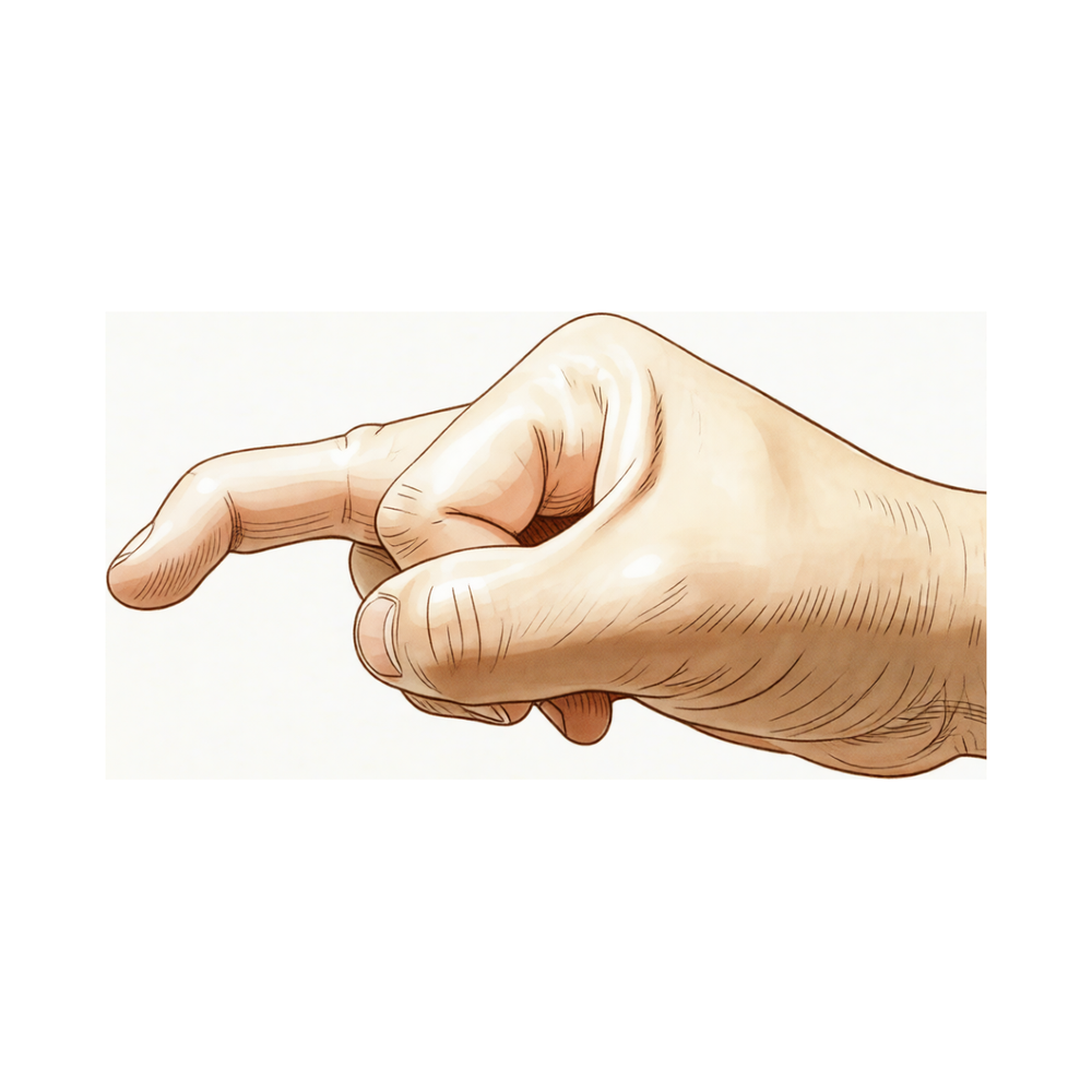
Em casos graves, você pode notar que a ponta do dedo fica caída para baixo. Essa queda pode dificultar a firmeza ao segurar objetos. Você também pode experimentar inchaço ao redor da articulação. Embora a maioria das lesões cicatrize bem com o uso de uma tala, alguns casos exigem cuidados mais complexos. Seu cirurgião determinará se você precisa de cirurgia com base no tamanho de qualquer fragmento ósseo ou na posição da articulação.
Se você tiver um fragmento ósseo grande ou se a articulação sair do lugar, seu cirurgião pode recomendar uma operação. Isso é frequentemente feito para restaurar o alinhamento e a função adequados. Tanto os tratamentos cirúrgicos quanto os não cirúrgicos geralmente levam a excelentes resultados. Você pode esperar recuperar o uso completo do dedo com os cuidados adequados.
O que está realmente acontecendo
A ponta do seu dedo possui um pequeno tendão chamado tendão terminal. Ele atua como uma corda que conecta o seu músculo ao osso na extremidade mais distal do dedo. Essa corda permite que você estenda o dedo. Quando você se lesiona nessa área, essa corda se rompe ou arranca um pequeno fragmento ósseo da articulação. Isso impede que o sinal de extensão chegue à ponta do seu dedo.
A causa mais comum é uma força súbita que dobra o seu dedo estendido para trás. Imagine que seu dedo ficou preso em uma bola ou na moldura de uma porta enquanto estava estendido. A articulação se flexiona rapidamente demais. Esse estiramento súbito arranca o tendão do osso. Em alguns casos, um pequeno fragmento ósseo se desprende junto com o tendão. A aparência clínica é semelhante, seja o tendão rompido ou um fragmento ósseo arrancado. Em ambos os casos, a conexão está interrompida.
Como a "corda" de extensão não está mais conectada, o seu dedo repousa em uma posição flexionada. Você não consegue ativamente levantar a ponta do seu dedo para estendê-lo. Isso é chamado de deformidade em dedo em martelo. No entanto, você ainda pode estendê-lo passivamente com a outra mão. A cápsula articular e os ligamentos permanecem intactos, portanto, a articulação em si não se subluxa. A posição flexionada é simplesmente o resultado da tração muscular desoposta do lado oposto do dedo.
Esta lesão afeta mais frequentemente os dedos mínimo, anelar ou médio da sua mão dominante. É mais comum em homens. Embora geralmente ocorra por trauma, alguns pacientes idosos com artrise por desgaste podem desenvolver essa postura sem uma lesão específica. Em crianças, a lesão pode envolver a placa de crescimento em vez do tendão. Independentemente da causa, o resultado é o mesmo: o mecanismo que levanta a ponta do seu dedo está desconectado, deixando-a pendente.
O que podemos fazer a respeito
A maioria das lesões de dedo em martelo cicatriza sem cirurgia. Provavelmente você usará uma tala para manter a ponta do dedo reta. Isso permite que o tendão cicatrize na posição correta. Um terapeuta da mão pode tratá-lo com a mesma eficácia de um cirurgião em casos simples. Eles utilizam métodos que raramente causam problemas de pele. Você também pode tentar o uso de talas noturnas, mas isso não melhora o resultado final. Alguns pacientes realizam exercícios em conjunto com o uso de talas, embora as evidências sobre isso sejam mistas. Você deve usar sua tala continuamente durante o tempo recomendado pelo seu cirurgião. Não a remova para lavar ou dobrar o dedo. A consistência é a chave para o sucesso.
O manejo da dor foca no conforto enquanto o tendão cicatriza. Você pode tomar analgésicos ou anti-inflamatórios sem prescrição médica conforme necessário. Eles ajudam você a gerenciar as atividades diárias enquanto o dedo está imobilizado. Alguns pacientes perguntam sobre injeções. No entanto, as evidências não apoiam injeções de cortisona, ácido hialurônico ou PRP para esta lesão específica. Esses tratamentos não fazem parte do plano de cuidado padrão para dedo em martelo. Seu foco deve permanecer em manter a articulação reta com sua tala. Se você tiver dor significativa, discuta opções seguras com seu cirurgião. Evite massagem ou alongamento agressivos, pois podem interromper a cicatrização.
A cirurgia só é considerada se o tratamento conservador falhar ou se a lesão for grave. Seu cirurgião pode recomendar uma operação se você tiver uma fratura grande envolvendo mais de um terço da superfície articular. A cirurgia também está indicada se o fragmento ósseo tiver se deslocado. Alguns pacientes escolhem a cirurgia porque não podem trabalhar com a tala no lugar. Se você tiver uma lesão crônica que não cicatrizou após meses de uso de tala, a cirurgia pode ser uma opção. O procedimento repara o tendão danificado para restaurar sua capacidade de estender o dedo. Esta é uma operação de menor porte com alta taxa de sucesso. Seu cirurgião discutirá os riscos e benefícios específicos com você se esse caminho se tornar necessário.
O que esperar
A maioria das lesões de dedo em martelo cicatriza bem sem cirurgia. Seu cirurgião provavelmente recomendará o uso de uma tala para manter a ponta do dedo reta. Isso permite que o tendão ou o osso se reconectem. Tanto os tratamentos cirúrgicos quanto os não cirúrgicos levam a excelentes resultados clínicos para a maioria dos pacientes. Você pode esperar um alto nível de satisfação com o resultado do seu tratamento.
O caminho para a recuperação depende da gravidade da sua lesão. Para casos simples, uma tala simples ou uma tala dorsal colada geralmente é suficiente. Se a sua lesão envolver um grande fragmento ósseo ou a articulação tiver se deslocado, a cirurgia pode ser recomendada. A cirurgia também é uma opção para casos crônicos ou se tratamentos anteriores falharam. Em crianças, a necessidade de cirurgia é menos clara, mas o manejo não cirúrgico permanece eficaz para a maioria.
Você deve saber que a correção completa da posição do dedo nem sempre é garantida. Se você tiver um dedo em martelo crônico grave com uma curvatura significativa, a correção completa é menos consistente. No entanto, mesmo nesses casos complexos, os resultados a longo prazo são frequentemente bons a excelentes. As taxas de complicação para o manejo conservador são baixas. Problemas graves, como infecção ou deformidade da unha, são raros, especialmente com as técnicas cirúrgicas modernas.
A recuperação é um processo gradual. Você pode notar algum rigidez ou uma pequena curvatura restante na ponta do seu dedo. Isso é comum e muitas vezes não afeta significativamente sua função diária. O uso suplementar de talas noturnas não melhora seus resultados em termos de incapacidade ou satisfação, portanto, você pode não precisar usá-las após sua fase inicial de cicatrização.
Se você tiver um tipo específico de fratura que envolve mais de um terço da superfície articular, há cerca de 50% de chance de que ela não progrida para subluxação articular (deslocamento). O tamanho da fratura e a rapidez com que você começa a usar sua tala são fatores-chave na sua cicatrização. Atrasar o tratamento pode aumentar o risco de complicações, portanto, seguir as orientações do seu cirurgião sobre o momento é importante.
No geral, você pode ter confiança de que o dedo em martelo é uma condição altamente tratável. Seja você escolher o uso de talas ou cirurgia, o objetivo é restaurar a função e minimizar a dor. A maioria dos pacientes retorna às suas atividades normais com um dedo funcional. Seu cirurgião o guiará pelo melhor caminho para a sua lesão específica, a fim de garantir o melhor prognóstico possível.
Quando procurar ajuda
Consulte o seu médico de família se tiver dor persistente que não melhora com o repouso. Solicite uma avaliação especializada se notar fraqueza ou instabilidade no dedo. Procure atendimento se a articulação bloquear ou ceder. Entre em contato com o seu médico se os sintomas interferirem no seu sono ou no trabalho. Procure ajuda para qualquer piora súbita da sua condição. As lesões do dedo mallet podem ocorrer simultaneamente em ambas as mãos. Raramente, essas lesões envolvem uma combinação de dano tendinoso e fratura óssea em pacientes mais jovens. Alterações bioquímicas também podem desempenhar um papel no desenvolvimento dessas lesões. A avaliação precoce ajuda a garantir o melhor resultado para o seu dedo.
Evidence & references
Overview
- Absolute indications for surgical intervention for mallet fingers in pediatric populations remain unclear [1].
- Large-fragment mallet finger cases can be effectively managed conservatively with low complication rates [2].
- Most mallet finger injuries can be managed non-surgically with splinting [3].
- Surgery is occasionally recommended for acute or chronic mallet finger cases or for salvage of failed prior treatment [3].
- Both surgical and nonsurgical treatments of mallet finger injuries lead to excellent clinical outcomes [4].
- All cases of mallet finger are proposed to be treated with a dorsal glued splint except for stage IV mallet finger, which is treated with extra-articular pinning [5].
- A simple splint is recommended as an alternative means of treating mallet finger [8].
- Surgery is generally indicated in mallet fractures involving more than one-third of the articular surface [9].
- Surgery is generally indicated in all patients who develop volar subluxation of the distal phalanx [9].
- A significant advantage of surgical management over conservative management in complicated cases (large fragment or volar subluxation) has yet to be clearly proven [9].
- The majority of hand fractures can be treated without surgery, though surgery offers distinct advantages in properly selected cases [12].
- Pullout wire fixation together with distal interphalangeal joint Kirschner wire stabilization for acute combined tendon and bone (double level) mallet finger injury reports good to excellent long-term results with no reported complications such as infection, nonunion, or nail deformity [13].
- Central slip tenotomy with distal repair is a technique described for severe chronic mallet fingers with deformities exceeding 36 degrees [18].
- Full correction of extensor lag is less consistent with greater degrees of preoperative flexion deformity in severe chronic mallet fingers treated with central slip tenotomy and distal repair [18].
- The Ishiguro extension block technique produces satisfactory results and is effective and minimally invasive for mallet finger fractures when properly applied [54].
Anatomy & Pathophysiology
- Mallet finger deformity is characterized by a loss of active distal interphalangeal (DIP) joint extension with full passive range of motion evident [19].
- The deformity reflects the loss of normal extensor force transmission via the terminal tendon insertion onto the distal phalanx [19].
- The unopposed flexor digitorum profundus pulls the distal joint into flexion [19].
- The usual mechanism of injury involves sudden passive flexion of an actively extended DIP joint [19].
- Disruption of the terminal tendon may be entirely confined to the tendon or may involve an avulsed fracture fragment from the dorsal lip of the distal phalanx proximal articular surface [19].
- The clinical appearance of soft tissue and bony mallet fingers is similar because the avulsed fragment includes the terminal tendon insertion [19].
- The distal joint rests in flexion, a posture that cannot be actively changed [19].
- Full passive extension of the DIP joint is possible in mallet finger [19].
- Mallet finger most commonly involves a closed rupture of the terminal tendon with or without associated fracture of the distal phalanx [37].
- Snagging the extending finger on an object that suddenly flexes the DIP joint is a frequent cause of mallet finger [37].
- Less commonly, a forceful hyperextension injury of the DIP joint may result in a large fracture of the base of the distal phalanx involving one-third or more of the articular surface [37].
- Elderly patients with osteoarthritis of the DIP joint may have mallet deformities that are not related to trauma [37].
- Individuals with hyperlax joints may have multiple pseudomallet swan neck postures that are unrelated to trauma [37].
- Open mallet injuries are uncommon [37].
- The most frequently involved digits are the small, ring, and middle fingers of the dominant hand [37].
- There is a male predominance in mallet finger incidence [37].
- Tendinous mallet fingers have been reported to occur from age 11 onward [37].
- In skeletally immature individuals, a transepiphyseal plate fracture may be seen instead of tendon rupture [37].
- There may be a familial predisposition to mallet fingers [37].
- Displacement of the epiphysis of the distal phalanx can cause the digit to assume a mallet finger posture [14].
- Hyperextension of the phalanx usually affords satisfactory reduction of a displaced epiphysis [14].
- The extensor apparatus of the fingers includes the interosseous muscle, extensor digitorum communis tendon, lumbrical muscle, flexor tendon sheath, sagittal bands, transverse metacarpal ligament, interosseous hood, interosseous hood oblique fibers, extensor lateral band, extensor middle band, interosseous middle band, interosseous lateral band, oblique retinacular ligament, central extensor lateral, spiral fibers, transverse retinacular ligament, lateral extensor tendon, triangular lamina, and terminal extensor tendon [14].
- Injuries to the finger extensor apparatus are very common and may produce chronic deformity and loss of function [34].
Classification
- Absolute indications for surgical intervention for mallet fingers in pediatric populations remain unclear [1].
- Doyle type 4c mallet finger cases can be effectively managed conservatively with low complication rates [2].
- Most mallet finger injuries can be managed non-surgically with splinting, although surgery is occasionally recommended for acute or chronic cases or for salvage of failed prior treatment [3].
- Both surgical and nonsurgical treatments of mallet finger injuries lead to excellent clinical outcomes [4].
- All cases of mallet finger are proposed to be treated with a dorsal glued splint except for stage IV mallet finger, which is treated with extra-articular pinning [5].
- Approximately 50% of patients with a mallet fracture involving more than one-third of the articular surface of the distal phalanx do not progress to subluxation of the DIP joint [6].
- Fracture size and time to application of finger immobilizer are independent risk factors for the development of DIP joint subluxation in mallet fracture [6].
- A simple splint is recommended as an alternative means of treating mallet finger [8].
- Deepithelialised pedicled skin flap technique is a new reliable alternative in the treatment of chronic mallet finger [11].
- A hand therapist can treat mallet finger injuries of type 1 as effectively as a surgeon, with a method of immobilisation that offers practically no complications regarding skin condition [16].
- A modification to the Doyle classification is proposed to make it more encompassing and less prone to interobserver error [35].
- The interrater reliability of the Kellgren & Lawrence and OARSI classification systems for post-traumatic osteoarthritis in the distal interphalangeal joint after mallet finger fractures is considerably lower than initially assumed [41].
- Non-operative management of mallet fractures, regardless of fracture classification, joint congruence or pre-existing degenerate change in the DIP joint, is safe and yields predictably good outcomes in most patients [46].
- Most acute mallet fingers can be treated by continuous splinting, while fracture dislocations require open reduction and internal fixation [50].
Clinical Presentation
- Mallet finger injuries can present as bilateral cases [21].
- Biochemical abnormalities may play a role in the etiology of mallet fingers [21].
- Mallet finger injuries can involve a rare combination of tendon avulsion and fracture in juvenile patients [26].
Investigations
- Mallet finger deformity is characterized by a loss of active distal interphalangeal (DIP) joint extension with full passive range of motion evident [19].
- The deformity reflects the loss of normal extensor force transmission via the terminal tendon insertion onto the distal phalanx [19].
- The unopposed flexor digitorum profundus pulls the distal joint into flexion [19].
- The usual mechanism of injury involves sudden passive flexion of the actively extended distal interphalangeal joint [19].
- Disruption of the terminal tendon may be entirely confined to the tendon or may involve an avulsed fracture fragment from the dorsal lip of the distal phalanx proximal articular surface [19].
- Because the avulsed fragment includes the terminal tendon insertion, the clinical appearance of soft tissue and bony mallet fingers is similar [19].
- The distal joint rests in flexion, a posture that cannot be actively changed [19].
- Full passive extension of the distal interphalangeal joint is possible [19].
- A radiograph should be obtained to determine whether a fracture is present [19].
- If a fracture is present, radiographs should assess whether the dorsal fragment is large and whether the distal phalanx is subluxed palmarward [19].
Treatment
Non-Operative Management
- Most mallet finger injuries can be managed non-surgically with splinting [3].
- Both surgical and nonsurgical treatments of mallet finger injuries lead to excellent clinical outcomes [4].
- A dorsal glued splint is proposed for the treatment of all cases of mallet finger except stage IV injuries [5].
- A simple splint is recommended as an alternative means of treating mallet finger [8].
- Supplemental night splinting does not improve the outcome of mallet finger in terms of extensor lag, disability, or satisfaction with treatment [15].
- A hand therapist can treat type 1 mallet finger injuries as effectively as a surgeon [16].
- A hand therapist can treat type 1 mallet finger injuries as effectively as a surgeon, with a method of immobilisation that offers practically no complications regarding skin condition [39].
- Conservative therapeutic management of acute, closed mallet finger is diverse and varied, with exercises and interventions supplementary to splinting commonly utilised [23].
- An alternative simple and custom-made orthosis manages the mallet finger by allowing for PIP flexion while inhibiting full extension or hyperextension [25].
- The clinical efficacy of elastic taping for the treatment of mallet finger injuries remains to be tested vigorously [32].
Operative Management
- Surgery is occasionally recommended for acute or chronic mallet cases or for salvage of failed prior treatment [3].
- Surgical management may be considered for acute and chronic mallet lesions in patients who have failed nonsurgical treatment [10].
- Surgical management may be considered for acute and chronic mallet lesions in patients who are unable to work with the splint in position [10].
- Surgical management may be considered for acute and chronic mallet lesions in patients who have a fracture involving more than one third of the joint surface [10].
- Surgery is generally indicated in the case of mallet fractures involving more than one-third of the articular surface [9].
- Surgery is generally indicated in all patients who develop volar subluxation of the distal phalanx [9].
- A significant advantage of surgical management even in complicated cases (fracture >1/3 articular surface or volar subluxation) has yet to be clearly proven [9].
- Complication rates for conservative management of Doyle type 4c mallet finger are low, suggesting large-fragment cases can be effectively managed conservatively [2].
- Absolute indications for surgical intervention for mallet fingers in the pediatric population remain unclear [1].
Surgical Techniques
- The Thompson procedure is an effective technique for the salvage of a closed mallet injury with an associated swan neck deformity following failed treatment [27].
- A deepithelialised pedicled skin flap technique is a new reliable alternative in the treatment of chronic mallet finger [11].
- Scar overlapping suture for treating chronic tendinous mallet finger in children is safe and effective [36].
- For chronic mallet finger, if the distal phalanx droops severely but passive extension in the distal interphalangeal joint is still satisfactory, surgery may be indicated depending on the patient’s needs [14].
- Surgical treatment for chronic mallet finger involves making a small V-shaped or U-shaped incision on the dorsum of the finger, convex distally, with the tip no closer than 5 mm proximal to the nail base [14].
- The surgical technique for chronic mallet finger involves developing a flap between the tendon and subcutaneous fat to expose the extensor tendon with intervening scar [14].
- The surgical technique for chronic mallet finger involves identifying the junction of normal tendon with scar, severing the tendon transversely proximal to the joint, and resecting sufficient scar or tendon to allow closure with the finger in maximal extension [14].
- The surgical repair of chronic mallet finger is supported by a transarticular 0.045-inch Kirschner wire [14].
- The extensor tendon in chronic mallet finger surgery is repaired with 4-0 monofilament nylon or 4-0 monofilament wire as a pull-out roll stitch [14].
- Postoperative care for chronic mallet finger surgery involves removing sutures at 10 to 14 days and maintaining the distal joint in extension with Kirschner wire protection for 4 weeks [14].
- The Kirschner wire is removed after 4 to 6 weeks in chronic mallet finger surgery, with the repair protected by a splint for an additional 8 weeks [14].
Complications
- Absolute indications for surgical intervention for mallet fingers in pediatric populations remain unclear [1].
- Complication rates for conservative management of Doyle type 4c mallet fingers are low [2].
- Surgery is occasionally recommended for acute or chronic cases or for salvage of failed prior treatment [3].
- Both surgical and nonsurgical treatments of mallet finger injuries lead to excellent clinical outcomes [4].
- Stage IV mallet finger is treated with extra-articular pinning rather than dorsal glued splinting [5].
- Approximately 50% of patients with a mallet fracture involving more than one-third of the articular surface of the distal phalanx do not progress to subluxation of the DIP joint [6].
- Fracture size is an independent risk factor for the development of DIP joint subluxation in mallet fracture [6].
- Time to application of finger immobilizer is an independent risk factor for the development of DIP joint subluxation in mallet fracture [6].
- Pullout wire fixation together with distal interphalangeal joint Kirschner wire stabilization for acute combined tendon and bone mallet finger injuries reports no complications such as infection, nonunion, or nail deformity [13].
- Supplemental night splinting does not improve outcomes in terms of extensor lag, disability, or satisfaction with treatment [15].
- Full correction of extensor lag is less consistent with greater degrees of preoperative flexion deformity in severe chronic mallet fingers treated with central slip tenotomy and distal repair [18].
- The Thompson procedure is an effective technique for the salvage of a closed mallet injury with an associated swan neck deformity following failed treatment [27].
- The most common cause of procedural failure in closed reductions using an extension-block pin for bony mallet finger is inaccurate insertion of the K-wire to fix the distal interphalangeal joint [56].
Recovery
- Most mallet finger injuries can be managed non-surgically with splinting [3].
- Surgery is occasionally recommended for acute or chronic cases or for salvage of failed prior treatment [3].
- Both surgical and nonsurgical treatments of mallet finger injuries lead to excellent clinical outcomes [4].
- Complication rates for conservative management of large-fragment mallet finger cases are low [2].
- Supplemental night splinting does not improve the outcome of mallet finger in terms of extensor lag, disability, or satisfaction with treatment [15].
- LIPUS therapy may be recommended as an option to treat type I mallet finger fracture in children for whom initiation of treatment was delayed up to 8 weeks [29].
- Approximately 50% of patients with a mallet fracture involving more than one-third of the articular surface of the distal phalanx do not progress to subluxation of the DIP joint [6].
- Fracture size and time to application of finger immobilizer are independent risk factors for the development of DIP joint subluxation in mallet fracture [6].
- Full correction of extensor lag is less consistent with greater degrees of preoperative flexion deformity in severe chronic mallet fingers treated with central slip tenotomy with distal repair [18].
- The surgical technique of pullout wire fixation together with distal interphalangeal joint Kirschner wire stabilization for acute combined tendon and bone mallet finger injuries reports good to excellent long-term results [13].
- No reported complications such as infection, nonunion, or nail deformity were observed in the series of acute combined tendon and bone mallet finger injuries treated with pullout wire fixation and DIP joint Kirschner wire stabilization [13].
Key Evidence
- [L4] Absolute indications for surgical intervention for mallet fingers in this population remain unclear. [1] (10.1016/j.jhsa.2018.03.037)
- [L4] Complication rates were low, suggesting that large-fragment mallet finger cases can be effectively managed conservatively. [2] (10.1186/s12891-026-09787-w)
- [L5] Most mallet finger injuries can be managed non-surgically with splinting, although surgery is occasionally recommended for acute or chronic cases or for salvage of failed prior treatment. [3] (10.1007/s11552-014-9609-y)
- [L4] Both surgical and nonsurgical treatments of mallet finger injuries lead to excellent clinical outcomes. [4] (10.1016/j.jhsa.2017.10.004)
- [L5] The authors propose to treat all cases of mallet finger with a dorsal glued splint except for stage IV mallet finger, which they treat with extra-articular pinning. [5] (10.5999/aps.2016.43.2.134)
- [L2] Approximately 50% of patients with a mallet fracture involving more than one-third of the articular surface of the distal phalanx do not progress to subluxation of the DIP joint; fracture size and time to application of finger immobilizer are independent risk factors for the development of DIP joint subluxation in mallet fracture. [6] (10.1177/1753193414554556)
- [L2] The study recommends this splint as an alternative means of treating mallet finger. [8] (10.1136/emj.10.3.244)
- [L4] Although surgery is generally indicated in the case of mallet fractures involving more than one-third of the articular surface as well as in all patients who develop volar subluxation of the distal phalanx, a significant advantage of surgical management even in those complicated cases has yet to be clearly proven. [9] (10.1177/1558944716642763)
- [L5] Surgical management may be considered for acute and chronic mallet lesions in patients who have failed nonsurgical treatment, are unable to work with the splint in position, or have a fracture involving more than one third of the joint surface. [10] (10.5435/00124635-200509000-00007)
- [Paper] This method seems to be a new reliable alternative in the treatment of chronic mallet finger. [11] (10.1016/j.injury.2013.01.013)
- [L5] The majority of hand fractures can be treated without surgery, though surgery offers distinct advantages in properly selected cases. [12] (10.1016/j.jhsa.2013.02.017)
- [L4] The study describes a surgical technique for acute combined tendon and bone mallet fingers and reports good to excellent long-term results with no reported complications such as infection, nonunion, or nail deformity in the series. [13] (10.1016/j.jhsa.2014.11.011)
- [L1] Supplemental night splinting does not improve the outcome of mallet finger in terms of extensor lag, disability, or satisfaction with treatment. [15] (10.1007/s11552-013-9600-z)
- [L4] A hand therapist can treat mallet finger injuries of type 1 as effectively as a surgeon, with a method of immobilisation that offers practically no complications regarding skin condition. [16] (10.1177/175899830501000103)
- [L4] The article describes a technique combining central slip tenotomy with distal repair for severe chronic mallet fingers with deformities exceeding 36 degrees, noting that full correction of extensor lag is less consistent with greater degrees of preoperative flexion deformity. [18] (10.1016/j.jhsa.2014.01.040)
- [L4] This case raises a question regarding the possible role of biochemical abnormalities causing mallet fingers. [21] (10.1177/175899830400900103)
- [L4] Conservative therapeutic management of acute, closed mallet finger is diverse and varied, with exercises and interventions supplementary to splinting commonly utilised. [23] (10.1177/1758998316664822)
- [L5] An alternative simple and custom-made orthosis has been presented to manage the mallet finger, allowing for PIP flexion while inhibiting full extension or hyperextension. [25] (10.1016/j.jht.2016.01.001)
- [L4] The authors describe a rare combination of tendon avulsion and fracture in a juvenile mallet finger, noting that while few mallet fingers require open treatment, this combined injury may be more common than thought. [26] (10.1177/1753193415571777)
- [L4] It is an effective technique for the salvage of a closed mallet injury with an associated swan neck deformity following failed treatment. [27] (10.1016/j.jhsa.2013.04.011)
- [L3] LIPUS therapy may be recommended as an option to treat type I mallet finger fracture in children for whom initiation of treatment was delayed up to 8 weeks. [29] (10.1177/1558944717692095)
- [L4] The clinical efficacy of the proposed method of elastic taping for the treatment of mallet finger injuries remains to be tested vigorously. [32] (10.1016/j.jht.2014.02.005)
- [L5] Injuries to the finger extensor apparatus are very common and may produce chronic deformity and loss of function. [34] (10.1016/j.hcl.2013.03.003)
- [L4] This article provides a topical review of the contemporary literature concerning acute mallet finger injuries and proposes a modification to the Doyle classification to make it more encompassing and less prone to interobserver error. [35] (10.1016/j.jhsa.2022.10.013)
- [L4] Scar overlapping suture for treating chronic tendinous mallet finger in children is safe and effective. [36] (10.1186/s13018-019-1106-0)
- [L4] A hand therapist can treat type 1 mallet finger injuries as effectively as a surgeon. [39] (10.1197/j.jht.2008.04.002)
- [L4] The interrater reliability of the Kellgren & Lawrence and OARSI classification systems for post-traumatic osteoarthritis in the distal interphalangeal joint after mallet finger fractures is considerably lower than initially assumed. [41] (10.1016/j.jhsa.2024.03.012)
- [L3] Non-operative management of mallet fractures, regardless of fracture classification, joint congruence or pre-existing degenerate change in the DIP joint, is safe and yields predictably good outcomes in most patients. [46] (10.1177/1753193421992986)
- [L5] Most acute mallet fingers can be treated by continuous splinting, while fracture dislocations require open reduction and internal fixation. [50] (10.1097/01.blo.0000205903.51727.62)
- [L4] The extension block technique, when properly applied, produces satisfactory results and is effective and minimally invasive for mallet finger fractures. [54] (10.1054/jhsb.2001.0733)
- [Paper] The most common cause of procedural failure was inaccurate insertion of the K-wire to fix the distal interphalangeal joint. [56] (10.1055/s-0040-1701318)
References
[1] Outcomes of Splinting in Pediatric Mallet Finger. The Journal of Hand Surgery. 2018. DOI: 10.1016/j.jhsa.2018.03.037 [2] Surgical versus conservative management of Doyle type 4c mallet finger: a comparative study. BMC Musculoskeletal Disorders. 2026. DOI: 10.1186/s12891-026-09787-w [3] Current Concepts: Mallet Finger. HAND. 2014. DOI: 10.1007/s11552-014-9609-y [4] Surgical and Nonsurgical Management of Mallet Finger: A Systematic Review. The Journal of Hand Surgery. 2018. DOI: 10.1016/j.jhsa.2017.10.004 [5] Review of Acute Traumatic Closed Mallet Finger Injuries in Adults. Archives of Plastic Surgery. 2016. DOI: 10.5999/aps.2016.43.2.134 [6] The risk factors associated with subluxation of the distal interphalangeal joint in mallet fracture. Journal of Hand Surgery (European Volume). 2014. DOI: 10.1177/1753193414554556 [8] The conservative treatment of mallet finger with a simple splint: a case report.. Emergency Medicine Journal. 1993. DOI: 10.1136/emj.10.3.244 [9] The Diagnosis and Management of Mallet Finger Injuries. HAND. 2016. DOI: 10.1177/1558944716642763 [10] Mallet Finger. Journal of the American Academy of Orthopaedic Surgeons. 2005. DOI: 10.5435/00124635-200509000-00007 [11] A new surgical treatment for mallet finger deformity: Deepithelialised pedicled skin flap technique. Injury. 2013. DOI: 10.1016/j.injury.2013.01.013 [12] Hand Fractures: A Review of Current Treatment Strategies. The Journal of Hand Surgery. 2013. DOI: 10.1016/j.jhsa.2013.02.017 [13] Pullout Wire Fixation Together With Distal Interphalangeal Joint Kirschner Wire Stabilization for Acute Combined Tendon and Bone (Double Level) Mallet Finger Injury. The Journal of Hand Surgery. 2015. DOI: 10.1016/j.jhsa.2014.11.011 [14] Campbell S Operative Orthopaedics 4 Volume Set. RECONSTRUCTION OF FINGER FLEXORS: SINGLE-STAGE TENDON GRAFT > CHRONIC MALLET FINGER (SECONDARY REPAIR). [15] A Prospective Randomized Controlled Trial Comparing Night Splinting with No Splinting after Treatment of Mallet Finger. HAND. 2014. DOI: 10.1007/s11552-013-9600-z [16] Hand Therapist-led Management of Mallet Finger. The British Journal of Hand Therapy. 2005. DOI: 10.1177/175899830501000103 [18] Central Slip Tenotomy With Distal Repair in the Treatment of Severe Chronic Mallet Fingers. The Journal of Hand Surgery. 2014. DOI: 10.1016/j.jhsa.2014.01.040 [19] A Lange Medical Book Current Diagnosis Treatment In Orthopedics Fifth Edition. 9Hand Surgery > 3. Mallet Finger. [21] Bilateral Mallet Fingers: A Case Study. The British Journal of Hand Therapy. 2004. DOI: 10.1177/175899830400900103 [23] How do hand therapists conservatively manage acute, closed mallet finger? A survey of members of the British Association of Hand Therapists. Hand Therapy. 2016. DOI: 10.1177/1758998316664822 [25] The challenge of the mallet orthosis: A simple solution. Journal of Hand Therapy. 2016. DOI: 10.1016/j.jht.2016.01.001 [26] Combined tendon avulsion and fracture in a mallet finger injury in a juvenile. Journal of Hand Surgery (European Volume). 2015. DOI: 10.1177/1753193415571777 [27] The Thompson Procedure for Chronic Mallet Finger Deformity. The Journal of Hand Surgery. 2013. DOI: 10.1016/j.jhsa.2013.04.011 [29] Comparison of Treatment Results for Mallet Finger Fractures in Children Between Low-Intensity Pulsed Ultrasound Stimulation and Ishiguro’s Method. HAND. 2017. DOI: 10.1177/1558944717692095 [32] A novel way of treating mallet finger injuries. Journal of Hand Therapy. 2014. DOI: 10.1016/j.jht.2014.02.005 [34] Diagnosis and Treatment of Finger Deformities Following Injuries to the Extensor Tendon Mechanism. Hand Clinics. 2013. DOI: 10.1016/j.hcl.2013.03.003 [35] Acute Mallet Finger Injuries—A Review. The Journal of Hand Surgery. 2023. DOI: 10.1016/j.jhsa.2022.10.013 [36] Scar overlapping suture for treating chronic tendinous mallet finger in children. Journal of Orthopaedic Surgery and Research. 2019. DOI: 10.1186/s13018-019-1106-0 [37] Green S Operative Hand Surgery. CASE STUDY 5.2 Unusual Mallet Finger Presentation. [39] Hand Therapist-led Management of Mallet Finger. Journal of Hand Therapy. 2008. DOI: 10.1197/j.jht.2008.04.002 [41] Rater Agreement of Post-Traumatic Osteoarthritis of the Distal Interphalangeal Joint 12 Years After a Mallet Finger Fracture. The Journal of Hand Surgery. 2024. DOI: 10.1016/j.jhsa.2024.03.012 [46] The non-operative management of bony mallet injuries. Journal of Hand Surgery (European Volume). 2021. DOI: 10.1177/1753193421992986 [50] Tendon Avulsion Injuries of the Distal Phalanx. Clinical Orthopaedics and Related Research. 2006. DOI: 10.1097/01.blo.0000205903.51727.62 [54] The Ishiguro Extension Block Technique for the Treatment of Mallet Finger Fracture: Indications and Clinical Results. Journal of Hand Surgery. 2003. DOI: 10.1054/jhsb.2001.0733 [56] Causes of Procedural Failures of Closed Reductions using an Extension-Block Pin for Bony Mallet Finger. Journal of Hand and Microsurgery. 2021. DOI: 10.1055/s-0040-1701318


