Mallet Finger Info Evidence
Last reviewed
Also on YouTube.
Video transcript
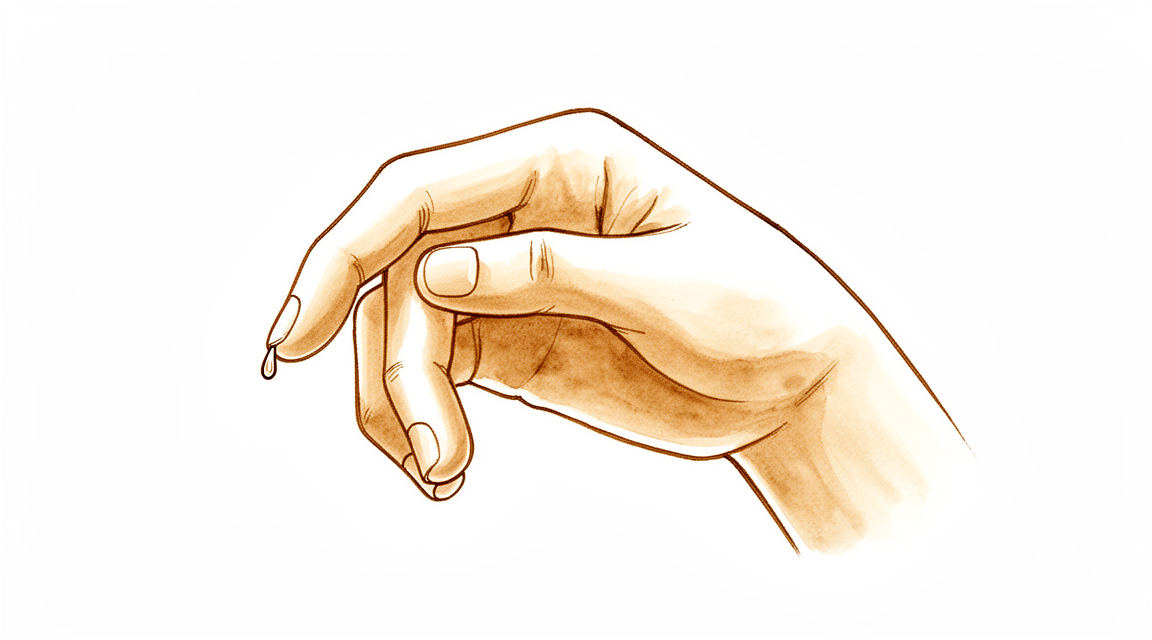
You might notice your fingertip drooping after a ball strikes the tip of your finger during sport or your fingertip catches on a sheet or sleeve. The most noticeable change is that the tip hangs downward and you cannot straighten it yourself, even though bending it remains perfectly normal. Day to day, this often means struggling to button a shirt, type on a keyboard, or pick up small flat items. Many people delay getting it checked because the discomfort is mild and the finger still handles most tasks. Starting treatment early makes a real difference to how well the finger recovers. The swelling and bruising usually appear over the back of the last joint, while the pain tends to stay quite mild. You will likely find that the finger still works for everyday activities, which is why some patients wait before seeking advice. The most striking sign remains the downward droop of the fingertip that refuses to lift without help. Catching these changes early helps protect the delicate structures inside the tip. A thin band of tissue called the extensor tendon sits along the back of your finger and normally pulls the tip straight. When the injury happens, this tendon either tears near the nail or pulls a small piece of bone away with it. Because the connection is broken, muscle force cannot reach the last joint, leaving the fingertip to hang down. The damaged ends must stay perfectly aligned for several weeks so they can knit back together. Allowing the tip to bend even briefly during this time stretches the healing tissue and can make the droop permanent. Most cases are managed without surgery using a small plastic splint that holds the tip joint completely straight around the clock for six to eight weeks. The splint must never be removed, even for washing, because letting the fingertip bend resets the healing process. Many patients then wear the device only at night for a few extra weeks once the continuous phase finishes. Surgery is sometimes recommended when a large bone fragment has shifted out of place or when the injury breaks the skin. The bone piece may be pinned back into position during the procedure. Even injuries that have been present for more than several weeks are still worth treating, though the recovery tends to be less complete. Once the splinting period ends, the finger often feels quite stiff and may show a slight droop at first. Gentle hand therapy exercises are introduced gradually to help restore normal movement and strength. Most people end up with a finger that functions normally, though a few degrees of permanent droop can sometimes remain. It is best to show the droopy fingertip to a clinician within a few days of the injury so treatment can begin promptly. You should seek same day attention if there is an open wound near the joint, or if you notice numbness, tingling, coldness, or a dusky colour in the finger. Continuing home splinting is fine, but you will need a professional review if the droop has not improved by the end of the recommended period.
Mallet finger causes fingertip drooping after extensor tendon injury; splinting is key, surgery occasionally needed.
What you're feeling
You may notice pain at the very tip of your finger. This discomfort often centers on the last joint, where the tendon connects to the bone. In some cases, you might feel this pain in both hands at the same time. The injury can involve a tear of the tendon or a small fracture where it attaches.
The pain typically worsens when you try to straighten your finger. You might find it difficult to push against resistance or lift heavy objects. Simple tasks like tucking in a shirt or reaching behind your back to fasten a bra can become challenging. Your finger may feel stiff, especially when you first wake up in the morning.
Many people find that resting the finger helps ease the ache. However, keeping the finger straight is crucial for healing. If you bend the tip, you may feel a sharp pull or increased pain. This is because the tendon is trying to pull the joint out of alignment.
In severe cases, you might notice the tip of your finger drooping downward. This droop can make it hard to grip objects firmly. You may also experience swelling around the joint. While most injuries heal well with a splint, some cases require more complex care. Your surgeon will determine if you need surgery based on the size of any bone fragment or the position of the joint.
If you have a large bone fragment or if the joint shifts out of place, your surgeon might recommend an operation. This is often done to restore proper alignment and function. Both surgical and non-surgical treatments generally lead to excellent outcomes. You can expect to regain full use of your finger with proper care.
What's actually happening
Your fingertip has a small tendon called the terminal tendon. It acts like a rope that connects your muscle to the bone at the very tip of your finger. This rope allows you to straighten your finger. When you injure this area, that rope tears or pulls a small piece of bone away from the joint. This stops the straightening signal from reaching your fingertip.
The most common cause is a sudden force that bends your straight finger backward. Imagine snagging your finger on a ball or a door frame while it was extended. The joint snaps into a bent position too quickly. This sudden stretch rips the tendon from the bone. In some cases, a small fragment of bone breaks off along with the tendon. The clinical look is similar whether the tendon itself tears or a bone chip pulls away. In both cases, the connection is broken.
Because the straightening rope is no longer attached, your finger rests in a bent posture. You cannot actively lift the tip of your finger to straighten it. This is called a mallet finger deformity. However, you can still passively straighten it with your other hand. The joint capsule and ligaments remain intact, so the joint itself does not dislocate. The bent position is simply the result of unopposed muscle pull from the other side of the finger.
This injury most often affects the small, ring, or middle fingers of your dominant hand. It is more common in men. While it usually happens from trauma, some elderly patients with wear-and-tear arthritis may develop this posture without a specific injury. In children, the injury might involve the growth plate rather than the tendon. Regardless of the cause, the result is the same: the mechanism that lifts your fingertip is disconnected, leaving it drooping.
What we can do about it
Most mallet finger injuries heal without surgery. You will likely wear a splint to keep the tip of your finger straight. This allows the tendon to heal in the correct position. A hand therapist can treat you as effectively as a surgeon for simple cases. They use methods that rarely cause skin problems. You might also try night splinting, but this does not improve your final result. Some patients use exercises alongside splinting, though evidence for this is mixed. You must wear your splint continuously for the time your surgeon recommends. Do not remove it to wash or bend the finger. Consistency is the key to success.
Pain management focuses on comfort while the tendon heals. You can take over-the-counter pain relievers or anti-inflammatories as needed. These help you manage daily activities while your finger is immobilized. Some patients ask about injections. However, the evidence does not support cortisone, hyaluronic acid, or PRP injections for this specific injury. These treatments are not part of the standard care plan for mallet finger. Your focus should remain on keeping the joint straight with your splint. If you have significant pain, discuss safe options with your surgeon. Avoid aggressive massage or stretching, which can disrupt healing.
Surgery is only considered if conservative care fails or if the injury is severe. Your surgeon may recommend an operation if you have a large fracture involving more than one-third of the joint surface. Surgery is also indicated if the bone fragment has shifted out of place. Some patients choose surgery because they cannot work with a splint in place. If you have a chronic injury that has not healed after months of splinting, surgery may be an option. The procedure repairs the damaged tendon to restore your ability to straighten the finger. This is a minor operation with a high success rate. Your surgeon will discuss the specific risks and benefits with you if this path becomes necessary.
What to expect
Most mallet finger injuries heal well without surgery. Your surgeon will likely recommend splinting to keep the tip of your finger straight. This allows the tendon or bone to reconnect. Both surgical and non-surgical treatments lead to excellent clinical outcomes for the majority of patients. You can expect a high level of satisfaction with your treatment result.
The path to recovery depends on the severity of your injury. For simple cases, a simple splint or a dorsal glued splint is usually sufficient. If your injury involves a large bone fragment or the joint has shifted out of place, surgery may be recommended. Surgery is also an option for chronic cases or if previous treatments have failed. In children, the need for surgery is less clear, but non-surgical management remains effective for most.
You should know that full correction of the finger’s position is not always guaranteed. If you have a severe chronic mallet finger with a significant bend, full correction is less consistent. However, even in these complex cases, long-term results are often good to excellent. Complication rates for conservative management are low. Serious issues like infection or nail deformity are rare, especially with modern surgical techniques.
Recovery is a gradual process. You may notice some stiffness or a small remaining bend in your finger tip. This is common and often does not affect your daily function significantly. Supplemental night splinting does not improve your outcome in terms of disability or satisfaction, so you may not need to wear it after your initial healing phase.
If you have a specific type of fracture involving more than one-third of the joint surface, there is about a 50% chance it will not progress to joint subluxation (slipping out of place). The size of the fracture and how quickly you start wearing your splint are key factors in your healing. Delaying treatment can increase the risk of complications, so following your surgeon’s advice on timing is important.
Overall, you can feel confident that mallet finger is a highly treatable condition. Whether you choose splinting or surgery, the goal is to restore function and minimize pain. Most patients return to their normal activities with a functional finger. Your surgeon will guide you through the best path for your specific injury to ensure the best possible outlook.
When to see someone
See your GP if you have persistent pain that does not improve with rest. Ask for a specialist review if you notice weakness or instability in the finger. Seek care if the joint locks or gives way. Contact your doctor if symptoms interfere with your sleep or work. Get help for any sudden worsening of your condition. Mallet finger injuries can sometimes happen on both hands at once. Rarely, these injuries involve a combination of tendon damage and bone fracture in younger patients. Biochemical changes may also play a role in how these injuries develop. Early assessment helps ensure the best outcome for your finger.
Evidence & references
title: "Mallet Finger" slug: mallet-finger region: hand audience: patient mesh_terms: ["Finger Injuries", "Finger Joint", "Hand Deformities, Acquired", "Finger Phalanges", "Splints", "Orthotic Devices", "Joint Dislocations", "Hand Injuries"] article_count: 218 model_used: Qwen3.6-35B-A3B-Q8_0.gguf generated_at: '2026-06-13T10:25:10+00:00' key_articles: - title: "Outcomes of Splinting in Pediatric Mallet Finger" ref_num: 1 evidence_tier: paper evidence_level: 4 doi: 10.1016/j.jhsa.2018.03.037 year: 2018 - title: "Surgical versus conservative management of Doyle type 4c mallet finger: a comparative study" ref_num: 2 evidence_tier: paper evidence_level: 4 doi: 10.1186/s12891-026-09787-w year: 2026 - title: "Current Concepts: Mallet Finger" ref_num: 3 evidence_tier: paper evidence_level: 5 doi: 10.1007/s11552-014-9609-y year: 2014 - title: "Surgical and Nonsurgical Management of Mallet Finger: A Systematic Review" ref_num: 4 evidence_tier: paper evidence_level: 4 doi: 10.1016/j.jhsa.2017.10.004 year: 2018 - title: "Review of Acute Traumatic Closed Mallet Finger Injuries in Adults" ref_num: 5 evidence_tier: paper evidence_level: 5 doi: 10.5999/aps.2016.43.2.134 year: 2016 - title: "The risk factors associated with subluxation of the distal interphalangeal joint in mallet fracture" ref_num: 6 evidence_tier: paper evidence_level: 2 doi: 10.1177/1753193414554556 year: 2014 - title: "The conservative treatment of mallet finger with a simple splint: a case report." ref_num: 8 evidence_tier: paper evidence_level: 2 doi: 10.1136/emj.10.3.244 year: 1993 - title: "The Diagnosis and Management of Mallet Finger Injuries" ref_num: 9 evidence_tier: paper evidence_level: 4 doi: 10.1177/1558944716642763 year: 2016 - title: "Mallet Finger" ref_num: 10 evidence_tier: paper evidence_level: 5 doi: 10.5435/00124635-200509000-00007 year: 2005 - title: "A new surgical treatment for mallet finger deformity: Deepithelialised pedicled skin flap technique" ref_num: 11 evidence_tier: paper doi: 10.1016/j.injury.2013.01.013 year: 2013 - title: "Hand Fractures: A Review of Current Treatment Strategies" ref_num: 12 evidence_tier: paper evidence_level: 5 doi: 10.1016/j.jhsa.2013.02.017 year: 2013 - title: "Pullout Wire Fixation Together With Distal Interphalangeal Joint Kirschner Wire Stabilization for Acute Combined Tendon and Bone (Double Level) Mallet Finger Injury" ref_num: 13 evidence_tier: paper evidence_level: 4 doi: 10.1016/j.jhsa.2014.11.011 year: 2015 - title: "A Prospective Randomized Controlled Trial Comparing Night Splinting with No Splinting after Treatment of Mallet Finger" ref_num: 15 evidence_tier: paper evidence_level: 1 doi: 10.1007/s11552-013-9600-z year: 2014 - title: "Hand Therapist-led Management of Mallet Finger" ref_num: 16 evidence_tier: paper evidence_level: 4 doi: 10.1177/175899830501000103 year: 2005 - title: "Central Slip Tenotomy With Distal Repair in the Treatment of Severe Chronic Mallet Fingers" ref_num: 18 evidence_tier: paper evidence_level: 4 doi: 10.1016/j.jhsa.2014.01.040 year: 2014 - title: "Bilateral Mallet Fingers: A Case Study" ref_num: 21 evidence_tier: paper evidence_level: 4 doi: 10.1177/175899830400900103 year: 2004 - title: "How do hand therapists conservatively manage acute, closed mallet finger? A survey of members of the British Association of Hand Therapists" ref_num: 23 evidence_tier: paper evidence_level: 4 doi: 10.1177/1758998316664822 year: 2016 - title: "The challenge of the mallet orthosis: A simple solution" ref_num: 25 evidence_tier: paper evidence_level: 5 doi: 10.1016/j.jht.2016.01.001 year: 2016 - title: "Combined tendon avulsion and fracture in a mallet finger injury in a juvenile" ref_num: 26 evidence_tier: paper evidence_level: 4 doi: 10.1177/1753193415571777 year: 2015 - title: "The Thompson Procedure for Chronic Mallet Finger Deformity" ref_num: 27 evidence_tier: paper evidence_level: 4 doi: 10.1016/j.jhsa.2013.04.011 year: 2013 - title: "Comparison of Treatment Results for Mallet Finger Fractures in Children Between Low-Intensity Pulsed Ultrasound Stimulation and Ishiguro’s Method" ref_num: 29 evidence_tier: paper evidence_level: 3 doi: 10.1177/1558944717692095 year: 2017 - title: "A novel way of treating mallet finger injuries" ref_num: 32 evidence_tier: paper evidence_level: 4 doi: 10.1016/j.jht.2014.02.005 year: 2014 - title: "Diagnosis and Treatment of Finger Deformities Following Injuries to the Extensor Tendon Mechanism" ref_num: 34 evidence_tier: paper evidence_level: 5 doi: 10.1016/j.hcl.2013.03.003 year: 2013 - title: "Acute Mallet Finger Injuries—A Review" ref_num: 35 evidence_tier: paper evidence_level: 4 doi: 10.1016/j.jhsa.2022.10.013 year: 2023 - title: "Scar overlapping suture for treating chronic tendinous mallet finger in children" ref_num: 36 evidence_tier: paper evidence_level: 4 doi: 10.1186/s13018-019-1106-0 year: 2019 - title: "Hand Therapist-led Management of Mallet Finger" ref_num: 39 evidence_tier: paper evidence_level: 4 doi: 10.1197/j.jht.2008.04.002 year: 2008 - title: "Rater Agreement of Post-Traumatic Osteoarthritis of the Distal Interphalangeal Joint 12 Years After a Mallet Finger Fracture" ref_num: 41 evidence_tier: paper evidence_level: 4 doi: 10.1016/j.jhsa.2024.03.012 year: 2024 - title: "The non-operative management of bony mallet injuries" ref_num: 46 evidence_tier: paper evidence_level: 3 doi: 10.1177/1753193421992986 year: 2021 - title: "Tendon Avulsion Injuries of the Distal Phalanx" ref_num: 50 evidence_tier: paper evidence_level: 5 doi: 10.1097/01.blo.0000205903.51727.62 year: 2006 - title: "The Ishiguro Extension Block Technique for the Treatment of Mallet Finger Fracture: Indications and Clinical Results" ref_num: 54 evidence_tier: paper evidence_level: 4 doi: 10.1054/jhsb.2001.0733 year: 2003 - title: "Causes of Procedural Failures of Closed Reductions using an Extension-Block Pin for Bony Mallet Finger" ref_num: 56 evidence_tier: paper doi: 10.1055/s-0040-1701318 year: 2021 synthesis_version: "v2" verifier_status: skipped
Overview
- Absolute indications for surgical intervention for mallet fingers in pediatric populations remain unclear [1].
- Large-fragment mallet finger cases can be effectively managed conservatively with low complication rates [2].
- Most mallet finger injuries can be managed non-surgically with splinting [3].
- Surgery is occasionally recommended for acute or chronic mallet finger cases or for salvage of failed prior treatment [3].
- Both surgical and nonsurgical treatments of mallet finger injuries lead to excellent clinical outcomes [4].
- All cases of mallet finger are proposed to be treated with a dorsal glued splint except for stage IV mallet finger, which is treated with extra-articular pinning [5].
- A simple splint is recommended as an alternative means of treating mallet finger [8].
- Surgery is generally indicated in mallet fractures involving more than one-third of the articular surface [9].
- Surgery is generally indicated in all patients who develop volar subluxation of the distal phalanx [9].
- A significant advantage of surgical management over conservative management in complicated cases (large fragment or volar subluxation) has yet to be clearly proven [9].
- The majority of hand fractures can be treated without surgery, though surgery offers distinct advantages in properly selected cases [12].
- Pullout wire fixation together with distal interphalangeal joint Kirschner wire stabilization for acute combined tendon and bone (double level) mallet finger injury reports good to excellent long-term results with no reported complications such as infection, nonunion, or nail deformity [13].
- Central slip tenotomy with distal repair is a technique described for severe chronic mallet fingers with deformities exceeding 36 degrees [18].
- Full correction of extensor lag is less consistent with greater degrees of preoperative flexion deformity in severe chronic mallet fingers treated with central slip tenotomy and distal repair [18].
- The Ishiguro extension block technique produces satisfactory results and is effective and minimally invasive for mallet finger fractures when properly applied [54].
Anatomy & Pathophysiology
- Mallet finger deformity is characterized by a loss of active distal interphalangeal (DIP) joint extension with full passive range of motion evident [19].
- The deformity reflects the loss of normal extensor force transmission via the terminal tendon insertion onto the distal phalanx [19].
- The unopposed flexor digitorum profundus pulls the distal joint into flexion [19].
- The usual mechanism of injury involves sudden passive flexion of an actively extended DIP joint [19].
- Disruption of the terminal tendon may be entirely confined to the tendon or may involve an avulsed fracture fragment from the dorsal lip of the distal phalanx proximal articular surface [19].
- The clinical appearance of soft tissue and bony mallet fingers is similar because the avulsed fragment includes the terminal tendon insertion [19].
- The distal joint rests in flexion, a posture that cannot be actively changed [19].
- Full passive extension of the DIP joint is possible in mallet finger [19].
- Mallet finger most commonly involves a closed rupture of the terminal tendon with or without associated fracture of the distal phalanx [37].
- Snagging the extending finger on an object that suddenly flexes the DIP joint is a frequent cause of mallet finger [37].
- Less commonly, a forceful hyperextension injury of the DIP joint may result in a large fracture of the base of the distal phalanx involving one-third or more of the articular surface [37].
- Elderly patients with osteoarthritis of the DIP joint may have mallet deformities that are not related to trauma [37].
- Individuals with hyperlax joints may have multiple pseudomallet swan neck postures that are unrelated to trauma [37].
- Open mallet injuries are uncommon [37].
- The most frequently involved digits are the small, ring, and middle fingers of the dominant hand [37].
- There is a male predominance in mallet finger incidence [37].
- Tendinous mallet fingers have been reported to occur from age 11 onward [37].
- In skeletally immature individuals, a transepiphyseal plate fracture may be seen instead of tendon rupture [37].
- There may be a familial predisposition to mallet fingers [37].
- Displacement of the epiphysis of the distal phalanx can cause the digit to assume a mallet finger posture [14].
- Hyperextension of the phalanx usually affords satisfactory reduction of a displaced epiphysis [14].
- The extensor apparatus of the fingers includes the interosseous muscle, extensor digitorum communis tendon, lumbrical muscle, flexor tendon sheath, sagittal bands, transverse metacarpal ligament, interosseous hood, interosseous hood oblique fibers, extensor lateral band, extensor middle band, interosseous middle band, interosseous lateral band, oblique retinacular ligament, central extensor lateral, spiral fibers, transverse retinacular ligament, lateral extensor tendon, triangular lamina, and terminal extensor tendon [14].
- Injuries to the finger extensor apparatus are very common and may produce chronic deformity and loss of function [34].
Classification
- Absolute indications for surgical intervention for mallet fingers in pediatric populations remain unclear [1].
- Doyle type 4c mallet finger cases can be effectively managed conservatively with low complication rates [2].
- Most mallet finger injuries can be managed non-surgically with splinting, although surgery is occasionally recommended for acute or chronic cases or for salvage of failed prior treatment [3].
- Both surgical and nonsurgical treatments of mallet finger injuries lead to excellent clinical outcomes [4].
- All cases of mallet finger are proposed to be treated with a dorsal glued splint except for stage IV mallet finger, which is treated with extra-articular pinning [5].
- Approximately 50% of patients with a mallet fracture involving more than one-third of the articular surface of the distal phalanx do not progress to subluxation of the DIP joint [6].
- Fracture size and time to application of finger immobilizer are independent risk factors for the development of DIP joint subluxation in mallet fracture [6].
- A simple splint is recommended as an alternative means of treating mallet finger [8].
- Deepithelialised pedicled skin flap technique is a new reliable alternative in the treatment of chronic mallet finger [11].
- A hand therapist can treat mallet finger injuries of type 1 as effectively as a surgeon, with a method of immobilisation that offers practically no complications regarding skin condition [16].
- A modification to the Doyle classification is proposed to make it more encompassing and less prone to interobserver error [35].
- The interrater reliability of the Kellgren & Lawrence and OARSI classification systems for post-traumatic osteoarthritis in the distal interphalangeal joint after mallet finger fractures is considerably lower than initially assumed [41].
- Non-operative management of mallet fractures, regardless of fracture classification, joint congruence or pre-existing degenerate change in the DIP joint, is safe and yields predictably good outcomes in most patients [46].
- Most acute mallet fingers can be treated by continuous splinting, while fracture dislocations require open reduction and internal fixation [50].
Clinical Presentation
- Mallet finger injuries can present as bilateral cases [21].
- Biochemical abnormalities may play a role in the etiology of mallet fingers [21].
- Mallet finger injuries can involve a rare combination of tendon avulsion and fracture in juvenile patients [26].
Investigations
- Mallet finger deformity is characterized by a loss of active distal interphalangeal (DIP) joint extension with full passive range of motion evident [19].
- The deformity reflects the loss of normal extensor force transmission via the terminal tendon insertion onto the distal phalanx [19].
- The unopposed flexor digitorum profundus pulls the distal joint into flexion [19].
- The usual mechanism of injury involves sudden passive flexion of the actively extended distal interphalangeal joint [19].
- Disruption of the terminal tendon may be entirely confined to the tendon or may involve an avulsed fracture fragment from the dorsal lip of the distal phalanx proximal articular surface [19].
- Because the avulsed fragment includes the terminal tendon insertion, the clinical appearance of soft tissue and bony mallet fingers is similar [19].
- The distal joint rests in flexion, a posture that cannot be actively changed [19].
- Full passive extension of the distal interphalangeal joint is possible [19].
- A radiograph should be obtained to determine whether a fracture is present [19].
- If a fracture is present, radiographs should assess whether the dorsal fragment is large and whether the distal phalanx is subluxed palmarward [19].
Treatment
Non-Operative Management
- Most mallet finger injuries can be managed non-surgically with splinting [3].
- Both surgical and nonsurgical treatments of mallet finger injuries lead to excellent clinical outcomes [4].
- A dorsal glued splint is proposed for the treatment of all cases of mallet finger except stage IV injuries [5].
- A simple splint is recommended as an alternative means of treating mallet finger [8].
- Supplemental night splinting does not improve the outcome of mallet finger in terms of extensor lag, disability, or satisfaction with treatment [15].
- A hand therapist can treat type 1 mallet finger injuries as effectively as a surgeon [16].
- A hand therapist can treat type 1 mallet finger injuries as effectively as a surgeon, with a method of immobilisation that offers practically no complications regarding skin condition [39].
- Conservative therapeutic management of acute, closed mallet finger is diverse and varied, with exercises and interventions supplementary to splinting commonly utilised [23].
- An alternative simple and custom-made orthosis manages the mallet finger by allowing for PIP flexion while inhibiting full extension or hyperextension [25].
- The clinical efficacy of elastic taping for the treatment of mallet finger injuries remains to be tested vigorously [32].
Operative Management
- Surgery is occasionally recommended for acute or chronic mallet cases or for salvage of failed prior treatment [3].
- Surgical management may be considered for acute and chronic mallet lesions in patients who have failed nonsurgical treatment [10].
- Surgical management may be considered for acute and chronic mallet lesions in patients who are unable to work with the splint in position [10].
- Surgical management may be considered for acute and chronic mallet lesions in patients who have a fracture involving more than one third of the joint surface [10].
- Surgery is generally indicated in the case of mallet fractures involving more than one-third of the articular surface [9].
- Surgery is generally indicated in all patients who develop volar subluxation of the distal phalanx [9].
- A significant advantage of surgical management even in complicated cases (fracture >1/3 articular surface or volar subluxation) has yet to be clearly proven [9].
- Complication rates for conservative management of Doyle type 4c mallet finger are low, suggesting large-fragment cases can be effectively managed conservatively [2].
- Absolute indications for surgical intervention for mallet fingers in the pediatric population remain unclear [1].
Surgical Techniques
- The Thompson procedure is an effective technique for the salvage of a closed mallet injury with an associated swan neck deformity following failed treatment [27].
- A deepithelialised pedicled skin flap technique is a new reliable alternative in the treatment of chronic mallet finger [11].
- Scar overlapping suture for treating chronic tendinous mallet finger in children is safe and effective [36].
- For chronic mallet finger, if the distal phalanx droops severely but passive extension in the distal interphalangeal joint is still satisfactory, surgery may be indicated depending on the patient’s needs [14].
- Surgical treatment for chronic mallet finger involves making a small V-shaped or U-shaped incision on the dorsum of the finger, convex distally, with the tip no closer than 5 mm proximal to the nail base [14].
- The surgical technique for chronic mallet finger involves developing a flap between the tendon and subcutaneous fat to expose the extensor tendon with intervening scar [14].
- The surgical technique for chronic mallet finger involves identifying the junction of normal tendon with scar, severing the tendon transversely proximal to the joint, and resecting sufficient scar or tendon to allow closure with the finger in maximal extension [14].
- The surgical repair of chronic mallet finger is supported by a transarticular 0.045-inch Kirschner wire [14].
- The extensor tendon in chronic mallet finger surgery is repaired with 4-0 monofilament nylon or 4-0 monofilament wire as a pull-out roll stitch [14].
- Postoperative care for chronic mallet finger surgery involves removing sutures at 10 to 14 days and maintaining the distal joint in extension with Kirschner wire protection for 4 weeks [14].
- The Kirschner wire is removed after 4 to 6 weeks in chronic mallet finger surgery, with the repair protected by a splint for an additional 8 weeks [14].
Complications
- Absolute indications for surgical intervention for mallet fingers in pediatric populations remain unclear [1].
- Complication rates for conservative management of Doyle type 4c mallet fingers are low [2].
- Surgery is occasionally recommended for acute or chronic cases or for salvage of failed prior treatment [3].
- Both surgical and nonsurgical treatments of mallet finger injuries lead to excellent clinical outcomes [4].
- Stage IV mallet finger is treated with extra-articular pinning rather than dorsal glued splinting [5].
- Approximately 50% of patients with a mallet fracture involving more than one-third of the articular surface of the distal phalanx do not progress to subluxation of the DIP joint [6].
- Fracture size is an independent risk factor for the development of DIP joint subluxation in mallet fracture [6].
- Time to application of finger immobilizer is an independent risk factor for the development of DIP joint subluxation in mallet fracture [6].
- Pullout wire fixation together with distal interphalangeal joint Kirschner wire stabilization for acute combined tendon and bone mallet finger injuries reports no complications such as infection, nonunion, or nail deformity [13].
- Supplemental night splinting does not improve outcomes in terms of extensor lag, disability, or satisfaction with treatment [15].
- Full correction of extensor lag is less consistent with greater degrees of preoperative flexion deformity in severe chronic mallet fingers treated with central slip tenotomy and distal repair [18].
- The Thompson procedure is an effective technique for the salvage of a closed mallet injury with an associated swan neck deformity following failed treatment [27].
- The most common cause of procedural failure in closed reductions using an extension-block pin for bony mallet finger is inaccurate insertion of the K-wire to fix the distal interphalangeal joint [56].
Recovery
- Most mallet finger injuries can be managed non-surgically with splinting [3].
- Surgery is occasionally recommended for acute or chronic cases or for salvage of failed prior treatment [3].
- Both surgical and nonsurgical treatments of mallet finger injuries lead to excellent clinical outcomes [4].
- Complication rates for conservative management of large-fragment mallet finger cases are low [2].
- Supplemental night splinting does not improve the outcome of mallet finger in terms of extensor lag, disability, or satisfaction with treatment [15].
- LIPUS therapy may be recommended as an option to treat type I mallet finger fracture in children for whom initiation of treatment was delayed up to 8 weeks [29].
- Approximately 50% of patients with a mallet fracture involving more than one-third of the articular surface of the distal phalanx do not progress to subluxation of the DIP joint [6].
- Fracture size and time to application of finger immobilizer are independent risk factors for the development of DIP joint subluxation in mallet fracture [6].
- Full correction of extensor lag is less consistent with greater degrees of preoperative flexion deformity in severe chronic mallet fingers treated with central slip tenotomy with distal repair [18].
- The surgical technique of pullout wire fixation together with distal interphalangeal joint Kirschner wire stabilization for acute combined tendon and bone mallet finger injuries reports good to excellent long-term results [13].
- No reported complications such as infection, nonunion, or nail deformity were observed in the series of acute combined tendon and bone mallet finger injuries treated with pullout wire fixation and DIP joint Kirschner wire stabilization [13].
Key Evidence
- [L4] Absolute indications for surgical intervention for mallet fingers in this population remain unclear. (10.1016/j.jhsa.2018.03.037)
- [L4] Complication rates were low, suggesting that large-fragment mallet finger cases can be effectively managed conservatively. (10.1186/s12891-026-09787-w)
- [L5] Most mallet finger injuries can be managed non-surgically with splinting, although surgery is occasionally recommended for acute or chronic cases or for salvage of failed prior treatment. (10.1007/s11552-014-9609-y)
- [L4] Both surgical and nonsurgical treatments of mallet finger injuries lead to excellent clinical outcomes. (10.1016/j.jhsa.2017.10.004)
- [L5] The authors propose to treat all cases of mallet finger with a dorsal glued splint except for stage IV mallet finger, which they treat with extra-articular pinning. (10.5999/aps.2016.43.2.134)
- [L2] Approximately 50% of patients with a mallet fracture involving more than one-third of the articular surface of the distal phalanx do not progress to subluxation of the DIP joint; fracture size and time to application of finger immobilizer are independent risk factors for the development of DIP joint subluxation in mallet fracture. (10.1177/1753193414554556)
- [L2] The study recommends this splint as an alternative means of treating mallet finger. (10.1136/emj.10.3.244)
- [L4] Although surgery is generally indicated in the case of mallet fractures involving more than one-third of the articular surface as well as in all patients who develop volar subluxation of the distal phalanx, a significant advantage of surgical management even in those complicated cases has yet to be clearly proven. (10.1177/1558944716642763)
- [L5] Surgical management may be considered for acute and chronic mallet lesions in patients who have failed nonsurgical treatment, are unable to work with the splint in position, or have a fracture involving more than one third of the joint surface. (10.5435/00124635-200509000-00007)
- [Paper] This method seems to be a new reliable alternative in the treatment of chronic mallet finger. (10.1016/j.injury.2013.01.013)
- [L5] The majority of hand fractures can be treated without surgery, though surgery offers distinct advantages in properly selected cases. (10.1016/j.jhsa.2013.02.017)
- [L4] The study describes a surgical technique for acute combined tendon and bone mallet fingers and reports good to excellent long-term results with no reported complications such as infection, nonunion, or nail deformity in the series. (10.1016/j.jhsa.2014.11.011)
- [L1] Supplemental night splinting does not improve the outcome of mallet finger in terms of extensor lag, disability, or satisfaction with treatment. (10.1007/s11552-013-9600-z)
- [L4] A hand therapist can treat mallet finger injuries of type 1 as effectively as a surgeon, with a method of immobilisation that offers practically no complications regarding skin condition. (10.1177/175899830501000103)
- [L4] The article describes a technique combining central slip tenotomy with distal repair for severe chronic mallet fingers with deformities exceeding 36 degrees, noting that full correction of extensor lag is less consistent with greater degrees of preoperative flexion deformity. (10.1016/j.jhsa.2014.01.040)
- [L4] This case raises a question regarding the possible role of biochemical abnormalities causing mallet fingers. (10.1177/175899830400900103)
- [L4] Conservative therapeutic management of acute, closed mallet finger is diverse and varied, with exercises and interventions supplementary to splinting commonly utilised. (10.1177/1758998316664822)
- [L5] An alternative simple and custom-made orthosis has been presented to manage the mallet finger, allowing for PIP flexion while inhibiting full extension or hyperextension. (10.1016/j.jht.2016.01.001)
- [L4] The authors describe a rare combination of tendon avulsion and fracture in a juvenile mallet finger, noting that while few mallet fingers require open treatment, this combined injury may be more common than thought. (10.1177/1753193415571777)
- [L4] It is an effective technique for the salvage of a closed mallet injury with an associated swan neck deformity following failed treatment. (10.1016/j.jhsa.2013.04.011)
- [L3] LIPUS therapy may be recommended as an option to treat type I mallet finger fracture in children for whom initiation of treatment was delayed up to 8 weeks. (10.1177/1558944717692095)
- [L4] The clinical efficacy of the proposed method of elastic taping for the treatment of mallet finger injuries remains to be tested vigorously. (10.1016/j.jht.2014.02.005)
- [L5] Injuries to the finger extensor apparatus are very common and may produce chronic deformity and loss of function. (10.1016/j.hcl.2013.03.003)
- [L4] This article provides a topical review of the contemporary literature concerning acute mallet finger injuries and proposes a modification to the Doyle classification to make it more encompassing and less prone to interobserver error. (10.1016/j.jhsa.2022.10.013)
- [L4] Scar overlapping suture for treating chronic tendinous mallet finger in children is safe and effective. (10.1186/s13018-019-1106-0)
- [L4] A hand therapist can treat type 1 mallet finger injuries as effectively as a surgeon. (10.1197/j.jht.2008.04.002)
- [L4] The interrater reliability of the Kellgren & Lawrence and OARSI classification systems for post-traumatic osteoarthritis in the distal interphalangeal joint after mallet finger fractures is considerably lower than initially assumed. (10.1016/j.jhsa.2024.03.012)
- [L3] Non-operative management of mallet fractures, regardless of fracture classification, joint congruence or pre-existing degenerate change in the DIP joint, is safe and yields predictably good outcomes in most patients. (10.1177/1753193421992986)
- [L5] Most acute mallet fingers can be treated by continuous splinting, while fracture dislocations require open reduction and internal fixation. (10.1097/01.blo.0000205903.51727.62)
- [L4] The extension block technique, when properly applied, produces satisfactory results and is effective and minimally invasive for mallet finger fractures. (10.1054/jhsb.2001.0733)
- [Paper] The most common cause of procedural failure was inaccurate insertion of the K-wire to fix the distal interphalangeal joint. (10.1055/s-0040-1701318)
References
[1] Outcomes of Splinting in Pediatric Mallet Finger. The Journal of Hand Surgery. 2018. DOI: 10.1016/j.jhsa.2018.03.037 [2] Surgical versus conservative management of Doyle type 4c mallet finger: a comparative study. BMC Musculoskeletal Disorders. 2026. DOI: 10.1186/s12891-026-09787-w [3] Current Concepts: Mallet Finger. HAND. 2014. DOI: 10.1007/s11552-014-9609-y [4] Surgical and Nonsurgical Management of Mallet Finger: A Systematic Review. The Journal of Hand Surgery. 2018. DOI: 10.1016/j.jhsa.2017.10.004 [5] Review of Acute Traumatic Closed Mallet Finger Injuries in Adults. Archives of Plastic Surgery. 2016. DOI: 10.5999/aps.2016.43.2.134 [6] The risk factors associated with subluxation of the distal interphalangeal joint in mallet fracture. Journal of Hand Surgery (European Volume). 2014. DOI: 10.1177/1753193414554556 [8] The conservative treatment of mallet finger with a simple splint: a case report.. Emergency Medicine Journal. 1993. DOI: 10.1136/emj.10.3.244 [9] The Diagnosis and Management of Mallet Finger Injuries. HAND. 2016. DOI: 10.1177/1558944716642763 [10] Mallet Finger. Journal of the American Academy of Orthopaedic Surgeons. 2005. DOI: 10.5435/00124635-200509000-00007 [11] A new surgical treatment for mallet finger deformity: Deepithelialised pedicled skin flap technique. Injury. 2013. DOI: 10.1016/j.injury.2013.01.013 [12] Hand Fractures: A Review of Current Treatment Strategies. The Journal of Hand Surgery. 2013. DOI: 10.1016/j.jhsa.2013.02.017 [13] Pullout Wire Fixation Together With Distal Interphalangeal Joint Kirschner Wire Stabilization for Acute Combined Tendon and Bone (Double Level) Mallet Finger Injury. The Journal of Hand Surgery. 2015. DOI: 10.1016/j.jhsa.2014.11.011 [14] Campbell S Operative Orthopaedics 4 Volume Set. RECONSTRUCTION OF FINGER FLEXORS: SINGLE-STAGE TENDON GRAFT > CHRONIC MALLET FINGER (SECONDARY REPAIR). [15] A Prospective Randomized Controlled Trial Comparing Night Splinting with No Splinting after Treatment of Mallet Finger. HAND. 2014. DOI: 10.1007/s11552-013-9600-z [16] Hand Therapist-led Management of Mallet Finger. The British Journal of Hand Therapy. 2005. DOI: 10.1177/175899830501000103 [18] Central Slip Tenotomy With Distal Repair in the Treatment of Severe Chronic Mallet Fingers. The Journal of Hand Surgery. 2014. DOI: 10.1016/j.jhsa.2014.01.040 [19] A Lange Medical Book Current Diagnosis Treatment In Orthopedics Fifth Edition. 9Hand Surgery > 3. Mallet Finger. [21] Bilateral Mallet Fingers: A Case Study. The British Journal of Hand Therapy. 2004. DOI: 10.1177/175899830400900103 [23] How do hand therapists conservatively manage acute, closed mallet finger? A survey of members of the British Association of Hand Therapists. Hand Therapy. 2016. DOI: 10.1177/1758998316664822 [25] The challenge of the mallet orthosis: A simple solution. Journal of Hand Therapy. 2016. DOI: 10.1016/j.jht.2016.01.001 [26] Combined tendon avulsion and fracture in a mallet finger injury in a juvenile. Journal of Hand Surgery (European Volume). 2015. DOI: 10.1177/1753193415571777 [27] The Thompson Procedure for Chronic Mallet Finger Deformity. The Journal of Hand Surgery. 2013. DOI: 10.1016/j.jhsa.2013.04.011 [29] Comparison of Treatment Results for Mallet Finger Fractures in Children Between Low-Intensity Pulsed Ultrasound Stimulation and Ishiguro’s Method. HAND. 2017. DOI: 10.1177/1558944717692095 [32] A novel way of treating mallet finger injuries. Journal of Hand Therapy. 2014. DOI: 10.1016/j.jht.2014.02.005 [34] Diagnosis and Treatment of Finger Deformities Following Injuries to the Extensor Tendon Mechanism. Hand Clinics. 2013. DOI: 10.1016/j.hcl.2013.03.003 [35] Acute Mallet Finger Injuries—A Review. The Journal of Hand Surgery. 2023. DOI: 10.1016/j.jhsa.2022.10.013 [36] Scar overlapping suture for treating chronic tendinous mallet finger in children. Journal of Orthopaedic Surgery and Research. 2019. DOI: 10.1186/s13018-019-1106-0 [37] Green S Operative Hand Surgery. CASE STUDY 5.2 Unusual Mallet Finger Presentation. [39] Hand Therapist-led Management of Mallet Finger. Journal of Hand Therapy. 2008. DOI: 10.1197/j.jht.2008.04.002 [41] Rater Agreement of Post-Traumatic Osteoarthritis of the Distal Interphalangeal Joint 12 Years After a Mallet Finger Fracture. The Journal of Hand Surgery. 2024. DOI: 10.1016/j.jhsa.2024.03.012 [46] The non-operative management of bony mallet injuries. Journal of Hand Surgery (European Volume). 2021. DOI: 10.1177/1753193421992986 [50] Tendon Avulsion Injuries of the Distal Phalanx. Clinical Orthopaedics and Related Research. 2006. DOI: 10.1097/01.blo.0000205903.51727.62 [54] The Ishiguro Extension Block Technique for the Treatment of Mallet Finger Fracture: Indications and Clinical Results. Journal of Hand Surgery. 2003. DOI: 10.1054/jhsb.2001.0733 [56] Causes of Procedural Failures of Closed Reductions using an Extension-Block Pin for Bony Mallet Finger. Journal of Hand and Microsurgery. 2021. DOI: 10.1055/s-0040-1701318


