锤状指
Patients › Hand
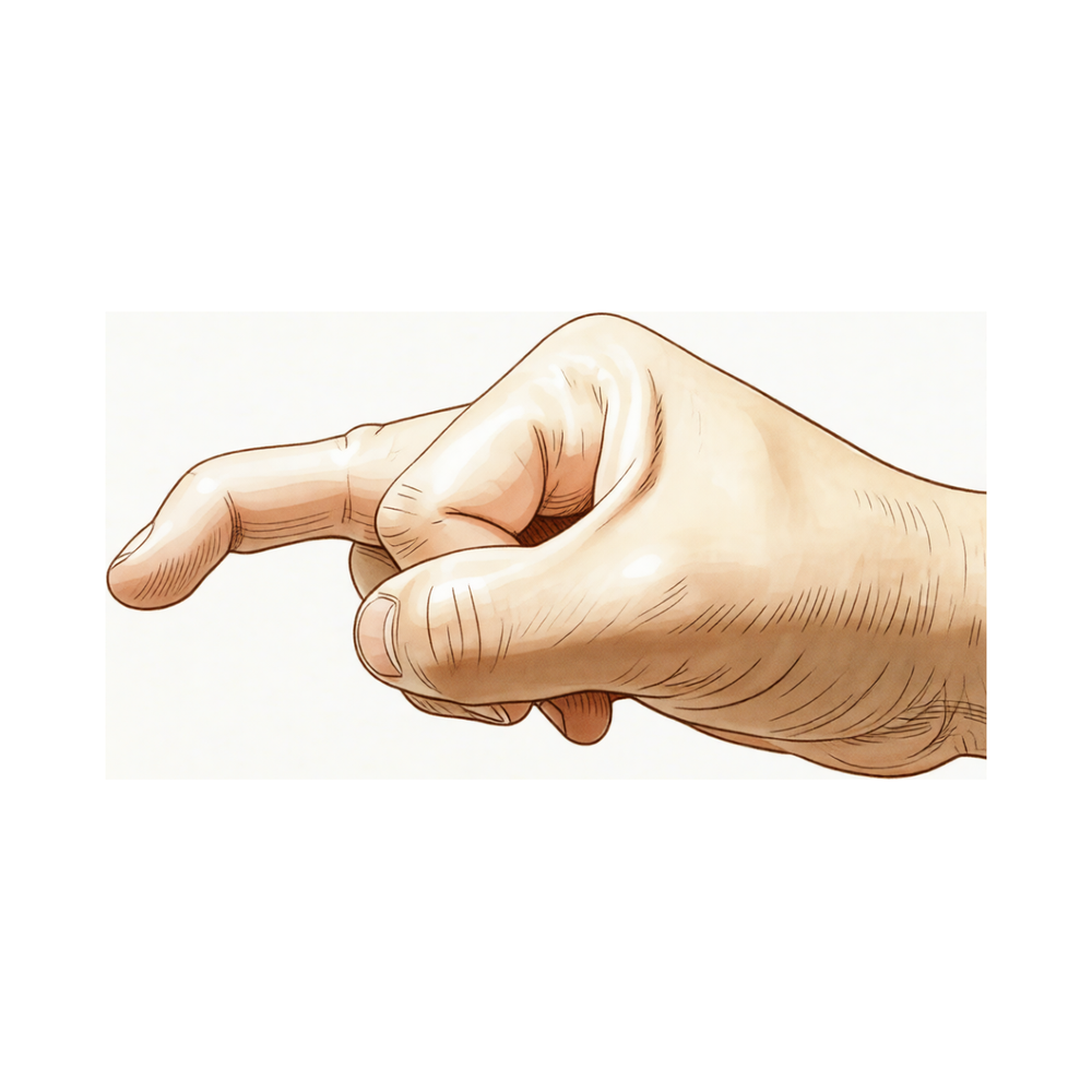
Mallet finger causes fingertip drooping after extensor tendon injury; splinting is key, surgery occasionally needed.
您的感受
您可能会注意到指尖出现疼痛。这种不适通常集中在末节指间关节,即肌腱附着于骨骼的位置。在某些情况下,您可能会感到双手同时出现疼痛。该损伤可能涉及肌腱撕裂或肌腱附着处的微小骨折。
当您尝试伸直手指时,疼痛通常会加剧。您可能会发现难以对抗阻力或提起重物。简单的任务,如将衬衫塞进裤腰或伸手到背后扣内衣,可能会变得困难。您的手指可能会感到僵硬,尤其是在早晨刚醒来时。
许多人发现休息手指有助于缓解酸痛。然而,保持手指伸直对于愈合至关重要。如果您弯曲指尖,您可能会感到剧烈的牵拉感或疼痛加剧。这是因为肌腱试图将关节拉离其正常对位。
在严重情况下,您可能会注意到指尖下垂。这种下垂会使您难以牢固地抓握物体。您还可能会经历关节周围的肿胀。虽然大多数损伤通过夹板固定即可良好愈合,但某些情况需要更复杂的治疗。您的外科医生会根据骨块的大小或关节的位置来决定您是否需要手术。
如果您有较大的骨块或关节发生脱位,您的外科医生可能会建议进行手术。这通常是为了恢复正常的对位和功能。手术和非手术治疗通常都能取得良好的效果。在适当的治疗下,您可以期望恢复手指的全部功能。
实际发生了什么
您的指尖有一根名为终腱的小肌腱。它像一根绳索,将您的肌肉连接到指尖末端的骨骼上。这根绳索使您能够伸直手指。当该区域受伤时,这根绳索会撕裂或将一小块骨骼从关节处撕脱。这会阻止伸直信号传递到指尖。
最常见的原因是突然施加的力量使伸直的手指向后弯曲。想象一下,当手指处于伸直状态时,指尖勾到了球或门框上。关节过快陷入弯曲位置。这种突然的拉伸将肌腱从骨骼上撕脱。在某些情况下,一小块骨骼碎片会随肌腱一起撕脱。无论是肌腱本身撕裂还是骨片撕脱,临床表现相似。在这两种情况下,连接均被破坏。
由于伸直的“绳索”不再附着,您的手指会保持弯曲姿势。您无法主动抬起指尖以伸直手指。这称为锤状指畸形。但是,您仍可用另一只手被动地将其伸直。关节囊和韧带保持完整,因此关节本身不会脱位。弯曲位置仅仅是由于手指另一侧肌肉无对抗牵拉所致。
这种损伤最常影响优势手的小指、无名指或中指。男性更为常见。虽然通常由创伤引起,但一些患有磨损性关节炎的老年患者可能在没有特定损伤的情况下出现这种姿势。在儿童中,损伤可能涉及生长板而非肌腱。无论原因如何,结果相同:抬起指尖的机制断开,导致其下垂。
我们能采取的措施
大多数锤状指损伤无需手术即可愈合。您可能需要佩戴夹板以保持手指末端伸直。这有助于肌腱在正确的位置愈合。对于简单病例,手部治疗师的治疗效果与外科医生相当。他们使用的方法很少引起皮肤问题。您也可以尝试夜间夹板固定,但这并不能改善最终结果。一些患者会结合夹板固定进行锻炼,但相关证据不一。您必须按照外科医生的建议连续佩戴夹板。不要为了洗手或弯曲手指而取下夹板。坚持是成功的关键。
疼痛管理侧重于在肌腱愈合期间提供舒适感。您可以根据需要服用非处方止痛药或抗炎药。这些药物有助于您在手指固定期间管理日常活动。一些患者会询问关于注射的问题。然而,证据并不支持针对这种特定损伤使用可的松、透明质酸或富血小板血浆(PRP)注射。这些治疗不属于锤状指的标准护理方案。您的重点应始终是使用夹板保持关节伸直。如果您有剧烈疼痛,请与您的外科医生讨论安全的选项。避免剧烈按摩或拉伸,因为这会干扰愈合。
仅在保守治疗失败或损伤严重时才会考虑手术。如果您的骨折较大,涉及关节表面超过三分之一,您的外科医生可能会建议手术。如果骨碎片移位,也需进行手术。一些患者选择手术是因为他们无法在佩戴夹板的情况下工作。如果您有慢性损伤,经过数月的夹板固定后仍未愈合,手术可能是一个选项。该手术修复受损的肌腱,以恢复您伸直手指的能力。这是一项成功率较高的微创手术。如果确实需要采取这一途径,您的外科医生将与您讨论具体的风险和益处。
预期情况
大多数锤状指损伤无需手术即可良好愈合。您的外科医生可能会建议佩戴夹板,以保持手指末端伸直。这有助于肌腱或骨骼重新连接。对于大多数患者而言,手术和非手术治疗均能带来良好的临床结果。您对治疗结果通常会感到高度满意。
康复过程取决于损伤的严重程度。对于简单病例,通常只需使用简单的夹板或背侧胶合夹板即可。如果损伤涉及较大的骨碎片或关节发生移位,则可能需要手术。对于慢性病例或既往治疗失败的情况,手术也是一种选择。在儿童中,手术指征尚不明确,但非手术治疗对大多数患儿仍然有效。
您需要了解,手指位置的完全矫正并不总是能保证。如果您患有伴有明显弯曲的重度慢性锤状指,完全矫正的效果往往不太一致。然而,即使在这些复杂病例中,长期结果通常也良好至优秀。保守治疗的并发症发生率较低。感染或指甲畸形等严重问题较为罕见,尤其是在采用现代外科技术的情况下。
康复是一个渐进的过程。您可能会发现手指末端存在一些僵硬或残留的轻微弯曲。这很常见,通常不会对您的日常功能产生显著影响。补充夜间夹板固定并不能改善残疾程度或满意度方面的结果,因此在初始愈合期后您可能无需继续佩戴。
如果您患有涉及关节面超过三分之一的特定类型骨折,其进展为关节半脱位(移位)的概率约为 50%。骨折的大小以及您开始佩戴夹板的速度是愈合的关键因素。延迟治疗会增加并发症的风险,因此遵循外科医生关于治疗时机的建议非常重要。
总体而言,您可以放心,锤状指是一种高度可治疗的疾病。无论您选择夹板固定还是手术,目标都是恢复功能并最大限度地减轻疼痛。大多数患者能够恢复使用功能性的手指进行正常活动。您的外科医生将指导您针对特定损伤选择最佳的治疗路径,以确保获得最佳预后。
何时就医
若休息后疼痛持续不缓解,请咨询全科医生。若发现手指出现无力或不稳,请要求专科医生评估。若关节出现交锁或无力感,请及时就医。若症状影响睡眠或工作,请联系医生。若病情突然加重,请寻求医疗帮助。锤状指损伤有时可双侧同时发生。在年轻患者中,此类损伤罕见地表现为肌腱损伤合并骨折。生化改变也可能在这些损伤的发生发展中发挥作用。早期评估有助于确保手指获得最佳预后。
Evidence & references
Overview
- Absolute indications for surgical intervention for mallet fingers in pediatric populations remain unclear [1].
- Large-fragment mallet finger cases can be effectively managed conservatively with low complication rates [2].
- Most mallet finger injuries can be managed non-surgically with splinting [3].
- Surgery is occasionally recommended for acute or chronic mallet finger cases or for salvage of failed prior treatment [3].
- Both surgical and nonsurgical treatments of mallet finger injuries lead to excellent clinical outcomes [4].
- All cases of mallet finger are proposed to be treated with a dorsal glued splint except for stage IV mallet finger, which is treated with extra-articular pinning [5].
- A simple splint is recommended as an alternative means of treating mallet finger [8].
- Surgery is generally indicated in mallet fractures involving more than one-third of the articular surface [9].
- Surgery is generally indicated in all patients who develop volar subluxation of the distal phalanx [9].
- A significant advantage of surgical management over conservative management in complicated cases (large fragment or volar subluxation) has yet to be clearly proven [9].
- The majority of hand fractures can be treated without surgery, though surgery offers distinct advantages in properly selected cases [12].
- Pullout wire fixation together with distal interphalangeal joint Kirschner wire stabilization for acute combined tendon and bone (double level) mallet finger injury reports good to excellent long-term results with no reported complications such as infection, nonunion, or nail deformity [13].
- Central slip tenotomy with distal repair is a technique described for severe chronic mallet fingers with deformities exceeding 36 degrees [18].
- Full correction of extensor lag is less consistent with greater degrees of preoperative flexion deformity in severe chronic mallet fingers treated with central slip tenotomy and distal repair [18].
- The Ishiguro extension block technique produces satisfactory results and is effective and minimally invasive for mallet finger fractures when properly applied [54].
Anatomy & Pathophysiology
- Mallet finger deformity is characterized by a loss of active distal interphalangeal (DIP) joint extension with full passive range of motion evident [19].
- The deformity reflects the loss of normal extensor force transmission via the terminal tendon insertion onto the distal phalanx [19].
- The unopposed flexor digitorum profundus pulls the distal joint into flexion [19].
- The usual mechanism of injury involves sudden passive flexion of an actively extended DIP joint [19].
- Disruption of the terminal tendon may be entirely confined to the tendon or may involve an avulsed fracture fragment from the dorsal lip of the distal phalanx proximal articular surface [19].
- The clinical appearance of soft tissue and bony mallet fingers is similar because the avulsed fragment includes the terminal tendon insertion [19].
- The distal joint rests in flexion, a posture that cannot be actively changed [19].
- Full passive extension of the DIP joint is possible in mallet finger [19].
- Mallet finger most commonly involves a closed rupture of the terminal tendon with or without associated fracture of the distal phalanx [37].
- Snagging the extending finger on an object that suddenly flexes the DIP joint is a frequent cause of mallet finger [37].
- Less commonly, a forceful hyperextension injury of the DIP joint may result in a large fracture of the base of the distal phalanx involving one-third or more of the articular surface [37].
- Elderly patients with osteoarthritis of the DIP joint may have mallet deformities that are not related to trauma [37].
- Individuals with hyperlax joints may have multiple pseudomallet swan neck postures that are unrelated to trauma [37].
- Open mallet injuries are uncommon [37].
- The most frequently involved digits are the small, ring, and middle fingers of the dominant hand [37].
- There is a male predominance in mallet finger incidence [37].
- Tendinous mallet fingers have been reported to occur from age 11 onward [37].
- In skeletally immature individuals, a transepiphyseal plate fracture may be seen instead of tendon rupture [37].
- There may be a familial predisposition to mallet fingers [37].
- Displacement of the epiphysis of the distal phalanx can cause the digit to assume a mallet finger posture [14].
- Hyperextension of the phalanx usually affords satisfactory reduction of a displaced epiphysis [14].
- The extensor apparatus of the fingers includes the interosseous muscle, extensor digitorum communis tendon, lumbrical muscle, flexor tendon sheath, sagittal bands, transverse metacarpal ligament, interosseous hood, interosseous hood oblique fibers, extensor lateral band, extensor middle band, interosseous middle band, interosseous lateral band, oblique retinacular ligament, central extensor lateral, spiral fibers, transverse retinacular ligament, lateral extensor tendon, triangular lamina, and terminal extensor tendon [14].
- Injuries to the finger extensor apparatus are very common and may produce chronic deformity and loss of function [34].
Classification
- Absolute indications for surgical intervention for mallet fingers in pediatric populations remain unclear [1].
- Doyle type 4c mallet finger cases can be effectively managed conservatively with low complication rates [2].
- Most mallet finger injuries can be managed non-surgically with splinting, although surgery is occasionally recommended for acute or chronic cases or for salvage of failed prior treatment [3].
- Both surgical and nonsurgical treatments of mallet finger injuries lead to excellent clinical outcomes [4].
- All cases of mallet finger are proposed to be treated with a dorsal glued splint except for stage IV mallet finger, which is treated with extra-articular pinning [5].
- Approximately 50% of patients with a mallet fracture involving more than one-third of the articular surface of the distal phalanx do not progress to subluxation of the DIP joint [6].
- Fracture size and time to application of finger immobilizer are independent risk factors for the development of DIP joint subluxation in mallet fracture [6].
- A simple splint is recommended as an alternative means of treating mallet finger [8].
- Deepithelialised pedicled skin flap technique is a new reliable alternative in the treatment of chronic mallet finger [11].
- A hand therapist can treat mallet finger injuries of type 1 as effectively as a surgeon, with a method of immobilisation that offers practically no complications regarding skin condition [16].
- A modification to the Doyle classification is proposed to make it more encompassing and less prone to interobserver error [35].
- The interrater reliability of the Kellgren & Lawrence and OARSI classification systems for post-traumatic osteoarthritis in the distal interphalangeal joint after mallet finger fractures is considerably lower than initially assumed [41].
- Non-operative management of mallet fractures, regardless of fracture classification, joint congruence or pre-existing degenerate change in the DIP joint, is safe and yields predictably good outcomes in most patients [46].
- Most acute mallet fingers can be treated by continuous splinting, while fracture dislocations require open reduction and internal fixation [50].
Clinical Presentation
- Mallet finger injuries can present as bilateral cases [21].
- Biochemical abnormalities may play a role in the etiology of mallet fingers [21].
- Mallet finger injuries can involve a rare combination of tendon avulsion and fracture in juvenile patients [26].
Investigations
- Mallet finger deformity is characterized by a loss of active distal interphalangeal (DIP) joint extension with full passive range of motion evident [19].
- The deformity reflects the loss of normal extensor force transmission via the terminal tendon insertion onto the distal phalanx [19].
- The unopposed flexor digitorum profundus pulls the distal joint into flexion [19].
- The usual mechanism of injury involves sudden passive flexion of the actively extended distal interphalangeal joint [19].
- Disruption of the terminal tendon may be entirely confined to the tendon or may involve an avulsed fracture fragment from the dorsal lip of the distal phalanx proximal articular surface [19].
- Because the avulsed fragment includes the terminal tendon insertion, the clinical appearance of soft tissue and bony mallet fingers is similar [19].
- The distal joint rests in flexion, a posture that cannot be actively changed [19].
- Full passive extension of the distal interphalangeal joint is possible [19].
- A radiograph should be obtained to determine whether a fracture is present [19].
- If a fracture is present, radiographs should assess whether the dorsal fragment is large and whether the distal phalanx is subluxed palmarward [19].
Treatment
Non-Operative Management
- Most mallet finger injuries can be managed non-surgically with splinting [3].
- Both surgical and nonsurgical treatments of mallet finger injuries lead to excellent clinical outcomes [4].
- A dorsal glued splint is proposed for the treatment of all cases of mallet finger except stage IV injuries [5].
- A simple splint is recommended as an alternative means of treating mallet finger [8].
- Supplemental night splinting does not improve the outcome of mallet finger in terms of extensor lag, disability, or satisfaction with treatment [15].
- A hand therapist can treat type 1 mallet finger injuries as effectively as a surgeon [16].
- A hand therapist can treat type 1 mallet finger injuries as effectively as a surgeon, with a method of immobilisation that offers practically no complications regarding skin condition [39].
- Conservative therapeutic management of acute, closed mallet finger is diverse and varied, with exercises and interventions supplementary to splinting commonly utilised [23].
- An alternative simple and custom-made orthosis manages the mallet finger by allowing for PIP flexion while inhibiting full extension or hyperextension [25].
- The clinical efficacy of elastic taping for the treatment of mallet finger injuries remains to be tested vigorously [32].
Operative Management
- Surgery is occasionally recommended for acute or chronic mallet cases or for salvage of failed prior treatment [3].
- Surgical management may be considered for acute and chronic mallet lesions in patients who have failed nonsurgical treatment [10].
- Surgical management may be considered for acute and chronic mallet lesions in patients who are unable to work with the splint in position [10].
- Surgical management may be considered for acute and chronic mallet lesions in patients who have a fracture involving more than one third of the joint surface [10].
- Surgery is generally indicated in the case of mallet fractures involving more than one-third of the articular surface [9].
- Surgery is generally indicated in all patients who develop volar subluxation of the distal phalanx [9].
- A significant advantage of surgical management even in complicated cases (fracture >1/3 articular surface or volar subluxation) has yet to be clearly proven [9].
- Complication rates for conservative management of Doyle type 4c mallet finger are low, suggesting large-fragment cases can be effectively managed conservatively [2].
- Absolute indications for surgical intervention for mallet fingers in the pediatric population remain unclear [1].
Surgical Techniques
- The Thompson procedure is an effective technique for the salvage of a closed mallet injury with an associated swan neck deformity following failed treatment [27].
- A deepithelialised pedicled skin flap technique is a new reliable alternative in the treatment of chronic mallet finger [11].
- Scar overlapping suture for treating chronic tendinous mallet finger in children is safe and effective [36].
- For chronic mallet finger, if the distal phalanx droops severely but passive extension in the distal interphalangeal joint is still satisfactory, surgery may be indicated depending on the patient’s needs [14].
- Surgical treatment for chronic mallet finger involves making a small V-shaped or U-shaped incision on the dorsum of the finger, convex distally, with the tip no closer than 5 mm proximal to the nail base [14].
- The surgical technique for chronic mallet finger involves developing a flap between the tendon and subcutaneous fat to expose the extensor tendon with intervening scar [14].
- The surgical technique for chronic mallet finger involves identifying the junction of normal tendon with scar, severing the tendon transversely proximal to the joint, and resecting sufficient scar or tendon to allow closure with the finger in maximal extension [14].
- The surgical repair of chronic mallet finger is supported by a transarticular 0.045-inch Kirschner wire [14].
- The extensor tendon in chronic mallet finger surgery is repaired with 4-0 monofilament nylon or 4-0 monofilament wire as a pull-out roll stitch [14].
- Postoperative care for chronic mallet finger surgery involves removing sutures at 10 to 14 days and maintaining the distal joint in extension with Kirschner wire protection for 4 weeks [14].
- The Kirschner wire is removed after 4 to 6 weeks in chronic mallet finger surgery, with the repair protected by a splint for an additional 8 weeks [14].
Complications
- Absolute indications for surgical intervention for mallet fingers in pediatric populations remain unclear [1].
- Complication rates for conservative management of Doyle type 4c mallet fingers are low [2].
- Surgery is occasionally recommended for acute or chronic cases or for salvage of failed prior treatment [3].
- Both surgical and nonsurgical treatments of mallet finger injuries lead to excellent clinical outcomes [4].
- Stage IV mallet finger is treated with extra-articular pinning rather than dorsal glued splinting [5].
- Approximately 50% of patients with a mallet fracture involving more than one-third of the articular surface of the distal phalanx do not progress to subluxation of the DIP joint [6].
- Fracture size is an independent risk factor for the development of DIP joint subluxation in mallet fracture [6].
- Time to application of finger immobilizer is an independent risk factor for the development of DIP joint subluxation in mallet fracture [6].
- Pullout wire fixation together with distal interphalangeal joint Kirschner wire stabilization for acute combined tendon and bone mallet finger injuries reports no complications such as infection, nonunion, or nail deformity [13].
- Supplemental night splinting does not improve outcomes in terms of extensor lag, disability, or satisfaction with treatment [15].
- Full correction of extensor lag is less consistent with greater degrees of preoperative flexion deformity in severe chronic mallet fingers treated with central slip tenotomy and distal repair [18].
- The Thompson procedure is an effective technique for the salvage of a closed mallet injury with an associated swan neck deformity following failed treatment [27].
- The most common cause of procedural failure in closed reductions using an extension-block pin for bony mallet finger is inaccurate insertion of the K-wire to fix the distal interphalangeal joint [56].
Recovery
- Most mallet finger injuries can be managed non-surgically with splinting [3].
- Surgery is occasionally recommended for acute or chronic cases or for salvage of failed prior treatment [3].
- Both surgical and nonsurgical treatments of mallet finger injuries lead to excellent clinical outcomes [4].
- Complication rates for conservative management of large-fragment mallet finger cases are low [2].
- Supplemental night splinting does not improve the outcome of mallet finger in terms of extensor lag, disability, or satisfaction with treatment [15].
- LIPUS therapy may be recommended as an option to treat type I mallet finger fracture in children for whom initiation of treatment was delayed up to 8 weeks [29].
- Approximately 50% of patients with a mallet fracture involving more than one-third of the articular surface of the distal phalanx do not progress to subluxation of the DIP joint [6].
- Fracture size and time to application of finger immobilizer are independent risk factors for the development of DIP joint subluxation in mallet fracture [6].
- Full correction of extensor lag is less consistent with greater degrees of preoperative flexion deformity in severe chronic mallet fingers treated with central slip tenotomy with distal repair [18].
- The surgical technique of pullout wire fixation together with distal interphalangeal joint Kirschner wire stabilization for acute combined tendon and bone mallet finger injuries reports good to excellent long-term results [13].
- No reported complications such as infection, nonunion, or nail deformity were observed in the series of acute combined tendon and bone mallet finger injuries treated with pullout wire fixation and DIP joint Kirschner wire stabilization [13].
Key Evidence
- [L4] Absolute indications for surgical intervention for mallet fingers in this population remain unclear. [1] (10.1016/j.jhsa.2018.03.037)
- [L4] Complication rates were low, suggesting that large-fragment mallet finger cases can be effectively managed conservatively. [2] (10.1186/s12891-026-09787-w)
- [L5] Most mallet finger injuries can be managed non-surgically with splinting, although surgery is occasionally recommended for acute or chronic cases or for salvage of failed prior treatment. [3] (10.1007/s11552-014-9609-y)
- [L4] Both surgical and nonsurgical treatments of mallet finger injuries lead to excellent clinical outcomes. [4] (10.1016/j.jhsa.2017.10.004)
- [L5] The authors propose to treat all cases of mallet finger with a dorsal glued splint except for stage IV mallet finger, which they treat with extra-articular pinning. [5] (10.5999/aps.2016.43.2.134)
- [L2] Approximately 50% of patients with a mallet fracture involving more than one-third of the articular surface of the distal phalanx do not progress to subluxation of the DIP joint; fracture size and time to application of finger immobilizer are independent risk factors for the development of DIP joint subluxation in mallet fracture. [6] (10.1177/1753193414554556)
- [L2] The study recommends this splint as an alternative means of treating mallet finger. [8] (10.1136/emj.10.3.244)
- [L4] Although surgery is generally indicated in the case of mallet fractures involving more than one-third of the articular surface as well as in all patients who develop volar subluxation of the distal phalanx, a significant advantage of surgical management even in those complicated cases has yet to be clearly proven. [9] (10.1177/1558944716642763)
- [L5] Surgical management may be considered for acute and chronic mallet lesions in patients who have failed nonsurgical treatment, are unable to work with the splint in position, or have a fracture involving more than one third of the joint surface. [10] (10.5435/00124635-200509000-00007)
- [Paper] This method seems to be a new reliable alternative in the treatment of chronic mallet finger. [11] (10.1016/j.injury.2013.01.013)
- [L5] The majority of hand fractures can be treated without surgery, though surgery offers distinct advantages in properly selected cases. [12] (10.1016/j.jhsa.2013.02.017)
- [L4] The study describes a surgical technique for acute combined tendon and bone mallet fingers and reports good to excellent long-term results with no reported complications such as infection, nonunion, or nail deformity in the series. [13] (10.1016/j.jhsa.2014.11.011)
- [L1] Supplemental night splinting does not improve the outcome of mallet finger in terms of extensor lag, disability, or satisfaction with treatment. [15] (10.1007/s11552-013-9600-z)
- [L4] A hand therapist can treat mallet finger injuries of type 1 as effectively as a surgeon, with a method of immobilisation that offers practically no complications regarding skin condition. [16] (10.1177/175899830501000103)
- [L4] The article describes a technique combining central slip tenotomy with distal repair for severe chronic mallet fingers with deformities exceeding 36 degrees, noting that full correction of extensor lag is less consistent with greater degrees of preoperative flexion deformity. [18] (10.1016/j.jhsa.2014.01.040)
- [L4] This case raises a question regarding the possible role of biochemical abnormalities causing mallet fingers. [21] (10.1177/175899830400900103)
- [L4] Conservative therapeutic management of acute, closed mallet finger is diverse and varied, with exercises and interventions supplementary to splinting commonly utilised. [23] (10.1177/1758998316664822)
- [L5] An alternative simple and custom-made orthosis has been presented to manage the mallet finger, allowing for PIP flexion while inhibiting full extension or hyperextension. [25] (10.1016/j.jht.2016.01.001)
- [L4] The authors describe a rare combination of tendon avulsion and fracture in a juvenile mallet finger, noting that while few mallet fingers require open treatment, this combined injury may be more common than thought. [26] (10.1177/1753193415571777)
- [L4] It is an effective technique for the salvage of a closed mallet injury with an associated swan neck deformity following failed treatment. [27] (10.1016/j.jhsa.2013.04.011)
- [L3] LIPUS therapy may be recommended as an option to treat type I mallet finger fracture in children for whom initiation of treatment was delayed up to 8 weeks. [29] (10.1177/1558944717692095)
- [L4] The clinical efficacy of the proposed method of elastic taping for the treatment of mallet finger injuries remains to be tested vigorously. [32] (10.1016/j.jht.2014.02.005)
- [L5] Injuries to the finger extensor apparatus are very common and may produce chronic deformity and loss of function. [34] (10.1016/j.hcl.2013.03.003)
- [L4] This article provides a topical review of the contemporary literature concerning acute mallet finger injuries and proposes a modification to the Doyle classification to make it more encompassing and less prone to interobserver error. [35] (10.1016/j.jhsa.2022.10.013)
- [L4] Scar overlapping suture for treating chronic tendinous mallet finger in children is safe and effective. [36] (10.1186/s13018-019-1106-0)
- [L4] A hand therapist can treat type 1 mallet finger injuries as effectively as a surgeon. [39] (10.1197/j.jht.2008.04.002)
- [L4] The interrater reliability of the Kellgren & Lawrence and OARSI classification systems for post-traumatic osteoarthritis in the distal interphalangeal joint after mallet finger fractures is considerably lower than initially assumed. [41] (10.1016/j.jhsa.2024.03.012)
- [L3] Non-operative management of mallet fractures, regardless of fracture classification, joint congruence or pre-existing degenerate change in the DIP joint, is safe and yields predictably good outcomes in most patients. [46] (10.1177/1753193421992986)
- [L5] Most acute mallet fingers can be treated by continuous splinting, while fracture dislocations require open reduction and internal fixation. [50] (10.1097/01.blo.0000205903.51727.62)
- [L4] The extension block technique, when properly applied, produces satisfactory results and is effective and minimally invasive for mallet finger fractures. [54] (10.1054/jhsb.2001.0733)
- [Paper] The most common cause of procedural failure was inaccurate insertion of the K-wire to fix the distal interphalangeal joint. [56] (10.1055/s-0040-1701318)
References
[1] Outcomes of Splinting in Pediatric Mallet Finger. The Journal of Hand Surgery. 2018. DOI: 10.1016/j.jhsa.2018.03.037 [2] Surgical versus conservative management of Doyle type 4c mallet finger: a comparative study. BMC Musculoskeletal Disorders. 2026. DOI: 10.1186/s12891-026-09787-w [3] Current Concepts: Mallet Finger. HAND. 2014. DOI: 10.1007/s11552-014-9609-y [4] Surgical and Nonsurgical Management of Mallet Finger: A Systematic Review. The Journal of Hand Surgery. 2018. DOI: 10.1016/j.jhsa.2017.10.004 [5] Review of Acute Traumatic Closed Mallet Finger Injuries in Adults. Archives of Plastic Surgery. 2016. DOI: 10.5999/aps.2016.43.2.134 [6] The risk factors associated with subluxation of the distal interphalangeal joint in mallet fracture. Journal of Hand Surgery (European Volume). 2014. DOI: 10.1177/1753193414554556 [8] The conservative treatment of mallet finger with a simple splint: a case report.. Emergency Medicine Journal. 1993. DOI: 10.1136/emj.10.3.244 [9] The Diagnosis and Management of Mallet Finger Injuries. HAND. 2016. DOI: 10.1177/1558944716642763 [10] Mallet Finger. Journal of the American Academy of Orthopaedic Surgeons. 2005. DOI: 10.5435/00124635-200509000-00007 [11] A new surgical treatment for mallet finger deformity: Deepithelialised pedicled skin flap technique. Injury. 2013. DOI: 10.1016/j.injury.2013.01.013 [12] Hand Fractures: A Review of Current Treatment Strategies. The Journal of Hand Surgery. 2013. DOI: 10.1016/j.jhsa.2013.02.017 [13] Pullout Wire Fixation Together With Distal Interphalangeal Joint Kirschner Wire Stabilization for Acute Combined Tendon and Bone (Double Level) Mallet Finger Injury. The Journal of Hand Surgery. 2015. DOI: 10.1016/j.jhsa.2014.11.011 [14] Campbell S Operative Orthopaedics 4 Volume Set. RECONSTRUCTION OF FINGER FLEXORS: SINGLE-STAGE TENDON GRAFT > CHRONIC MALLET FINGER (SECONDARY REPAIR). [15] A Prospective Randomized Controlled Trial Comparing Night Splinting with No Splinting after Treatment of Mallet Finger. HAND. 2014. DOI: 10.1007/s11552-013-9600-z [16] Hand Therapist-led Management of Mallet Finger. The British Journal of Hand Therapy. 2005. DOI: 10.1177/175899830501000103 [18] Central Slip Tenotomy With Distal Repair in the Treatment of Severe Chronic Mallet Fingers. The Journal of Hand Surgery. 2014. DOI: 10.1016/j.jhsa.2014.01.040 [19] A Lange Medical Book Current Diagnosis Treatment In Orthopedics Fifth Edition. 9Hand Surgery > 3. Mallet Finger. [21] Bilateral Mallet Fingers: A Case Study. The British Journal of Hand Therapy. 2004. DOI: 10.1177/175899830400900103 [23] How do hand therapists conservatively manage acute, closed mallet finger? A survey of members of the British Association of Hand Therapists. Hand Therapy. 2016. DOI: 10.1177/1758998316664822 [25] The challenge of the mallet orthosis: A simple solution. Journal of Hand Therapy. 2016. DOI: 10.1016/j.jht.2016.01.001 [26] Combined tendon avulsion and fracture in a mallet finger injury in a juvenile. Journal of Hand Surgery (European Volume). 2015. DOI: 10.1177/1753193415571777 [27] The Thompson Procedure for Chronic Mallet Finger Deformity. The Journal of Hand Surgery. 2013. DOI: 10.1016/j.jhsa.2013.04.011 [29] Comparison of Treatment Results for Mallet Finger Fractures in Children Between Low-Intensity Pulsed Ultrasound Stimulation and Ishiguro’s Method. HAND. 2017. DOI: 10.1177/1558944717692095 [32] A novel way of treating mallet finger injuries. Journal of Hand Therapy. 2014. DOI: 10.1016/j.jht.2014.02.005 [34] Diagnosis and Treatment of Finger Deformities Following Injuries to the Extensor Tendon Mechanism. Hand Clinics. 2013. DOI: 10.1016/j.hcl.2013.03.003 [35] Acute Mallet Finger Injuries—A Review. The Journal of Hand Surgery. 2023. DOI: 10.1016/j.jhsa.2022.10.013 [36] Scar overlapping suture for treating chronic tendinous mallet finger in children. Journal of Orthopaedic Surgery and Research. 2019. DOI: 10.1186/s13018-019-1106-0 [37] Green S Operative Hand Surgery. CASE STUDY 5.2 Unusual Mallet Finger Presentation. [39] Hand Therapist-led Management of Mallet Finger. Journal of Hand Therapy. 2008. DOI: 10.1197/j.jht.2008.04.002 [41] Rater Agreement of Post-Traumatic Osteoarthritis of the Distal Interphalangeal Joint 12 Years After a Mallet Finger Fracture. The Journal of Hand Surgery. 2024. DOI: 10.1016/j.jhsa.2024.03.012 [46] The non-operative management of bony mallet injuries. Journal of Hand Surgery (European Volume). 2021. DOI: 10.1177/1753193421992986 [50] Tendon Avulsion Injuries of the Distal Phalanx. Clinical Orthopaedics and Related Research. 2006. DOI: 10.1097/01.blo.0000205903.51727.62 [54] The Ishiguro Extension Block Technique for the Treatment of Mallet Finger Fracture: Indications and Clinical Results. Journal of Hand Surgery. 2003. DOI: 10.1054/jhsb.2001.0733 [56] Causes of Procedural Failures of Closed Reductions using an Extension-Block Pin for Bony Mallet Finger. Journal of Hand and Microsurgery. 2021. DOI: 10.1055/s-0040-1701318


