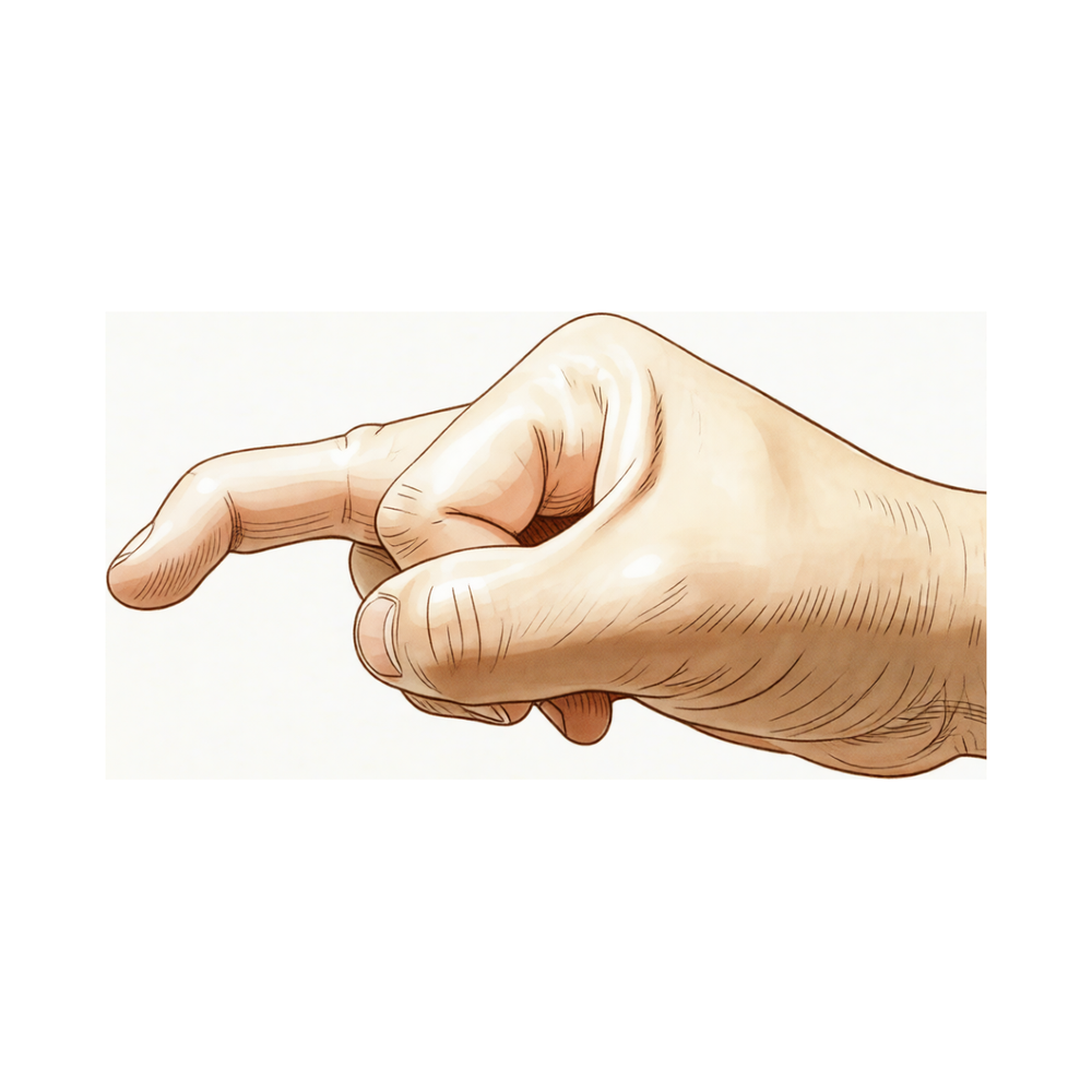
மால்ட் விரல்
Patients › Hand
Mallet finger causes fingertip drooping after extensor tendon injury; splinting is key, surgery occasionally needed.
நீங்கள் என்ன உணர்கிறீர்கள்
உங்கள் விரல் நுனியில் வலியை நீங்கள் உணரலாம். இந்த அசௌகரியம் பெரும்பாலும் கடைசி மூட்டு மையமாக உள்ளது, அங்கு தசை எலும்புடன் இணைகிறது. சில சந்தர்ப்பங்களில், இந்த வலியை நீங்கள் இரு கைகளிலும் ஒரே நேரத்தில் உணரலாம். காயம் தசைக் கண்ணீர் அல்லது அது இணைக்கும் இடத்தில் ஒரு சிறிய எலும்பு முறிவு ஆகியவற்றை உள்ளடக்கியிருக்கலாம்.
உங்கள் விரலை நேராக்க முயற்சிக்கும்போது வலி பொதுவாக மோசமடைகிறது. எதிர்ப்புக்கு எதிராக தள்ளுவது அல்லது கனமான பொருட்களை தூக்குவது உங்களுக்கு கடினமாக இருக்கலாம். சட்டைக்குள் நுழைவது அல்லது பிராவைப் பிணைக்க உங்கள் முதுகுக்குப் பின்னால் செல்வது போன்ற எளிய பணிகள் சவாலானதாக மாறக்கூடும். உங்கள் விரல் இறுக்கமாக உணரக்கூடும், குறிப்பாக நீங்கள் காலையில் எழுந்தவுடன்.
விரலை ஓய்வெடுப்பது வலியைக் குறைக்க உதவுகிறது என்று பலர் கண்டறிந்துள்ளனர். இருப்பினும், விரலை நேராக வைத்திருப்பது குணமடைவதற்கு மிகவும் முக்கியமானது. நீங்கள் நுனியை வளைத்தால், நீங்கள் ஒரு கூர்மையான இழுப்பை அல்லது அதிகரித்த வலியை உணரலாம். இது தண்டு மூட்டு சீரமைப்பிலிருந்து வெளியேற முயற்சிப்பதால் தான்.
கடுமையான நோய்களில், உங்கள் விரலின் நுனி கீழ்நோக்கி சாய்வதை நீங்கள் கவனிக்கலாம். இந்த சாய்வு பொருட்களை உறுதியாகப் பிடிப்பதை கடினமாக்குகிறது. நீங்கள் மூட்டுக்குச் சுற்றி வீக்கத்தையும் அனுபவிக்கலாம். பெரும்பாலான காயங்கள் ஒரு அடுக்குடன் நன்றாக குணமடைந்தாலும், சில சந்தர்ப்பங்களில் மிகவும் சிக்கலான கவனிப்பு தேவைப்படுகிறது. எந்தவொரு எலும்பு துண்டுகளின் அளவு அல்லது மூட்டு நிலை ஆகியவற்றின் அடிப்படையில் உங்களுக்கு அறுவை சிகிச்சை தேவைப்படுகிறதா என்பதை உங்கள் அறுவை சிகிச்சையாளர் தீர்மானிப்பார்.
உங்களுக்கு ஒரு பெரிய எலும்பு துண்டு இருந்தால் அல்லது மூட்டு இடத்திலிருந்து நகர்ந்தால், உங்கள் அறுவை சிகிச்சையாளர் ஒரு அறுவை சிகிச்சையை பரிந்துரைக்கலாம். இது பெரும்பாலும் சரியான சீரமைப்பு மற்றும் செயல்பாட்டை மீட்டெடுக்க செய்யப்படுகிறது. அறுவை சிகிச்சை மற்றும் அறுவை சிகிச்சை அல்லாத சிகிச்சைகள் இரண்டும் பொதுவாக சிறந்த முடிவுகளுக்கு வழிவகுக்கின்றன. சரியான கவனிப்புடன் உங்கள் விரலை முழுமையாகப் பயன்படுத்த நீங்கள் எதிர்பார்க்கலாம்.
உண்மையில் என்ன நடக்கிறது
உங்கள் விரல் நுனியில் டெர்மினல் டெண்டன் என்று அழைக்கப்படும் ஒரு சிறிய தசை உள்ளது. இது உங்கள் விரல் நுனியில் உள்ள எலும்புடன் உங்கள் தசைகளை இணைக்கும் ஒரு கயிறு போல செயல்படுகிறது. இந்த கயிறு உங்கள் விரலை நிமிர்ந்து நிற்க அனுமதிக்கிறது. இந்த பகுதியை நீங்கள் காயப்படுத்தும்போது, அந்த கயிறு கிழிகிறது அல்லது மூட்டுவிலிருந்து ஒரு சிறிய எலும்பு துண்டுகளை இழுக்கிறது. இது உங்கள் விரல் நுனியை அடைவதைத் தடுக்கிறது.
எலும்பு விறைப்பு நோய்க்கான பொதுவான காரணம் உங்கள் நேரான விரலை பின்னோக்கி வளைக்கும் திடீர் சக்தியாகும். உங்கள் விரல் ஒரு பந்து அல்லது ஒரு கதவு சட்டத்தில் நீட்டப்பட்டிருப்பதை கற்பனை செய்து பாருங்கள். மூட்டு மிக விரைவாக வளைந்த நிலையில் சிக்கிக் கொள்கிறது. இந்த திடீர் நீட்டிப்பு எலும்பிலிருந்து இடுப்பைக் கிழிக்கிறது. சில சந்தர்ப்பங்களில், எலும்பின் ஒரு சிறிய துண்டு இடுப்புடன் சேர்ந்து உடைந்துவிடும். இடுப்பு தன்னைக் கிழித்தாலும் அல்லது எலும்பு சிப் இழுத்தாலும் மருத்துவ தோற்றம் ஒத்ததாக இருக்கும். இரண்டு சந்தர்ப்பங்களிலும், இணைப்பு உடைக்கப்படுகிறது.
உங்கள் விரலின் நுனியை நேராக உயர்த்த முடியாது. இது ஒரு மல்லட் விரல் சிதைவு என்று அழைக்கப்படுகிறது. இருப்பினும், நீங்கள் இன்னும் அதை உங்கள் மற்ற கையால் செயலற்ற முறையில் நேராக்கலாம். மூட்டு காப்ஸ்யூல் மற்றும் இழைகள் அப்படியே இருக்கின்றன, எனவே மூட்டு தன்னை வெளியேற்றாது. வளைந்த நிலை வெறுமனே விரலின் மறுபக்கத்திலிருந்து எதிர்மறையான தசை இழுப்பின் விளைவாகும்.
இந்த காயம் பெரும்பாலும் உங்கள் ஆதிக்கம் செலுத்தும் கையின் சிறிய, மோதிரம் அல்லது நடுத்தர விரல்களை பாதிக்கிறது. இது ஆண்களில் மிகவும் பொதுவானது. இது வழக்கமாக அதிர்ச்சியால் ஏற்படுகையில், உடைந்துபோகும் மூட்டுவலி கொண்ட சில வயதான நோயாளிகள் குறிப்பிட்ட காயம் இல்லாமல் இந்த நிலைப்பாட்டை உருவாக்கலாம். குழந்தைகளில், காயம் தசைக்கு பதிலாக வளர்ச்சித் தகடு சம்பந்தப்பட்டிருக்கலாம். காரணம் எதுவாக இருந்தாலும், இதன் விளைவு ஒன்றேஃ உங்கள் விரல் நுனியை உயர்த்தும் பொறிமுறை துண்டிக்கப்பட்டு, அதை சாய்ந்து விடுகிறது.
நாம் என்ன செய்ய முடியும்
பெரும்பாலான மல்லட் விரல் காயங்கள் அறுவை சிகிச்சை இல்லாமல் குணமடைகின்றன. உங்கள் விரல் நுனியை நேராக வைத்திருக்க நீங்கள் ஒரு ஸ்பிளண்ட் அணிவீர்கள். இது சரியான நிலையில் இழை குணமடைய அனுமதிக்கிறது. ஒரு கை சிகிச்சையாளர் எளிய வழக்குகளுக்கு ஒரு அறுவை சிகிச்சையாளரைப் போலவே உங்களை திறம்பட சிகிச்சையளிக்க முடியும். அவர்கள் சரும பிரச்சினைகளை அரிதாகவே ஏற்படுத்தும் முறைகளைப் பயன்படுத்துகிறார்கள். நீங்கள் இரவு ஸ்பிளெண்ட்டையும் முயற்சி செய்யலாம், ஆனால் இது உங்கள் இறுதி முடிவை மேம்படுத்தாது. சில நோயாளிகள் ஸ்பிளெண்ட்டுடன் உடற்பயிற்சிகளைப் பயன்படுத்துகிறார்கள், இருப்பினும் இதற்கு ஆதாரங்கள் கலக்கப்பட்டுள்ளன. உங்கள் அறுவை சிகிச்சையாளர் பரிந்துரைக்கும் நேரத்திற்கு உங்கள் ஸ்பிளெண்ட்டை தொடர்ந்து அணிய வேண்டும். விரலைக் கழுவ அல்லது வளைக்க அதை அகற்ற வேண்டாம். நிலைத்தன்மை வெற்றிக்கு முக்கியமாகும்.
வலி மேலாண்மை இடுப்பு குணமடையும் போது ஆறுதலில் கவனம் செலுத்துகிறது. தேவைக்கேற்ப வலி நிவாரணிகள் அல்லது அழற்சி எதிர்ப்பு மருந்துகளை நீங்கள் எடுத்துக்கொள்ளலாம். உங்கள் விரல் இயங்காத நிலையில் தினசரி நடவடிக்கைகளை நிர்வகிக்க இவை உதவுகின்றன. சில நோயாளிகள் ஊசிகளைப் பற்றி கேட்கிறார்கள். இருப்பினும், இந்த குறிப்பிட்ட காயத்திற்கு கோர்டிசோன், ஹைலூரோனிக் அமிலம் அல்லது பிஆர்பி ஊசி மருந்துகளை ஆதாரங்கள் ஆதரிக்கவில்லை. இந்த சிகிச்சைகள் மல்லட் விரலுக்கான நிலையான பராமரிப்பு திட்டத்தின் ஒரு பகுதியாக இல்லை. உங்கள் அடுக்குடன் மூட்டு நேராக வைத்திருப்பதில் உங்கள் கவனம் இருக்க வேண்டும். உங்களுக்கு குறிப்பிடத்தக்க வலி இருந்தால், உங்கள் அறுவை சிகிச்சையாளருடன் பாதுகாப்பான விருப்பங்களைப் பற்றி விவாதிக்கவும். ஆக்கிரமிப்பு மசாஜ் அல்லது நீட்டினைத் தவிர்க்கவும், இது குணமடைவதைத் தடுக்கலாம்.
கன்சர்வேடிவ் பராமரிப்பு தோல்வியுற்றால் அல்லது காயம் கடுமையானதாக இருந்தால் மட்டுமே அறுவை சிகிச்சை பரிசீலிக்கப்படுகிறது. மூட்டு மேற்பரப்பில் மூன்றில் ஒரு பங்கை விட அதிகமான எலும்பு முறிவு இருந்தால் உங்கள் அறுவை சிகிச்சையாளர் ஒரு அறுவை சிகிச்சையை பரிந்துரைக்கலாம். எலும்பு துண்டு இடத்திலிருந்து நகர்ந்திருந்தால் அறுவை சிகிச்சை குறிக்கப்படுகிறது. சில நோயாளிகள் அறுவை சிகிச்சையைத் தேர்வு செய்கிறார்கள், ஏனென்றால் அவர்கள் ஒரு அடுக்குடன் வேலை செய்ய முடியாது. பல மாதங்கள் அடுக்குக்கு பிறகு குணமடையாத நாள்பட்ட காயம் இருந்தால், அறுவை சிகிச்சை ஒரு விருப்பமாக இருக்கலாம். விரலை நேராகச் செய்வதற்கான உங்கள் திறனை மீட்டெடுப்பதற்காக சேதமடைந்த இடுப்பை இந்த செயல்முறை சரிசெய்கிறது. இது ஒரு சிறிய அறுவை சிகிச்சை மற்றும் அதிக வெற்றி விகிதத்தைக் கொண்டுள்ளது. இந்த பாதை தேவைப்பட்டால் உங்கள் அறுவை சிகிச்சை நிபுணர்வான அபாயங்கள் மற்றும் நன்மைகளைப் பற்றி உங்களுடன் விவாதிக்கும்.
எதிர்பார்ப்பது என்ன
பெரும்பாலான மல்லெட் விரல் காயங்கள் அறுவை சிகிச்சை இல்லாமல் நன்றாக குணமடைகின்றன. உங்கள் அறுவை சிகிச்சை நிபுணர் உங்கள் விரலின் நுனியை நேராக வைத்திருக்க அடுக்குகளை பரிந்துரைக்கலாம். இது இடுப்பு அல்லது எலும்பு மீண்டும் இணைக்க அனுமதிக்கிறது. அறுவை சிகிச்சை மற்றும் அறுவை சிகிச்சை அல்லாத சிகிச்சைகள் இரண்டும் பெரும்பாலான நோயாளிகளுக்கு சிறந்த மருத்துவ விளைவுகளை ஏற்படுத்துகின்றன. உங்கள் சிகிச்சை முடிவில் அதிக அளவு திருப்தியை நீங்கள் எதிர்பார்க்கலாம்.
குணமடைவதற்கான பாதை உங்கள் காயத்தின் தீவிரத்தைப் பொறுத்தது. எளிய வழக்குகளுக்கு, ஒரு எளிய அடுக்கு அல்லது முதுகில் ஒட்டப்பட்ட அடுக்கு பொதுவாக போதுமானது. உங்கள் காயம் ஒரு பெரிய எலும்பு துண்டு அல்லது மூட்டு இடத்திலிருந்து நகர்ந்திருந்தால், அறுவை சிகிச்சை பரிந்துரைக்கப்படலாம். நாள்பட்ட வழக்குகளுக்கு அறுவை சிகிச்சை ஒரு விருப்பமாகும் அல்லது முந்தைய சிகிச்சைகள் தோல்வியுற்றிருந்தால். குழந்தைகளில், அறுவை சிகிச்சையின் தேவை குறைவாக தெளிவாக உள்ளது, ஆனால் அறுவை சிகிச்சை அல்லாத மேலாண்மை பெரும்பாலானவர்களுக்கு பயனுள்ளதாக இருக்கும்.
விரல்களின் நிலையை முழுமையாக சரிசெய்வது எப்போதும் உத்தரவாதம் அளிக்கப்படுவதில்லை என்பதை நீங்கள் அறிந்து கொள்ள வேண்டும். உங்களுக்கு கணிசமான வளைவுடன் கடுமையான நாள்பட்ட மல்லெட் விரல் இருந்தால், முழு திருத்தம் குறைவாக இருக்கும். இருப்பினும், இந்த சிக்கலான சந்தர்ப்பங்களில் கூட, நீண்ட கால முடிவுகள் பெரும்பாலும் சிறப்பாக இருக்கும். பழமைவாத நிர்வாகத்திற்கான சிக்கல் விகிதங்கள் குறைவாக உள்ளன. குறிப்பாக நவீன அறுவை சிகிச்சை நுட்பங்களுடன் தொற்று அல்லது நக விலகல் போன்ற கடுமையான பிரச்சினைகள் அரிதானவை.
குணமடைதல் என்பது ஒரு படிப்படியான செயல்முறையாகும். உங்கள் விரல் நுனியில் சில கடினத்தன்மை அல்லது ஒரு சிறிய மீதமுள்ள வளைவை நீங்கள் கவனிக்கலாம். இது பொதுவானது மற்றும் பெரும்பாலும் உங்கள் அன்றாட செயல்பாட்டை கணிசமாக பாதிக்காது. கூடுதல் இரவு அடுக்கு இயலாமை அல்லது திருப்தி ஆகியவற்றின் அடிப்படையில் உங்கள் முடிவை மேம்படுத்தாது, எனவே உங்கள் ஆரம்ப குணமடைந்த கட்டத்திற்குப் பிறகு அதை அணிய வேண்டிய அவசியமில்லை.
நீங்கள் ஒரு குறிப்பிட்ட வகையான எலும்பு முறிவு இருந்தால், அது மூட்டு மேற்பரப்பில் மூன்றில் ஒரு பங்கை விட அதிகமாக இருந்தால், அது மூட்டு சுப்ளூக்ஸேஷனுக்கு (இருப்பிலிருந்து விலகிச் செல்வது) முன்னேறாது என்பது சுமார் 50% வாய்ப்பு உள்ளது. எலும்பு முறிவின் அளவு மற்றும் உங்கள் அடுப்பை எவ்வளவு விரைவாக அணியத் தொடங்குவது என்பது உங்கள் குணமடைவதில் முக்கிய காரணிகளாகும். சிகிச்சையை தாமதப்படுத்துவது சிக்கல்களின் அபாயத்தை அதிகரிக்கக்கூடும், எனவே உங்கள் அறுவை சிகிச்சையாளரின் நேர ஆலோசனையைப் பின்பற்றுவது முக்கியம்.
ஒட்டுமொத்தமாக, மல்லெட் விரல் மிகவும் சிகிச்சையளிக்கக்கூடிய ஒரு நிலை என்று நீங்கள் நம்பிக்கையுடன் உணரலாம். நீங்கள் ஸ்பிளின்ட் அல்லது அறுவை சிகிச்சையைத் தேர்வுசெய்தாலும், நோக்கம் செயல்பாட்டை மீட்டெடுப்பதும் வலியைக் குறைப்பதும் ஆகும். பெரும்பாலான நோயாளிகள் ஒரு செயல்பாட்டு விரலுடன் தங்கள் சாதாரண நடவடிக்கைகளுக்குத் திரும்புகிறார்கள். உங்கள் அறுவை சிகிச்சையாளர் உங்கள் குறிப்பிட்ட காயத்திற்கான சிறந்த பாதையின் மூலம் உங்களுக்கு வழிகாட்டுவார்.
யாரையாவது எப்போது பார்க்க வேண்டும்
உங்கள் விரலில் பலவீனம் அல்லது நிலையற்ற தன்மையைக் கண்டால் ஒரு நிபுணர் மதிப்பாய்வைக் கேளுங்கள். மூட்டு பூட்டப்பட்டால் அல்லது வழிவகுத்தால் கவனிப்பைத் தேடுங்கள். உங்கள் தூக்கத்திலோ அல்லது வேலையிலோ அறிகுறிகள் தலையிட்டால் உங்கள் மருத்துவரைத் தொடர்பு கொள்ளுங்கள். உங்கள் நிலை திடீரென மோசமடைவதற்கு உதவி பெறுங்கள். மால்ட் விரல் காயங்கள் சில நேரங்களில் இரு கைகளிலும் ஒரே நேரத்தில் நிகழலாம். அரிதாகவே, இந்த காயங்கள் இடுப்பு சேதம் மற்றும் எலும்பு முறிவு ஆகியவற்றின் கலவையை இளம் நோயாளிகளில் உள்ளடக்குகின்றன. இந்த காயங்கள் எவ்வாறு உருவாகின்றன என்பதில் உயிர்வேதியியல் மாற்றங்களும் பங்கு வகிக்கக்கூடும். உங்கள் விரலுக்கான சிறந்த முடிவை உறுதிப்படுத்த ஆரம்ப மதிப்பீடு உதவுகிறது.
Evidence & references
Overview
- Absolute indications for surgical intervention for mallet fingers in pediatric populations remain unclear [1].
- Large-fragment mallet finger cases can be effectively managed conservatively with low complication rates [2].
- Most mallet finger injuries can be managed non-surgically with splinting [3].
- Surgery is occasionally recommended for acute or chronic mallet finger cases or for salvage of failed prior treatment [3].
- Both surgical and nonsurgical treatments of mallet finger injuries lead to excellent clinical outcomes [4].
- All cases of mallet finger are proposed to be treated with a dorsal glued splint except for stage IV mallet finger, which is treated with extra-articular pinning [5].
- A simple splint is recommended as an alternative means of treating mallet finger [8].
- Surgery is generally indicated in mallet fractures involving more than one-third of the articular surface [9].
- Surgery is generally indicated in all patients who develop volar subluxation of the distal phalanx [9].
- A significant advantage of surgical management over conservative management in complicated cases (large fragment or volar subluxation) has yet to be clearly proven [9].
- The majority of hand fractures can be treated without surgery, though surgery offers distinct advantages in properly selected cases [12].
- Pullout wire fixation together with distal interphalangeal joint Kirschner wire stabilization for acute combined tendon and bone (double level) mallet finger injury reports good to excellent long-term results with no reported complications such as infection, nonunion, or nail deformity [13].
- Central slip tenotomy with distal repair is a technique described for severe chronic mallet fingers with deformities exceeding 36 degrees [18].
- Full correction of extensor lag is less consistent with greater degrees of preoperative flexion deformity in severe chronic mallet fingers treated with central slip tenotomy and distal repair [18].
- The Ishiguro extension block technique produces satisfactory results and is effective and minimally invasive for mallet finger fractures when properly applied [54].
Anatomy & Pathophysiology
- Mallet finger deformity is characterized by a loss of active distal interphalangeal (DIP) joint extension with full passive range of motion evident [19].
- The deformity reflects the loss of normal extensor force transmission via the terminal tendon insertion onto the distal phalanx [19].
- The unopposed flexor digitorum profundus pulls the distal joint into flexion [19].
- The usual mechanism of injury involves sudden passive flexion of an actively extended DIP joint [19].
- Disruption of the terminal tendon may be entirely confined to the tendon or may involve an avulsed fracture fragment from the dorsal lip of the distal phalanx proximal articular surface [19].
- The clinical appearance of soft tissue and bony mallet fingers is similar because the avulsed fragment includes the terminal tendon insertion [19].
- The distal joint rests in flexion, a posture that cannot be actively changed [19].
- Full passive extension of the DIP joint is possible in mallet finger [19].
- Mallet finger most commonly involves a closed rupture of the terminal tendon with or without associated fracture of the distal phalanx [37].
- Snagging the extending finger on an object that suddenly flexes the DIP joint is a frequent cause of mallet finger [37].
- Less commonly, a forceful hyperextension injury of the DIP joint may result in a large fracture of the base of the distal phalanx involving one-third or more of the articular surface [37].
- Elderly patients with osteoarthritis of the DIP joint may have mallet deformities that are not related to trauma [37].
- Individuals with hyperlax joints may have multiple pseudomallet swan neck postures that are unrelated to trauma [37].
- Open mallet injuries are uncommon [37].
- The most frequently involved digits are the small, ring, and middle fingers of the dominant hand [37].
- There is a male predominance in mallet finger incidence [37].
- Tendinous mallet fingers have been reported to occur from age 11 onward [37].
- In skeletally immature individuals, a transepiphyseal plate fracture may be seen instead of tendon rupture [37].
- There may be a familial predisposition to mallet fingers [37].
- Displacement of the epiphysis of the distal phalanx can cause the digit to assume a mallet finger posture [14].
- Hyperextension of the phalanx usually affords satisfactory reduction of a displaced epiphysis [14].
- The extensor apparatus of the fingers includes the interosseous muscle, extensor digitorum communis tendon, lumbrical muscle, flexor tendon sheath, sagittal bands, transverse metacarpal ligament, interosseous hood, interosseous hood oblique fibers, extensor lateral band, extensor middle band, interosseous middle band, interosseous lateral band, oblique retinacular ligament, central extensor lateral, spiral fibers, transverse retinacular ligament, lateral extensor tendon, triangular lamina, and terminal extensor tendon [14].
- Injuries to the finger extensor apparatus are very common and may produce chronic deformity and loss of function [34].
Classification
- Absolute indications for surgical intervention for mallet fingers in pediatric populations remain unclear [1].
- Doyle type 4c mallet finger cases can be effectively managed conservatively with low complication rates [2].
- Most mallet finger injuries can be managed non-surgically with splinting, although surgery is occasionally recommended for acute or chronic cases or for salvage of failed prior treatment [3].
- Both surgical and nonsurgical treatments of mallet finger injuries lead to excellent clinical outcomes [4].
- All cases of mallet finger are proposed to be treated with a dorsal glued splint except for stage IV mallet finger, which is treated with extra-articular pinning [5].
- Approximately 50% of patients with a mallet fracture involving more than one-third of the articular surface of the distal phalanx do not progress to subluxation of the DIP joint [6].
- Fracture size and time to application of finger immobilizer are independent risk factors for the development of DIP joint subluxation in mallet fracture [6].
- A simple splint is recommended as an alternative means of treating mallet finger [8].
- Deepithelialised pedicled skin flap technique is a new reliable alternative in the treatment of chronic mallet finger [11].
- A hand therapist can treat mallet finger injuries of type 1 as effectively as a surgeon, with a method of immobilisation that offers practically no complications regarding skin condition [16].
- A modification to the Doyle classification is proposed to make it more encompassing and less prone to interobserver error [35].
- The interrater reliability of the Kellgren & Lawrence and OARSI classification systems for post-traumatic osteoarthritis in the distal interphalangeal joint after mallet finger fractures is considerably lower than initially assumed [41].
- Non-operative management of mallet fractures, regardless of fracture classification, joint congruence or pre-existing degenerate change in the DIP joint, is safe and yields predictably good outcomes in most patients [46].
- Most acute mallet fingers can be treated by continuous splinting, while fracture dislocations require open reduction and internal fixation [50].
Clinical Presentation
- Mallet finger injuries can present as bilateral cases [21].
- Biochemical abnormalities may play a role in the etiology of mallet fingers [21].
- Mallet finger injuries can involve a rare combination of tendon avulsion and fracture in juvenile patients [26].
Investigations
- Mallet finger deformity is characterized by a loss of active distal interphalangeal (DIP) joint extension with full passive range of motion evident [19].
- The deformity reflects the loss of normal extensor force transmission via the terminal tendon insertion onto the distal phalanx [19].
- The unopposed flexor digitorum profundus pulls the distal joint into flexion [19].
- The usual mechanism of injury involves sudden passive flexion of the actively extended distal interphalangeal joint [19].
- Disruption of the terminal tendon may be entirely confined to the tendon or may involve an avulsed fracture fragment from the dorsal lip of the distal phalanx proximal articular surface [19].
- Because the avulsed fragment includes the terminal tendon insertion, the clinical appearance of soft tissue and bony mallet fingers is similar [19].
- The distal joint rests in flexion, a posture that cannot be actively changed [19].
- Full passive extension of the distal interphalangeal joint is possible [19].
- A radiograph should be obtained to determine whether a fracture is present [19].
- If a fracture is present, radiographs should assess whether the dorsal fragment is large and whether the distal phalanx is subluxed palmarward [19].
Treatment
Non-Operative Management
- Most mallet finger injuries can be managed non-surgically with splinting [3].
- Both surgical and nonsurgical treatments of mallet finger injuries lead to excellent clinical outcomes [4].
- A dorsal glued splint is proposed for the treatment of all cases of mallet finger except stage IV injuries [5].
- A simple splint is recommended as an alternative means of treating mallet finger [8].
- Supplemental night splinting does not improve the outcome of mallet finger in terms of extensor lag, disability, or satisfaction with treatment [15].
- A hand therapist can treat type 1 mallet finger injuries as effectively as a surgeon [16].
- A hand therapist can treat type 1 mallet finger injuries as effectively as a surgeon, with a method of immobilisation that offers practically no complications regarding skin condition [39].
- Conservative therapeutic management of acute, closed mallet finger is diverse and varied, with exercises and interventions supplementary to splinting commonly utilised [23].
- An alternative simple and custom-made orthosis manages the mallet finger by allowing for PIP flexion while inhibiting full extension or hyperextension [25].
- The clinical efficacy of elastic taping for the treatment of mallet finger injuries remains to be tested vigorously [32].
Operative Management
- Surgery is occasionally recommended for acute or chronic mallet cases or for salvage of failed prior treatment [3].
- Surgical management may be considered for acute and chronic mallet lesions in patients who have failed nonsurgical treatment [10].
- Surgical management may be considered for acute and chronic mallet lesions in patients who are unable to work with the splint in position [10].
- Surgical management may be considered for acute and chronic mallet lesions in patients who have a fracture involving more than one third of the joint surface [10].
- Surgery is generally indicated in the case of mallet fractures involving more than one-third of the articular surface [9].
- Surgery is generally indicated in all patients who develop volar subluxation of the distal phalanx [9].
- A significant advantage of surgical management even in complicated cases (fracture >1/3 articular surface or volar subluxation) has yet to be clearly proven [9].
- Complication rates for conservative management of Doyle type 4c mallet finger are low, suggesting large-fragment cases can be effectively managed conservatively [2].
- Absolute indications for surgical intervention for mallet fingers in the pediatric population remain unclear [1].
Surgical Techniques
- The Thompson procedure is an effective technique for the salvage of a closed mallet injury with an associated swan neck deformity following failed treatment [27].
- A deepithelialised pedicled skin flap technique is a new reliable alternative in the treatment of chronic mallet finger [11].
- Scar overlapping suture for treating chronic tendinous mallet finger in children is safe and effective [36].
- For chronic mallet finger, if the distal phalanx droops severely but passive extension in the distal interphalangeal joint is still satisfactory, surgery may be indicated depending on the patient’s needs [14].
- Surgical treatment for chronic mallet finger involves making a small V-shaped or U-shaped incision on the dorsum of the finger, convex distally, with the tip no closer than 5 mm proximal to the nail base [14].
- The surgical technique for chronic mallet finger involves developing a flap between the tendon and subcutaneous fat to expose the extensor tendon with intervening scar [14].
- The surgical technique for chronic mallet finger involves identifying the junction of normal tendon with scar, severing the tendon transversely proximal to the joint, and resecting sufficient scar or tendon to allow closure with the finger in maximal extension [14].
- The surgical repair of chronic mallet finger is supported by a transarticular 0.045-inch Kirschner wire [14].
- The extensor tendon in chronic mallet finger surgery is repaired with 4-0 monofilament nylon or 4-0 monofilament wire as a pull-out roll stitch [14].
- Postoperative care for chronic mallet finger surgery involves removing sutures at 10 to 14 days and maintaining the distal joint in extension with Kirschner wire protection for 4 weeks [14].
- The Kirschner wire is removed after 4 to 6 weeks in chronic mallet finger surgery, with the repair protected by a splint for an additional 8 weeks [14].
Complications
- Absolute indications for surgical intervention for mallet fingers in pediatric populations remain unclear [1].
- Complication rates for conservative management of Doyle type 4c mallet fingers are low [2].
- Surgery is occasionally recommended for acute or chronic cases or for salvage of failed prior treatment [3].
- Both surgical and nonsurgical treatments of mallet finger injuries lead to excellent clinical outcomes [4].
- Stage IV mallet finger is treated with extra-articular pinning rather than dorsal glued splinting [5].
- Approximately 50% of patients with a mallet fracture involving more than one-third of the articular surface of the distal phalanx do not progress to subluxation of the DIP joint [6].
- Fracture size is an independent risk factor for the development of DIP joint subluxation in mallet fracture [6].
- Time to application of finger immobilizer is an independent risk factor for the development of DIP joint subluxation in mallet fracture [6].
- Pullout wire fixation together with distal interphalangeal joint Kirschner wire stabilization for acute combined tendon and bone mallet finger injuries reports no complications such as infection, nonunion, or nail deformity [13].
- Supplemental night splinting does not improve outcomes in terms of extensor lag, disability, or satisfaction with treatment [15].
- Full correction of extensor lag is less consistent with greater degrees of preoperative flexion deformity in severe chronic mallet fingers treated with central slip tenotomy and distal repair [18].
- The Thompson procedure is an effective technique for the salvage of a closed mallet injury with an associated swan neck deformity following failed treatment [27].
- The most common cause of procedural failure in closed reductions using an extension-block pin for bony mallet finger is inaccurate insertion of the K-wire to fix the distal interphalangeal joint [56].
Recovery
- Most mallet finger injuries can be managed non-surgically with splinting [3].
- Surgery is occasionally recommended for acute or chronic cases or for salvage of failed prior treatment [3].
- Both surgical and nonsurgical treatments of mallet finger injuries lead to excellent clinical outcomes [4].
- Complication rates for conservative management of large-fragment mallet finger cases are low [2].
- Supplemental night splinting does not improve the outcome of mallet finger in terms of extensor lag, disability, or satisfaction with treatment [15].
- LIPUS therapy may be recommended as an option to treat type I mallet finger fracture in children for whom initiation of treatment was delayed up to 8 weeks [29].
- Approximately 50% of patients with a mallet fracture involving more than one-third of the articular surface of the distal phalanx do not progress to subluxation of the DIP joint [6].
- Fracture size and time to application of finger immobilizer are independent risk factors for the development of DIP joint subluxation in mallet fracture [6].
- Full correction of extensor lag is less consistent with greater degrees of preoperative flexion deformity in severe chronic mallet fingers treated with central slip tenotomy with distal repair [18].
- The surgical technique of pullout wire fixation together with distal interphalangeal joint Kirschner wire stabilization for acute combined tendon and bone mallet finger injuries reports good to excellent long-term results [13].
- No reported complications such as infection, nonunion, or nail deformity were observed in the series of acute combined tendon and bone mallet finger injuries treated with pullout wire fixation and DIP joint Kirschner wire stabilization [13].
Key Evidence
- [L4] Absolute indications for surgical intervention for mallet fingers in this population remain unclear. [1] (10.1016/j.jhsa.2018.03.037)
- [L4] Complication rates were low, suggesting that large-fragment mallet finger cases can be effectively managed conservatively. [2] (10.1186/s12891-026-09787-w)
- [L5] Most mallet finger injuries can be managed non-surgically with splinting, although surgery is occasionally recommended for acute or chronic cases or for salvage of failed prior treatment. [3] (10.1007/s11552-014-9609-y)
- [L4] Both surgical and nonsurgical treatments of mallet finger injuries lead to excellent clinical outcomes. [4] (10.1016/j.jhsa.2017.10.004)
- [L5] The authors propose to treat all cases of mallet finger with a dorsal glued splint except for stage IV mallet finger, which they treat with extra-articular pinning. [5] (10.5999/aps.2016.43.2.134)
- [L2] Approximately 50% of patients with a mallet fracture involving more than one-third of the articular surface of the distal phalanx do not progress to subluxation of the DIP joint; fracture size and time to application of finger immobilizer are independent risk factors for the development of DIP joint subluxation in mallet fracture. [6] (10.1177/1753193414554556)
- [L2] The study recommends this splint as an alternative means of treating mallet finger. [8] (10.1136/emj.10.3.244)
- [L4] Although surgery is generally indicated in the case of mallet fractures involving more than one-third of the articular surface as well as in all patients who develop volar subluxation of the distal phalanx, a significant advantage of surgical management even in those complicated cases has yet to be clearly proven. [9] (10.1177/1558944716642763)
- [L5] Surgical management may be considered for acute and chronic mallet lesions in patients who have failed nonsurgical treatment, are unable to work with the splint in position, or have a fracture involving more than one third of the joint surface. [10] (10.5435/00124635-200509000-00007)
- [Paper] This method seems to be a new reliable alternative in the treatment of chronic mallet finger. [11] (10.1016/j.injury.2013.01.013)
- [L5] The majority of hand fractures can be treated without surgery, though surgery offers distinct advantages in properly selected cases. [12] (10.1016/j.jhsa.2013.02.017)
- [L4] The study describes a surgical technique for acute combined tendon and bone mallet fingers and reports good to excellent long-term results with no reported complications such as infection, nonunion, or nail deformity in the series. [13] (10.1016/j.jhsa.2014.11.011)
- [L1] Supplemental night splinting does not improve the outcome of mallet finger in terms of extensor lag, disability, or satisfaction with treatment. [15] (10.1007/s11552-013-9600-z)
- [L4] A hand therapist can treat mallet finger injuries of type 1 as effectively as a surgeon, with a method of immobilisation that offers practically no complications regarding skin condition. [16] (10.1177/175899830501000103)
- [L4] The article describes a technique combining central slip tenotomy with distal repair for severe chronic mallet fingers with deformities exceeding 36 degrees, noting that full correction of extensor lag is less consistent with greater degrees of preoperative flexion deformity. [18] (10.1016/j.jhsa.2014.01.040)
- [L4] This case raises a question regarding the possible role of biochemical abnormalities causing mallet fingers. [21] (10.1177/175899830400900103)
- [L4] Conservative therapeutic management of acute, closed mallet finger is diverse and varied, with exercises and interventions supplementary to splinting commonly utilised. [23] (10.1177/1758998316664822)
- [L5] An alternative simple and custom-made orthosis has been presented to manage the mallet finger, allowing for PIP flexion while inhibiting full extension or hyperextension. [25] (10.1016/j.jht.2016.01.001)
- [L4] The authors describe a rare combination of tendon avulsion and fracture in a juvenile mallet finger, noting that while few mallet fingers require open treatment, this combined injury may be more common than thought. [26] (10.1177/1753193415571777)
- [L4] It is an effective technique for the salvage of a closed mallet injury with an associated swan neck deformity following failed treatment. [27] (10.1016/j.jhsa.2013.04.011)
- [L3] LIPUS therapy may be recommended as an option to treat type I mallet finger fracture in children for whom initiation of treatment was delayed up to 8 weeks. [29] (10.1177/1558944717692095)
- [L4] The clinical efficacy of the proposed method of elastic taping for the treatment of mallet finger injuries remains to be tested vigorously. [32] (10.1016/j.jht.2014.02.005)
- [L5] Injuries to the finger extensor apparatus are very common and may produce chronic deformity and loss of function. [34] (10.1016/j.hcl.2013.03.003)
- [L4] This article provides a topical review of the contemporary literature concerning acute mallet finger injuries and proposes a modification to the Doyle classification to make it more encompassing and less prone to interobserver error. [35] (10.1016/j.jhsa.2022.10.013)
- [L4] Scar overlapping suture for treating chronic tendinous mallet finger in children is safe and effective. [36] (10.1186/s13018-019-1106-0)
- [L4] A hand therapist can treat type 1 mallet finger injuries as effectively as a surgeon. [39] (10.1197/j.jht.2008.04.002)
- [L4] The interrater reliability of the Kellgren & Lawrence and OARSI classification systems for post-traumatic osteoarthritis in the distal interphalangeal joint after mallet finger fractures is considerably lower than initially assumed. [41] (10.1016/j.jhsa.2024.03.012)
- [L3] Non-operative management of mallet fractures, regardless of fracture classification, joint congruence or pre-existing degenerate change in the DIP joint, is safe and yields predictably good outcomes in most patients. [46] (10.1177/1753193421992986)
- [L5] Most acute mallet fingers can be treated by continuous splinting, while fracture dislocations require open reduction and internal fixation. [50] (10.1097/01.blo.0000205903.51727.62)
- [L4] The extension block technique, when properly applied, produces satisfactory results and is effective and minimally invasive for mallet finger fractures. [54] (10.1054/jhsb.2001.0733)
- [Paper] The most common cause of procedural failure was inaccurate insertion of the K-wire to fix the distal interphalangeal joint. [56] (10.1055/s-0040-1701318)
References
[1] Outcomes of Splinting in Pediatric Mallet Finger. The Journal of Hand Surgery. 2018. DOI: 10.1016/j.jhsa.2018.03.037 [2] Surgical versus conservative management of Doyle type 4c mallet finger: a comparative study. BMC Musculoskeletal Disorders. 2026. DOI: 10.1186/s12891-026-09787-w [3] Current Concepts: Mallet Finger. HAND. 2014. DOI: 10.1007/s11552-014-9609-y [4] Surgical and Nonsurgical Management of Mallet Finger: A Systematic Review. The Journal of Hand Surgery. 2018. DOI: 10.1016/j.jhsa.2017.10.004 [5] Review of Acute Traumatic Closed Mallet Finger Injuries in Adults. Archives of Plastic Surgery. 2016. DOI: 10.5999/aps.2016.43.2.134 [6] The risk factors associated with subluxation of the distal interphalangeal joint in mallet fracture. Journal of Hand Surgery (European Volume). 2014. DOI: 10.1177/1753193414554556 [8] The conservative treatment of mallet finger with a simple splint: a case report.. Emergency Medicine Journal. 1993. DOI: 10.1136/emj.10.3.244 [9] The Diagnosis and Management of Mallet Finger Injuries. HAND. 2016. DOI: 10.1177/1558944716642763 [10] Mallet Finger. Journal of the American Academy of Orthopaedic Surgeons. 2005. DOI: 10.5435/00124635-200509000-00007 [11] A new surgical treatment for mallet finger deformity: Deepithelialised pedicled skin flap technique. Injury. 2013. DOI: 10.1016/j.injury.2013.01.013 [12] Hand Fractures: A Review of Current Treatment Strategies. The Journal of Hand Surgery. 2013. DOI: 10.1016/j.jhsa.2013.02.017 [13] Pullout Wire Fixation Together With Distal Interphalangeal Joint Kirschner Wire Stabilization for Acute Combined Tendon and Bone (Double Level) Mallet Finger Injury. The Journal of Hand Surgery. 2015. DOI: 10.1016/j.jhsa.2014.11.011 [14] Campbell S Operative Orthopaedics 4 Volume Set. RECONSTRUCTION OF FINGER FLEXORS: SINGLE-STAGE TENDON GRAFT > CHRONIC MALLET FINGER (SECONDARY REPAIR). [15] A Prospective Randomized Controlled Trial Comparing Night Splinting with No Splinting after Treatment of Mallet Finger. HAND. 2014. DOI: 10.1007/s11552-013-9600-z [16] Hand Therapist-led Management of Mallet Finger. The British Journal of Hand Therapy. 2005. DOI: 10.1177/175899830501000103 [18] Central Slip Tenotomy With Distal Repair in the Treatment of Severe Chronic Mallet Fingers. The Journal of Hand Surgery. 2014. DOI: 10.1016/j.jhsa.2014.01.040 [19] A Lange Medical Book Current Diagnosis Treatment In Orthopedics Fifth Edition. 9Hand Surgery > 3. Mallet Finger. [21] Bilateral Mallet Fingers: A Case Study. The British Journal of Hand Therapy. 2004. DOI: 10.1177/175899830400900103 [23] How do hand therapists conservatively manage acute, closed mallet finger? A survey of members of the British Association of Hand Therapists. Hand Therapy. 2016. DOI: 10.1177/1758998316664822 [25] The challenge of the mallet orthosis: A simple solution. Journal of Hand Therapy. 2016. DOI: 10.1016/j.jht.2016.01.001 [26] Combined tendon avulsion and fracture in a mallet finger injury in a juvenile. Journal of Hand Surgery (European Volume). 2015. DOI: 10.1177/1753193415571777 [27] The Thompson Procedure for Chronic Mallet Finger Deformity. The Journal of Hand Surgery. 2013. DOI: 10.1016/j.jhsa.2013.04.011 [29] Comparison of Treatment Results for Mallet Finger Fractures in Children Between Low-Intensity Pulsed Ultrasound Stimulation and Ishiguro’s Method. HAND. 2017. DOI: 10.1177/1558944717692095 [32] A novel way of treating mallet finger injuries. Journal of Hand Therapy. 2014. DOI: 10.1016/j.jht.2014.02.005 [34] Diagnosis and Treatment of Finger Deformities Following Injuries to the Extensor Tendon Mechanism. Hand Clinics. 2013. DOI: 10.1016/j.hcl.2013.03.003 [35] Acute Mallet Finger Injuries—A Review. The Journal of Hand Surgery. 2023. DOI: 10.1016/j.jhsa.2022.10.013 [36] Scar overlapping suture for treating chronic tendinous mallet finger in children. Journal of Orthopaedic Surgery and Research. 2019. DOI: 10.1186/s13018-019-1106-0 [37] Green S Operative Hand Surgery. CASE STUDY 5.2 Unusual Mallet Finger Presentation. [39] Hand Therapist-led Management of Mallet Finger. Journal of Hand Therapy. 2008. DOI: 10.1197/j.jht.2008.04.002 [41] Rater Agreement of Post-Traumatic Osteoarthritis of the Distal Interphalangeal Joint 12 Years After a Mallet Finger Fracture. The Journal of Hand Surgery. 2024. DOI: 10.1016/j.jhsa.2024.03.012 [46] The non-operative management of bony mallet injuries. Journal of Hand Surgery (European Volume). 2021. DOI: 10.1177/1753193421992986 [50] Tendon Avulsion Injuries of the Distal Phalanx. Clinical Orthopaedics and Related Research. 2006. DOI: 10.1097/01.blo.0000205903.51727.62 [54] The Ishiguro Extension Block Technique for the Treatment of Mallet Finger Fracture: Indications and Clinical Results. Journal of Hand Surgery. 2003. DOI: 10.1054/jhsb.2001.0733 [56] Causes of Procedural Failures of Closed Reductions using an Extension-Block Pin for Bony Mallet Finger. Journal of Hand and Microsurgery. 2021. DOI: 10.1055/s-0040-1701318


