Cisto Mucoso
Patients › Hand
Mucous cysts – common bumps near finger joints, often linked to arthritis, and treatment options.
O que você está sentindo
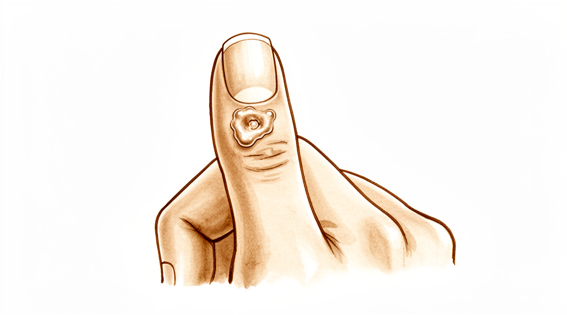
Você pode notar um caroço pequeno e firme na parte superior do seu dedo, geralmente próximo à última articulação. Esse caroço é um cisto mucoso. Ele frequentemente parece um pequeno grão de ervilha sob a pele. Você pode vê-lo claramente se a sua pele for fina nessa região. O cisto pode fazer com que a pele sobre ele pareça brilhante ou esticada. Em alguns casos, pode fazer com que a unha cresça em um padrão ondulado ou com sulcos.
A área ao redor do cisto pode parecer sensível ou dolorida. Você pode sentir dor ao pressioná-lo ou ao dobrar o dedo. As tarefas diárias podem se tornar difíceis. Alcançar objetos, digitar ou segurar ferramentas pode doer. Você pode achar difícil abotoar botões ou fechar o zíper de uma jaqueta. Algumas pessoas relatam que a dor é pior à noite ou após usar as mãos por um longo período. O desconforto pode interferir no sono se você apoiar a mão nessa região.
Seu cirurgião examinará o cisto e verificará como ele afeta o movimento do seu dedo. A dor geralmente vem das alterações na articulação subjacente, não apenas do caroço em si. Remover apenas o cisto pode não aliviar a dor se o esporão ósseo (osteófito) permanecer. Seu cirurgião pode discutir a remoção do esporão ósseo para ajudar a aliviar a pressão na articulação e na pele. Essa abordagem pode ajudar a pele a cicatrizar melhor e reduzir a chance de o cisto voltar.
Em muitos casos, tratar o esporão ósseo leva à resolução completa do cisto. Você pode não precisar de uma cirurgia complexa. A remoção simples do cisto combinada com a remoção do esporão ósseo tem uma taxa de recorrência extremamente rara. Opções ainda menos invasivas, como remover apenas o esporão ósseo, podem proporcionar resolução completa na maioria dos casos. Se for necessária uma retalho de pele, a satisfação do paciente em relação à cicatriz é alta. É provável que você fique satisfeito com a aparência e disposto a realizar o procedimento novamente.
Seu cirurgião adaptará o plano às suas necessidades específicas. O objetivo é aliviar sua dor e restaurar a função. A maioria dos pacientes percebe que, após o tratamento, seu dedo fica mais confortável e com melhor aparência. A pele tem bom potencial de recuperação assim que o problema principal é resolvido. Você pode esperar uma recuperação tranquila com cuidados adequados com a ferida.
O que realmente está acontecendo
Um cisto mucoso é uma pequena bolsa preenchida por líquido que se forma no seu dedo. Geralmente, aparece perto da ponta do dedo, próximo ao leito ungueal. O cisto situa-se sobre a cápsula articular, que é a capa fibrosa e resistente que envolve a sua articulação para mantê-la estável.
A causa subjacente é a artrite por desgaste na articulação. À medida que a cartilagem — o revestimento liso nas extremidades dos seus ossos — se degrada, o seu corpo tenta reparar o dano. Este processo frequentemente cria esporões ósseos, chamados osteófitos. Estes são pequenos saliências ásperas de osso extra que crescem a partir da superfície articular.
Pense na cápsula articular como uma junta ou vedação. Quando o esporão ósseo atrita contra esta vedação, irrita o tecido. Esta irritação faz com que o revestimento articular vaze líquido sinovial, que é o lubrificante natural que mantém o movimento da sua articulação suave. O líquido atravessa o ponto enfraquecido da cápsula, formando o inchaço visível na sua pele.
Como o cisto está conectado à articulação, ele é preenchido com o mesmo líquido lubrificante. A pressão do líquido pode tornar a pele sobre o cisto fina e frágil. Em alguns casos, o cisto pode pressionar os nervos próximos ou afetar a matriz ungueal, que é o tecido sob a cutícula que faz crescer a unha. Esta pressão é o que leva às ranhuras ou cristas que você pode ver na sua unha.
Remover apenas o cisto frequentemente leva ao seu retorno porque o esporão ósseo permanece. O esporão continua a irritar o revestimento articular, causando mais vazamento de líquido. Para interromper este ciclo, o esporão ósseo subjacente deve ser tratado. Quando o esporão é removido, a irritação cessa e o revestimento articular pode cicatrizar. É por isso que tratar o osso é tão importante quanto tratar o próprio cisto.
O que podemos fazer a respeito
A sua jornada geralmente começa com autocuidados simples e orientação profissional. Você pode tentar repousar o dedo e evitar atividades que exerçam pressão sobre o cisto. A fisioterapia ajuda a manter a mobilidade da articulação e mantém os músculos ao redor fortes. Essa abordagem visa reduzir a irritação e melhorar a função sem etapas invasivas. Você deve dar uma tentativa justa a esse tratamento conservador para ver se os sintomas se resolvem. Muitos pacientes descobrem que gerenciar os hábitos diários e seguir um plano de exercícios suaves proporciona alívio suficiente para evitar intervenções adicionais.
Se os autocuidados não forem suficientes, o seu cirurgião pode sugerir manejo médico para controlar a dor e a inflamação. Injeções de corticosteroides são uma opção comum para esses cistos. Uma injeção de corticosteroides volar permite uma colocação fácil e consistente da agulha na articulação. Esse método minimiza os riscos potenciais de dano aos tecidos moles e infecção em comparação com outras técnicas. A injeção ajuda a acalmar a inflamação que impulsiona o crescimento do cisto. Embora as evidências destaquem a segurança e a facilidade dessa abordagem, é importante entender que as injeções geralmente gerenciam os sintomas em vez de remover o cisto permanentemente. O efeito dura por um período de tempo, mas a artrite subjacente por desgaste permanece. Você e o seu cirurgião decidirão se esse alívio temporário é suficiente ou se são necessárias etapas adicionais.
A cirurgia é considerada quando o tratamento conservador atingiu seu limite e o cisto causa dor persistente, deformidade ou problemas funcionais. O seu cirurgião discutirá a melhor opção cirúrgica para o seu caso específico. O objetivo é remover o cisto e abordar a causa raiz, que são frequentemente esporões ósseos (osteófitos) provenientes da artrite. A remoção desses esporões ósseos é crucial, pois reduz significativamente a chance de o cisto retornar. Algumas técnicas envolvem a remoção do cisto junto com uma pequena pele retalho para garantir a cicatrização adequada, enquanto outras se concentram principalmente no osso. O seu cirurgião escolherá o método que oferece o melhor equilíbrio entre baixas taxas de recorrência e bons resultados estéticos para a sua mão.
O que esperar
Os cistos mucosos são pequenas protuberâncias cheias de líquido que frequentemente aparecem perto da articulação terminal do dedo ou do polegar. Eles estão intimamente ligados à osteoartrite (artrite por desgaste) nessa articulação. Como derivam dessa alteração articular subjacente, o cisto em si pode persistir ou retornar se a causa raiz não for tratada. No entanto, com o tratamento adequado, o prognóstico é geralmente muito positivo.
O seu cirurgião provavelmente recomendará a remoção do cisto juntamente com os esporões ósseos (osteófitos) que o causam. Esta abordagem erradica os cistos mucosos com recorrência extremamente rara. Se o seu cirurgião optar por uma técnica que utilize um retalho cutâneo de avanço local, a taxa de recorrência é baixa, de 1,4%. Em alguns casos, o seu cirurgião pode remover apenas o esporão ósseo sem retirar o cisto. Este método menos invasivo proporciona resolução completa na maioria dos casos. Independentemente do plano específico para os tecidos moles, o seu cirurgião removerá o esporão ósseo para evitar que o cisto volte.
Pode esperar uma elevada satisfação com os resultados, particularmente no que diz respeito à aparência da cicatriz. Muitos pacientes relatam que voltariam a submeter-se ao procedimento. As técnicas cirúrgicas utilizadas são concebidas para serem simples e fiáveis. Por exemplo, alguns métodos permitem a remoção da pele afinada sem adicionar risco à sua matriz ungueal. Outros utilizam enxertos cutâneos que proporcionam resultados cosméticos satisfatórios com taxas de recorrência aceitáveis. Mesmo casos complexos, como cistos que crescem dentro do nervo digital, podem resultar em resultados bem-sucedidos.
Se não for tratado, o cisto pode permanecer ou crescer, potencialmente afinando a pele e aumentando o risco de infeção. Ao tratar tanto o cisto como a artrite subjacente, o seu cirurgião visa uma solução duradoura. A maioria dos pacientes verifica que o seu dedo fica com melhor aspeto e sensação após o procedimento. O objetivo não é apenas remover a protuberância, mas impedir que esta volte. Pode esperar um processo de recuperação simples, focado na proteção do local cirúrgico enquanto permite que a sua articulação cicatrize.
Quando procurar ajuda médica
Consulte o seu médico de família se notar um pequeno nódulo no dedo, próximo à unha. Solicite uma avaliação especializada se o cisto causar dor persistente que não melhora com o repouso. Procure atendimento se experimentar fraqueza ou instabilidade na articulação. Esteja atento a sensações de bloqueio ou de "cedência" da articulação. Contacte o seu cirurgião se os sintomas interferirem com o sono ou com o trabalho. A piora súbita da área também justifica uma consulta. Uma avaliação precoce ajuda a prevenir complicações. O seu cirurgião pode discutir opções como a remoção do cisto e de eventuais osteófitos. Esta abordagem frequentemente resulta em elevada satisfação e baixas taxas de recorrência. Tratar a causa subjacente ajuda a pele a recuperar naturalmente.
Evidence & references
Overview
- Scientific data regarding mucous cysts consist almost entirely of retrospective studies [1].
- Much of the management or recommendations for mucous cysts is based on expert opinion [1].
- Total dorsal capsulectomy alone is a simple treatment for mucous cysts that does not lead to any recurrence [2].
- Excision of the cyst combined with complete removal of the marginal osteophyte eradicates mucous cysts with extremely rare recurrence [3].
- Osteophyte excision without cyst excision may be a good treatment choice for mucous cysts of the finger, providing a less invasive method with complete resolution in most cases [5].
- Osteophyte removal results in a low cyst recurrence rate, indicating it should be undertaken regardless of the surgeon's plan for the soft tissues [13].
- The Zitelli bilobed flap allows excision of the cyst and thinned skin with no added risk to the nail matrix [6].
- The use of a Wolfe graft for mucous cysts is simple, easy to perform, and provides satisfactory cosmesis with acceptable recurrence rates [7].
- Surgical excision with a local advancement skin flap is a reliable treatment for digital mucous cysts, demonstrating a low recurrence rate of 1.4% and high patient satisfaction regarding the scar and willingness to undergo the procedure again [9].
- A surgical technique involving excision of the cyst, synovectomy, and débridement of osteophytes with rotational flap closure resulted in no recurrences in thirty-six patients [10].
- Pathohistological analysis is useful in cases where doubts arise about the initial diagnosis of a benign tumorous lesion [4].
- Eccrine porocarcinomas have a substantial risk of metastasis, high risk of local recurrence, and are potentially fatal [8].
Anatomy & Pathophysiology
- Scientific data regarding mucous cysts consist almost entirely of retrospective studies [1].
- Much of the management or recommendations for mucous cysts is based on expert opinion [1].
- Mucous cysts are associated with marginal osteophytes at the distal interphalangeal joint [3].
- The primary pathology in mucous cysts involves osteophytes, and removal of these osteophytes allows for skin recovery potential [20].
- Ultrasound is a powerful modality for evaluating pathologic conditions in the hand and wrist [16].
- Ultrasound provides a cost-effective and expedient alternative or adjunct to MRI for hand and wrist evaluation [16].
- Ultrasound is best used when there is a specific clinical question regarding a well-localized abnormality [16].
- Pathohistological analysis is useful in cases where doubts arise about the initial diagnosis of a benign tumorous lesion [4].
- Subungual keratoacanthoma may show locally aggressive behaviour but does not metastasize [14].
Classification
- Scientific data regarding mucous cysts consist almost entirely of retrospective studies [1].
- Much of the management or recommendations for mucous cysts is based on expert opinion [1].
- Total dorsal capsulectomy alone is a simple treatment for mucous cysts that does not lead to any recurrence [2].
- Excision of the cyst combined with complete removal of the marginal osteophyte eradicates mucous cysts with extremely rare recurrence [3].
- Pathohistological analysis is useful in cases where doubts arise about the initial diagnosis of a benign tumorous lesion [4].
- Osteophyte excision without cyst excision may be a good treatment choice for mucous cyst of the finger, providing a less invasive method with complete resolution in most cases [5].
- The Zitelli bilobed flap allows excision of the cyst and thinned skin with no added risk to the nail matrix [6].
- The use of a Wolfe graft is simple, easy to perform, and provides satisfactory cosmesis with acceptable recurrence rates [7].
- Eccrine porocarcinomas have a substantial risk of metastasis, high risk of local recurrence, and are potentially fatal [8].
- Surgical excision with a local advancement skin flap is a reliable treatment for digital mucous cysts, demonstrating a low recurrence rate of 1.4% and high patient satisfaction regarding the scar and willingness to undergo the procedure again [9].
- A surgical technique involving excision of the cyst, synovectomy, and débridement of osteophytes with rotational flap closure resulted in no recurrences in thirty-six patients [10].
- There is a statistically significant difference in recurrence rates between Type I giant cell tumours of the tendon sheath (0%) and Type II tumours (38%) [11].
- Recurrence in Type II giant cell tumours of the tendon sheath is likely due to undetected satellite lesions or incomplete excision [11].
- Incomplete excision of a granular cell nerve tumor can lead to recurrence [12].
- Osteophyte removal results in a low cyst recurrence rate [13].
- Osteophyte removal should be undertaken regardless of the surgeon's plan for the soft tissues [13].
Clinical Presentation
- Scientific data regarding mucous cysts consist almost entirely of retrospective studies [1].
- Much of the management or recommendations for mucous cysts is based on expert opinion [1].
- Malignant natural-killer cell neoplasms can present as a mucous cyst on the distal interphalangeal joint of the finger [4].
- Eccrine porocarcinomas can present as a hand cyst [8].
- Subungual keratoacanthoma may present as a condition masquerading as flexor tenosynovitis in the finger [14].
- Ultrasound is a powerful modality for the evaluation of pathologic conditions in the hand and wrist [16].
- Ultrasound provides a cost-effective and expedient alternative and/or adjunct to MRI for hand and wrist evaluation [16].
- Ultrasound is best used when there is a specific clinical question regarding a well-localized abnormality in the hand or wrist [16].
Investigations
- Scientific data regarding mucous cysts consist almost entirely of retrospective studies [1].
- Much of the management of mucous cysts is based on expert opinion [1].
- Pathohistological analysis is useful when doubts arise about the initial diagnosis of a benign tumorous lesion [4].
- Eccrine porocarcinomas have a substantial risk of metastasis, high risk of local recurrence, and are potentially fatal [8].
- Ultrasound is a powerful modality for evaluation of pathologic conditions in the hand and wrist [16].
- Ultrasound provides a cost-effective and expedient alternative and/or adjunct to MRI [16].
- Ultrasound is best used when there is a specific clinical question regarding a well-localized abnormality [16].
Treatment
- Scientific data regarding mucous cysts consist almost entirely of retrospective studies, and much of what is done or recommended is based on expert opinion [1].
- Total dorsal capsulectomy alone is a simple treatment for mucous cysts that does not lead to any recurrence [2].
- Excision of the cyst and complete removal of the marginal osteophyte eradicates mucous cysts with extremely rare recurrence [3].
- Osteophyte excision without cyst excision may be a good treatment choice for mucous cyst of the finger, providing a less invasive method with complete resolution in most cases [5].
- Osteophyte removal results in a low cyst recurrence rate, indicating that it should be undertaken regardless of the surgeon's plan for the soft tissues [13].
- The Zitelli bilobed flap allows excision of the cyst and thinned skin with no added risk to the nail matrix [6].
- Use of Wolfe graft is simple, easy to perform, and provides satisfactory cosmesis with acceptable recurrence rates [7].
- Surgical excision with a local advancement skin flap is a reliable treatment for digital mucous cysts, demonstrating a low recurrence rate of 1.4% and high patient satisfaction regarding the scar and willingness to undergo the procedure again [9].
- A surgical technique involving excision of the cyst, synovectomy, and débridement of osteophytes with rotational flap closure resulted in no recurrences in thirty-six patients [10].
- Pathohistological analysis is useful in cases where doubts arise about the initial diagnosis of a benign tumorous lesion [4].
Complications
- Scientific data regarding mucous cysts consist almost entirely of retrospective studies, with many recommendations based on expert opinion [1].
- Total dorsal capsulectomy alone for mucous cysts did not lead to any recurrence [2].
- Excision of the cyst and complete removal of the marginal osteophyte eradicates mucous cysts with extremely rare recurrence [3].
- Osteophyte excision without cyst excision may provide complete resolution in most cases [5].
- The Zitelli bilobed flap allows excision of the cyst and thinned skin with no added risk to the nail matrix [6].
- Use of a Wolfe graft provides satisfactory cosmesis with acceptable recurrence rates [7].
- Surgical excision with a local advancement skin flap demonstrates a low recurrence rate of 1.4% and high patient satisfaction regarding the scar [9].
- A surgical technique involving excision of the cyst, synovectomy, and débridement of osteophytes with rotational flap closure resulted in no recurrences in thirty-six patients [10].
- Incomplete excision can lead to recurrence of granular cell nerve tumors [12].
- Type II giant cell tumors of the tendon sheath have a 38% recurrence rate, likely due to undetected satellite lesions or incomplete excision [11].
- Malignant natural-killer cell neoplasms can present as mucous cysts on the distal interphalangeal joint [4].
- Pathohistological analysis is useful in cases where doubts arise about the initial diagnosis of a benign tumorous lesion [4].
- Eccrine porocarcinomas have a substantial risk of metastasis, high risk of local recurrence, and are potentially fatal [8].
Recovery
- Scientific data regarding mucous cysts consist almost entirely of retrospective studies [1].
- Much of the treatment for mucous cysts is based on expert opinion [1].
- Total dorsal capsulectomy alone is a simple treatment for mucous cysts that does not lead to any recurrence [2].
- Excision of the cyst and complete removal of the marginal osteophyte eradicates mucous cysts with extremely rare recurrence [3].
- Osteophyte excision without cyst excision may be a good treatment choice for mucous cysts of the finger, providing a less invasive method with complete resolution in most cases [5].
- The Zitelli bilobed flap allows excision of the cyst and thinned skin with no added risk to the nail matrix [6].
- The use of a Wolfe graft is simple, easy to perform, and provides satisfactory cosmesis with acceptable recurrence rates [7].
- Surgical excision with a local advancement skin flap is a reliable treatment for digital mucous cysts, demonstrating a low recurrence rate of 1.4% and high patient satisfaction regarding the scar and willingness to undergo the procedure again [9].
- Pathohistological analysis is useful in cases where doubts arise about the initial diagnosis of a benign tumorous lesion [4].
- Eccrine porocarcinomas have a substantial risk of metastasis, high risk of local recurrence, and are potentially fatal [8].
- Incomplete excision can lead to recurrence in granular cell nerve tumors [12].
Key Evidence
- [L4] The scientific data regarding mucous cysts consist almost entirely of retrospective studies, and much of what is done or recommended is based on expert opinion. [1] (10.1016/j.jhsa.2010.01.029)
- [L4] A total dorsal capsulectomy alone was a simple treatment for mucous cysts and did not lead to any recurrence. [2] (10.1016/j.jhsa.2014.03.004)
- [L4] Excision of the cyst and complete removal of the marginal osteophyte eradicates mucous cysts with extremely rare recurrence. [3] (10.2106/00004623-197355030-00013)
- [L5] This case emphasizes the utility of a pathohistological analysis in cases where doubts arise about the initial diagnosis of a benign tumorous lesion. [4] (10.1007/s00402-008-0794-4)
- [L4] Osteophyte excision without cyst excision may be a good treatment choice for mucous cyst of the finger, providing a less invasive method with complete resolution in most cases. [5] (10.1177/1753193413478549)
- [L4] It allows excision of the cyst and thinned skin with no added risk to the nail matrix. [6] (10.1016/j.jhsa.2017.03.013)
- [L4] The technique is simple, easy to perform, and provides satisfactory cosmesis with acceptable recurrence rates. [7] (10.1177/1753193408103498)
- [L4] Prompt recognition and appropriate treatment are critical because eccrine porocarcinomas have a substantial risk of metastasis, high risk of local recurrence, and are potentially fatal. [8] (10.1016/j.jhsa.2016.07.112)
- [L4] Surgical excision with a local advancement skin flap is a reliable treatment for digital mucous cysts, demonstrating a low recurrence rate of 1.4% and high patient satisfaction regarding the scar and willingness to undergo the procedure again. [9] (10.1177/1753193413508540)
- [L4] A new surgical technique involving excision of the cyst, synovectomy, and débridement of osteophytes with rotational flap closure resulted in no recurrences in thirty-six patients. [10] (10.2106/00004623-197254070-00008)
- [L3] The study found a statistically significant difference in recurrence rates between Type I tumours (0%) and Type II tumours (38%), with recurrence in Type II likely due to undetected satellite lesions or incomplete excision. [11] (10.1054/jhsb.2000.0522)
- [Case_report] The author notes that while the true recurrence rate is unknown, incomplete excision can lead to recurrence. [12] (10.1016/j.jhsa.2009.05.011)
- [Commentary] The article shows that osteophyte removal results in a low cyst recurrence rate, indicating that it should be undertaken regardless of the surgeon's plan for the soft tissues. [13] (10.1177/1753193413510663)
- [L4] Subungual keratoacanthoma may show locally aggressive behaviour but does not metastasize. [14] (10.1177/1753193409360605)
- [L5] Ultrasound is a powerful modality for evaluation of pathologic conditions in the hand and wrist, providing a cost-effective and expedient alternative and/or adjunct to MRI, best used when there is a specific clinical question regarding a well-localized abnormality. [16] (10.1016/j.jhsa.2009.02.010)
- [L5] The authors of the original study believe that extensive damage to the skin is unnecessary and that the skin has recovery potential once the main problem (osteophytes) is removed, favoring a less invasive approach over techniques requiring skin flaps. [20] (10.1177/1753193414546443)
References
[1] Mucous Cysts. The Journal of Hand Surgery. 2010. DOI: 10.1016/j.jhsa.2010.01.029 [2] Total Dorsal Capsulectomy for the Treatment of Mucous Cysts. The Journal of Hand Surgery. 2014. DOI: 10.1016/j.jhsa.2014.03.004 [3] Marginal Osteophyte Excision in Treatment of Mucous Cysts. The Journal of Bone & Joint Surgery. 1973. DOI: 10.2106/00004623-197355030-00013 [4] Malignant Natural-Killer cell neoplasm presenting as a mucous cyst on the distal interphalangeal joint of the finger. Archives of Orthopaedic and Trauma Surgery. 2008. DOI: 10.1007/s00402-008-0794-4 [5] Osteophyte excision without cyst excision for a mucous cyst of the finger. Journal of Hand Surgery (European Volume). 2013. DOI: 10.1177/1753193413478549 [6] The Zitelli Bilobed Flap on Skin Coverage After Mucous Cyst Excision: A Retrospective Cohort of 33 Cases. The Journal of Hand Surgery. 2017. DOI: 10.1016/j.jhsa.2017.03.013 [7] Use of Wolfe Graft for the Treatment of Mucous Cysts. Journal of Hand Surgery (European Volume). 2009. DOI: 10.1177/1753193408103498 [8] Eccrine Porocarcinoma Presenting as a Hand Cyst. The Journal of Hand Surgery. 2016. DOI: 10.1016/j.jhsa.2016.07.112 [9] A reliable surgical treatment for digital mucous cysts. Journal of Hand Surgery (European Volume). 2013. DOI: 10.1177/1753193413508540 [10] Etiology and Treatment of the So-Called Mucous Cyst of the Finger. The Journal of Bone & Joint Surgery. 1972. DOI: 10.2106/00004623-197254070-00008 [11] Giant Cell Tumours of Tendon Sheath: Classification and Recurrence Rate. Journal of Hand Surgery. 2001. DOI: 10.1054/jhsb.2000.0522 [12] Granular Cell Nerve Tumor in the Hand: Case Report. The Journal of Hand Surgery. 2009. DOI: 10.1016/j.jhsa.2009.05.011 [13] Commentary on Lee et al. Osteophyte excision without cyst excision for a mucous cyst of the finger. Journal of Hand Surgery (European Volume). 2014. DOI: 10.1177/1753193413510663 [14] Metastases to the finger masquerading as flexor tenosynovitis. Journal of Hand Surgery (European Volume). 2010. DOI: 10.1177/1753193409360605 [16] Ultrasound of the Hand and Wrist. The Journal of Hand Surgery. 2009. DOI: 10.1016/j.jhsa.2009.02.010 [20] Re: Lee HJ, Kim PT, Jeon IH, et al. Osteophyte excision without cyst excision for a mucous cyst of the finger. J Hand Surg Eur. 2014, 39: 258–61. Journal of Hand Surgery (European Volume). 2014. DOI: 10.1177/1753193414546443


