U nang dịch nhầy
Patients › Hand
Mucous cysts – common bumps near finger joints, often linked to arthritis, and treatment options.
Những gì bạn đang cảm nhận
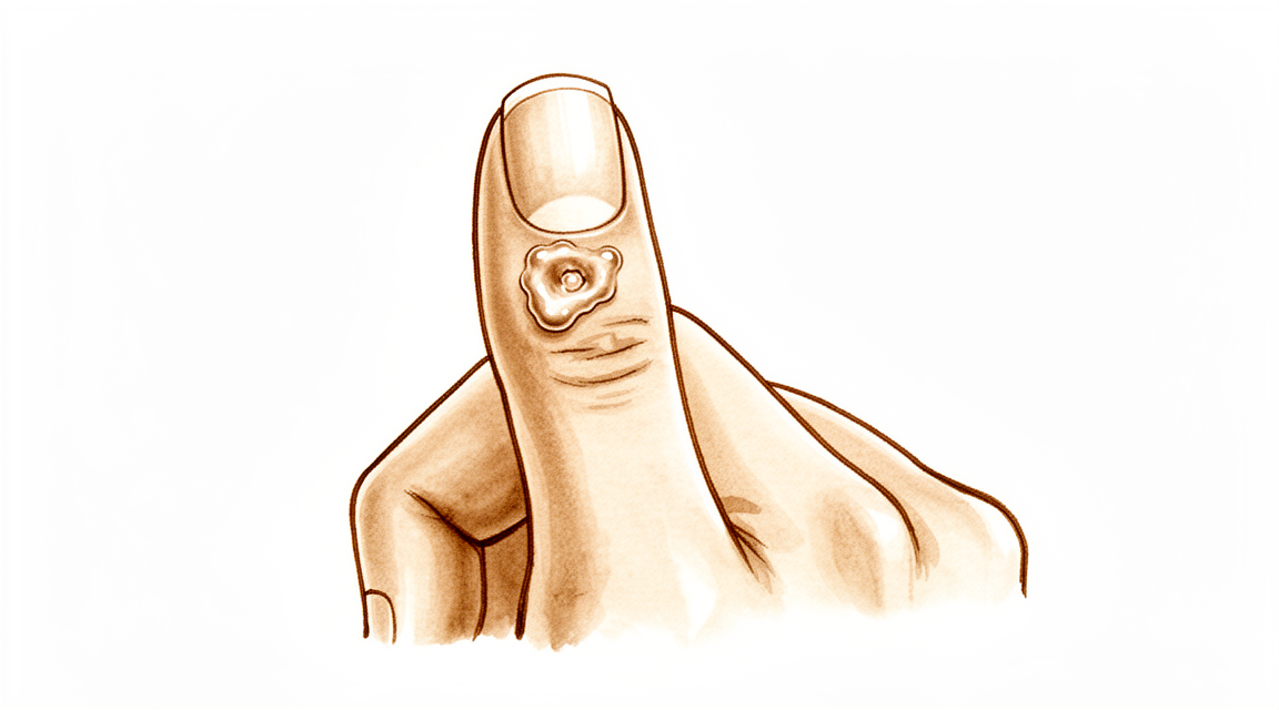
Bạn có thể nhận thấy một cục u nhỏ, chắc ở mặt trên của ngón tay, thường gần khớp cuối cùng. Cục u này là một nang dịch nhầy. Nó thường cảm giác như một hạt đậu nhỏ dưới da. Bạn có thể nhìn thấy rõ nếu da ở đó mỏng. Nang dịch có thể khiến lớp da bao phủ trông bóng hoặc căng. Trong một số trường hợp, nó có thể khiến móng tay mọc theo dạng gợn sóng hoặc có rãnh.
Vùng xung quanh nang dịch có thể cảm thấy nhạy cảm hoặc đau âm ỉ. Bạn có thể cảm thấy đau khi ấn vào nó hoặc khi uốn cong ngón tay. Các hoạt động hàng ngày có thể trở nên khó khăn. Với đồ vật, đánh máy hoặc cầm nắm dụng cụ có thể gây đau. Bạn có thể thấy khó cài khuy áo hoặc kéo khóa áo khoác. Một số người báo cáo rằng cơn đau nặng hơn vào ban đêm hoặc sau khi sử dụng tay trong thời gian dài. Sự khó chịu có thể cản trở giấc ngủ nếu bạn đặt tay đó xuống khi nghỉ ngơi.
Bác sĩ phẫu thuật của bạn sẽ kiểm tra nang dịch và đánh giá mức độ ảnh hưởng của nó đến khả năng vận động của ngón tay. Cơn đau thường bắt nguồn từ những thay đổi ở khớp nền, chứ không chỉ từ cục u. Việc loại bỏ riêng nang dịch có thể không làm giảm đau nếu gai xương (osteofyte) vẫn còn. Bác sĩ phẫu thuật của bạn có thể thảo luận về việc loại bỏ gai xương để giúp giảm áp lực lên khớp và da. Cách tiếp cận này có thể giúp da lành tốt hơn và giảm khả năng nang dịch tái phát.
Trong nhiều trường hợp, việc điều trị gai xương dẫn đến sự biến mất hoàn toàn của nang dịch. Bạn có thể không cần phẫu thuật phức tạp. Việc loại bỏ nang dịch đơn giản kết hợp với loại bỏ gai xương có tỷ lệ tái phát cực kỳ hiếm. Ngay cả các lựa chọn ít xâm lấn hơn, như chỉ loại bỏ gai xương, cũng có thể mang lại sự giải quyết hoàn toàn trong hầu hết các trường hợp. Nếu cần vá da, mức độ hài lòng của bệnh nhân về sẹo là cao. Bạn có khả năng sẽ hài lòng với vẻ ngoài và sẵn sàng thực hiện lại thủ thuật.
Bác sĩ phẫu thuật của bạn sẽ điều chỉnh kế hoạch theo nhu cầu cụ thể của bạn. Mục tiêu là giảm đau và khôi phục chức năng. Hầu hết bệnh nhân nhận thấy rằng sau khi điều trị, ngón tay của họ cảm thấy thoải mái hơn và trông đẹp hơn. Da có tiềm năng phục hồi tốt một khi vấn đề chính được giải quyết. Bạn có thể mong đợi quá trình hồi phục đơn giản với việc chăm sóc vết thương cẩn thận.
Những gì thực sự đang xảy ra
U nang dịch nhầy là một túi nhỏ chứa đầy dịch hình thành trên ngón tay của bạn. Nó thường xuất hiện gần đầu ngón tay, gần với giường móng. U nang nằm trên bao khớp, đây là lớp vỏ xơ dày bao quanh khớp của bạn để giữ cho khớp được ổn định.
Nguyên nhân gốc rễ là do viêm xương khớp do hao mòn ở khớp. Khi sụn—lớp phủ trơn láng ở đầu các xương của bạn—bị phá vỡ, cơ thể bạn cố gắng sửa chữa tổn thương. Quá trình này thường tạo ra các gai xương, gọi là gai xương (osteophytes). Đây là những gờ xương nhỏ, thô nhám phát triển ra từ bề mặt khớp.
Hãy tưởng tượng bao khớp giống như một vòng đệm hoặc một lớp niêm phong. Khi gai xương cọ xát vào lớp niêm phong này, nó gây kích ứng mô. Sự kích ứng này khiến lớp lót khớp rò rỉ dịch hoạt dịch, đây là chất bôi trơn tự nhiên giúp khớp của bạn chuyển động trơn tru. Dịch đẩy qua điểm yếu trên bao khớp, tạo thành khối u nổi rõ trên da của bạn.
Vì u nang được kết nối với khớp, nó chứa cùng loại dịch bôi trơn này. Áp lực từ dịch có thể làm cho da trên u nang trở nên mỏng và dễ vỡ. Trong một số trường hợp, u nang có thể chèn ép vào các dây thần kinh lân cận hoặc ảnh hưởng đến ma trận móng, đây là mô nằm dưới biểu bì móng giúp phát triển móng tay. Sự chèn ép này là nguyên nhân dẫn đến các rãnh hoặc gờ mà bạn có thể thấy trên móng tay.
Việc chỉ cắt bỏ u nang thường dẫn đến việc nó tái phát vì gai xương vẫn còn tồn tại. Gai xương tiếp tục kích thích lớp lót khớp, gây ra nhiều dịch rò rỉ hơn. Để ngăn chặn chu kỳ này, gai xương nền tảng phải được xử lý. Khi gai xương được loại bỏ, sự kích ứng sẽ dừng lại và lớp lót khớp có thể lành lại. Đây là lý do tại sao việc điều trị xương quan trọng không kém so với việc điều trị chính u nang.
Những gì chúng tôi có thể làm về vấn đề này
Hành trình của bạn thường bắt đầu với việc tự chăm sóc đơn giản và hướng dẫn từ chuyên gia. Bạn có thể thử nghỉ ngơi ngón tay và tránh các hoạt động gây áp lực lên khối u nang. Vật lý trị liệu giúp bạn duy trì khả năng vận động của khớp và giữ cho các cơ xung quanh khỏe mạnh. Phương pháp này nhằm mục đích giảm kích ứng và cải thiện chức năng mà không cần các bước xâm lấn. Bạn nên thử nghiệm phương pháp điều trị bảo tồn này một cách công bằng để xem liệu các triệu chứng có thuyên giảm hay không. Nhiều bệnh nhân nhận thấy rằng việc quản lý các thói quen hàng ngày và tuân theo một kế hoạch tập luyện nhẹ nhàng mang lại đủ sự giảm đau để tránh các can thiệp thêm.
Nếu việc tự chăm sóc không đủ hiệu quả, bác sĩ phẫu thuật của bạn có thể đề xuất quản lý bằng thuốc để kiểm soát đau và viêm. Tiêm corticosteroid là một lựa chọn phổ biến cho các khối u nang này. Tiêm corticosteroid ở mặt lòng bàn tay cho phép đặt kim vào khớp một cách dễ dàng và nhất quán. Phương pháp này giảm thiểu nguy cơ tổn thương mô mềm và nhiễm trùng so với các kỹ thuật khác. Tiêm giúp làm dịu tình trạng viêm thúc đẩy sự phát triển của khối u nang. Mặc dù bằng chứng nhấn mạnh tính an toàn và sự dễ dàng của phương pháp này, điều quan trọng là phải hiểu rằng các lần tiêm thường chỉ kiểm soát triệu chứng chứ không loại bỏ vĩnh viễn khối u nang. Hiệu quả kéo dài trong một khoảng thời gian, nhưng tình trạng viêm xương khớp do hao mòn vẫn còn. Bạn và bác sĩ phẫu thuật sẽ quyết định liệu sự giảm đau tạm thời này có đủ hay cần thực hiện các bước tiếp theo.
Phẫu thuật được xem xét khi việc điều trị bảo tồn đã đạt đến giới hạn và khối u nang gây đau dai dẳng, biến dạng hoặc các vấn đề về chức năng. Bác sĩ phẫu thuật của bạn sẽ thảo luận về lựa chọn phẫu thuật tốt nhất cho trường hợp cụ thể của bạn. Mục tiêu là loại bỏ khối u nang và giải quyết nguyên nhân gốc rễ, thường là các gai xương (osteophytes) do viêm xương khớp. Loại bỏ các gai xương này là rất quan trọng vì nó làm giảm đáng kể khả năng khối u nang tái phát. Một số kỹ thuật liên quan đến việc loại bỏ khối u nang cùng với một mảnh da nhỏ để đảm bảo quá trình lành bệnh đúng cách, trong khi những kỹ thuật khác tập trung chủ yếu vào xương. Bác sĩ phẫu thuật của bạn sẽ chọn phương pháp mang lại sự cân bằng tốt nhất giữa tỷ lệ tái phát thấp và kết quả thẩm mỹ tốt cho bàn tay của bạn.
Những điều cần biết
U nang nhầy là những khối u nhỏ, chứa dịch, thường xuất hiện gần khớp cuối của ngón tay hoặc ngón cái. Chúng có liên quan chặt chẽ với viêm xương khớp do hao mòn tại khớp đó. Vì chúng bắt nguồn từ những thay đổi khớp nền tảng này, u nang có thể tồn tại hoặc tái phát nếu nguyên nhân gốc rễ không được giải quyết. Tuy nhiên, với điều trị thích hợp, tiên lượng chung thường rất tích cực.
Bác sĩ phẫu thuật của bạn có thể sẽ khuyến nghị cắt bỏ u nang cùng với bất kỳ gai xương (osteophytes) nào gây ra nó. Phương pháp này loại bỏ u nang nhầy với tỷ lệ tái phát cực kỳ hiếm. Nếu bác sĩ phẫu thuật của bạn chọn kỹ thuật sử dụng vạt da tiến triển tại chỗ, tỷ lệ tái phát thấp ở mức 1,4%. Trong một số trường hợp, bác sĩ phẫu thuật của bạn có thể chỉ cắt bỏ gai xương mà không lấy bỏ u nang. Phương pháp ít xâm lấn này mang lại sự giải quyết hoàn toàn trong hầu hết các trường hợp. Bất kể kế hoạch cụ thể cho các mô mềm là gì, bác sĩ phẫu thuật của bạn sẽ loại bỏ gai xương để ngăn u nang tái phát.
Bạn có thể mong đợi mức độ hài lòng cao với kết quả, đặc biệt là về vẻ ngoài của vết sẹo. Nhiều bệnh nhân báo cáo rằng họ sẽ trải qua thủ thuật này một lần nữa. Các kỹ thuật phẫu thuật được thiết kế để đơn giản và đáng tin cậy. Ví dụ, một số phương pháp cho phép loại bỏ da mỏng mà không làm tăng nguy cơ cho ma trận móng của bạn. Những phương pháp khác sử dụng ghép da mang lại kết quả thẩm mỹ thỏa đáng với tỷ lệ tái phát chấp nhận được. Ngay cả các trường hợp phức tạp, chẳng hạn như u nang phát triển bên trong dây thần kinh ngón tay, cũng có thể dẫn đến kết quả thành công.
Nếu không được điều trị, u nang có thể tồn tại hoặc phát triển, có khả năng làm mỏng da và tăng nguy cơ nhiễm trùng. Bằng cách giải quyết cả u nang và viêm xương khớp nền tảng, bác sĩ phẫu thuật của bạn hướng tới một giải pháp lâu dài. Hầu hết bệnh nhân nhận thấy ngón tay của họ trông và cảm thấy tốt hơn sau thủ thuật. Mục tiêu không chỉ là loại bỏ khối u, mà là ngăn chặn nó tái phát. Bạn có thể mong đợi một quá trình phục hồi đơn giản, tập trung vào việc bảo vệ vị trí phẫu thuật trong khi cho phép khớp của bạn lành lại.
Khi nào cần gặp bác sĩ
Hãy gặp bác sĩ đa khoa nếu bạn nhận thấy một khối u nhỏ ở ngón tay gần móng tay. Yêu cầu đánh giá bởi chuyên gia nếu nang gây đau dai dẳng không cải thiện khi nghỉ ngơi. Hãy tìm kiếm chăm sóc y tế nếu bạn gặp phải tình trạng yếu hoặc mất ổn định ở khớp. Chú ý các cảm giác khóa khớp hoặc sập khớp. Liên hệ với bác sĩ phẫu thuật nếu các triệu chứng ảnh hưởng đến giấc ngủ hoặc công việc. Sự xấu đi đột ngột của vùng này cũng cần được kiểm tra. Đánh giá sớm giúp ngăn ngừa biến chứng. Bác sĩ phẫu thuật của bạn có thể thảo luận về các lựa chọn như loại bỏ nang và bất kỳ gai xương nào. Phương pháp này thường dẫn đến tỷ lệ hài lòng cao và tỷ lệ tái phát thấp. Điều trị nguyên nhân gốc rễ giúp da tự phục hồi.
Evidence & references
Overview
- Scientific data regarding mucous cysts consist almost entirely of retrospective studies [1].
- Much of the management or recommendations for mucous cysts is based on expert opinion [1].
- Total dorsal capsulectomy alone is a simple treatment for mucous cysts that does not lead to any recurrence [2].
- Excision of the cyst combined with complete removal of the marginal osteophyte eradicates mucous cysts with extremely rare recurrence [3].
- Osteophyte excision without cyst excision may be a good treatment choice for mucous cysts of the finger, providing a less invasive method with complete resolution in most cases [5].
- Osteophyte removal results in a low cyst recurrence rate, indicating it should be undertaken regardless of the surgeon's plan for the soft tissues [13].
- The Zitelli bilobed flap allows excision of the cyst and thinned skin with no added risk to the nail matrix [6].
- The use of a Wolfe graft for mucous cysts is simple, easy to perform, and provides satisfactory cosmesis with acceptable recurrence rates [7].
- Surgical excision with a local advancement skin flap is a reliable treatment for digital mucous cysts, demonstrating a low recurrence rate of 1.4% and high patient satisfaction regarding the scar and willingness to undergo the procedure again [9].
- A surgical technique involving excision of the cyst, synovectomy, and débridement of osteophytes with rotational flap closure resulted in no recurrences in thirty-six patients [10].
- Pathohistological analysis is useful in cases where doubts arise about the initial diagnosis of a benign tumorous lesion [4].
- Eccrine porocarcinomas have a substantial risk of metastasis, high risk of local recurrence, and are potentially fatal [8].
Anatomy & Pathophysiology
- Scientific data regarding mucous cysts consist almost entirely of retrospective studies [1].
- Much of the management or recommendations for mucous cysts is based on expert opinion [1].
- Mucous cysts are associated with marginal osteophytes at the distal interphalangeal joint [3].
- The primary pathology in mucous cysts involves osteophytes, and removal of these osteophytes allows for skin recovery potential [20].
- Ultrasound is a powerful modality for evaluating pathologic conditions in the hand and wrist [16].
- Ultrasound provides a cost-effective and expedient alternative or adjunct to MRI for hand and wrist evaluation [16].
- Ultrasound is best used when there is a specific clinical question regarding a well-localized abnormality [16].
- Pathohistological analysis is useful in cases where doubts arise about the initial diagnosis of a benign tumorous lesion [4].
- Subungual keratoacanthoma may show locally aggressive behaviour but does not metastasize [14].
Classification
- Scientific data regarding mucous cysts consist almost entirely of retrospective studies [1].
- Much of the management or recommendations for mucous cysts is based on expert opinion [1].
- Total dorsal capsulectomy alone is a simple treatment for mucous cysts that does not lead to any recurrence [2].
- Excision of the cyst combined with complete removal of the marginal osteophyte eradicates mucous cysts with extremely rare recurrence [3].
- Pathohistological analysis is useful in cases where doubts arise about the initial diagnosis of a benign tumorous lesion [4].
- Osteophyte excision without cyst excision may be a good treatment choice for mucous cyst of the finger, providing a less invasive method with complete resolution in most cases [5].
- The Zitelli bilobed flap allows excision of the cyst and thinned skin with no added risk to the nail matrix [6].
- The use of a Wolfe graft is simple, easy to perform, and provides satisfactory cosmesis with acceptable recurrence rates [7].
- Eccrine porocarcinomas have a substantial risk of metastasis, high risk of local recurrence, and are potentially fatal [8].
- Surgical excision with a local advancement skin flap is a reliable treatment for digital mucous cysts, demonstrating a low recurrence rate of 1.4% and high patient satisfaction regarding the scar and willingness to undergo the procedure again [9].
- A surgical technique involving excision of the cyst, synovectomy, and débridement of osteophytes with rotational flap closure resulted in no recurrences in thirty-six patients [10].
- There is a statistically significant difference in recurrence rates between Type I giant cell tumours of the tendon sheath (0%) and Type II tumours (38%) [11].
- Recurrence in Type II giant cell tumours of the tendon sheath is likely due to undetected satellite lesions or incomplete excision [11].
- Incomplete excision of a granular cell nerve tumor can lead to recurrence [12].
- Osteophyte removal results in a low cyst recurrence rate [13].
- Osteophyte removal should be undertaken regardless of the surgeon's plan for the soft tissues [13].
Clinical Presentation
- Scientific data regarding mucous cysts consist almost entirely of retrospective studies [1].
- Much of the management or recommendations for mucous cysts is based on expert opinion [1].
- Malignant natural-killer cell neoplasms can present as a mucous cyst on the distal interphalangeal joint of the finger [4].
- Eccrine porocarcinomas can present as a hand cyst [8].
- Subungual keratoacanthoma may present as a condition masquerading as flexor tenosynovitis in the finger [14].
- Ultrasound is a powerful modality for the evaluation of pathologic conditions in the hand and wrist [16].
- Ultrasound provides a cost-effective and expedient alternative and/or adjunct to MRI for hand and wrist evaluation [16].
- Ultrasound is best used when there is a specific clinical question regarding a well-localized abnormality in the hand or wrist [16].
Investigations
- Scientific data regarding mucous cysts consist almost entirely of retrospective studies [1].
- Much of the management of mucous cysts is based on expert opinion [1].
- Pathohistological analysis is useful when doubts arise about the initial diagnosis of a benign tumorous lesion [4].
- Eccrine porocarcinomas have a substantial risk of metastasis, high risk of local recurrence, and are potentially fatal [8].
- Ultrasound is a powerful modality for evaluation of pathologic conditions in the hand and wrist [16].
- Ultrasound provides a cost-effective and expedient alternative and/or adjunct to MRI [16].
- Ultrasound is best used when there is a specific clinical question regarding a well-localized abnormality [16].
Treatment
- Scientific data regarding mucous cysts consist almost entirely of retrospective studies, and much of what is done or recommended is based on expert opinion [1].
- Total dorsal capsulectomy alone is a simple treatment for mucous cysts that does not lead to any recurrence [2].
- Excision of the cyst and complete removal of the marginal osteophyte eradicates mucous cysts with extremely rare recurrence [3].
- Osteophyte excision without cyst excision may be a good treatment choice for mucous cyst of the finger, providing a less invasive method with complete resolution in most cases [5].
- Osteophyte removal results in a low cyst recurrence rate, indicating that it should be undertaken regardless of the surgeon's plan for the soft tissues [13].
- The Zitelli bilobed flap allows excision of the cyst and thinned skin with no added risk to the nail matrix [6].
- Use of Wolfe graft is simple, easy to perform, and provides satisfactory cosmesis with acceptable recurrence rates [7].
- Surgical excision with a local advancement skin flap is a reliable treatment for digital mucous cysts, demonstrating a low recurrence rate of 1.4% and high patient satisfaction regarding the scar and willingness to undergo the procedure again [9].
- A surgical technique involving excision of the cyst, synovectomy, and débridement of osteophytes with rotational flap closure resulted in no recurrences in thirty-six patients [10].
- Pathohistological analysis is useful in cases where doubts arise about the initial diagnosis of a benign tumorous lesion [4].
Complications
- Scientific data regarding mucous cysts consist almost entirely of retrospective studies, with many recommendations based on expert opinion [1].
- Total dorsal capsulectomy alone for mucous cysts did not lead to any recurrence [2].
- Excision of the cyst and complete removal of the marginal osteophyte eradicates mucous cysts with extremely rare recurrence [3].
- Osteophyte excision without cyst excision may provide complete resolution in most cases [5].
- The Zitelli bilobed flap allows excision of the cyst and thinned skin with no added risk to the nail matrix [6].
- Use of a Wolfe graft provides satisfactory cosmesis with acceptable recurrence rates [7].
- Surgical excision with a local advancement skin flap demonstrates a low recurrence rate of 1.4% and high patient satisfaction regarding the scar [9].
- A surgical technique involving excision of the cyst, synovectomy, and débridement of osteophytes with rotational flap closure resulted in no recurrences in thirty-six patients [10].
- Incomplete excision can lead to recurrence of granular cell nerve tumors [12].
- Type II giant cell tumors of the tendon sheath have a 38% recurrence rate, likely due to undetected satellite lesions or incomplete excision [11].
- Malignant natural-killer cell neoplasms can present as mucous cysts on the distal interphalangeal joint [4].
- Pathohistological analysis is useful in cases where doubts arise about the initial diagnosis of a benign tumorous lesion [4].
- Eccrine porocarcinomas have a substantial risk of metastasis, high risk of local recurrence, and are potentially fatal [8].
Recovery
- Scientific data regarding mucous cysts consist almost entirely of retrospective studies [1].
- Much of the treatment for mucous cysts is based on expert opinion [1].
- Total dorsal capsulectomy alone is a simple treatment for mucous cysts that does not lead to any recurrence [2].
- Excision of the cyst and complete removal of the marginal osteophyte eradicates mucous cysts with extremely rare recurrence [3].
- Osteophyte excision without cyst excision may be a good treatment choice for mucous cysts of the finger, providing a less invasive method with complete resolution in most cases [5].
- The Zitelli bilobed flap allows excision of the cyst and thinned skin with no added risk to the nail matrix [6].
- The use of a Wolfe graft is simple, easy to perform, and provides satisfactory cosmesis with acceptable recurrence rates [7].
- Surgical excision with a local advancement skin flap is a reliable treatment for digital mucous cysts, demonstrating a low recurrence rate of 1.4% and high patient satisfaction regarding the scar and willingness to undergo the procedure again [9].
- Pathohistological analysis is useful in cases where doubts arise about the initial diagnosis of a benign tumorous lesion [4].
- Eccrine porocarcinomas have a substantial risk of metastasis, high risk of local recurrence, and are potentially fatal [8].
- Incomplete excision can lead to recurrence in granular cell nerve tumors [12].
Key Evidence
- [L4] The scientific data regarding mucous cysts consist almost entirely of retrospective studies, and much of what is done or recommended is based on expert opinion. [1] (10.1016/j.jhsa.2010.01.029)
- [L4] A total dorsal capsulectomy alone was a simple treatment for mucous cysts and did not lead to any recurrence. [2] (10.1016/j.jhsa.2014.03.004)
- [L4] Excision of the cyst and complete removal of the marginal osteophyte eradicates mucous cysts with extremely rare recurrence. [3] (10.2106/00004623-197355030-00013)
- [L5] This case emphasizes the utility of a pathohistological analysis in cases where doubts arise about the initial diagnosis of a benign tumorous lesion. [4] (10.1007/s00402-008-0794-4)
- [L4] Osteophyte excision without cyst excision may be a good treatment choice for mucous cyst of the finger, providing a less invasive method with complete resolution in most cases. [5] (10.1177/1753193413478549)
- [L4] It allows excision of the cyst and thinned skin with no added risk to the nail matrix. [6] (10.1016/j.jhsa.2017.03.013)
- [L4] The technique is simple, easy to perform, and provides satisfactory cosmesis with acceptable recurrence rates. [7] (10.1177/1753193408103498)
- [L4] Prompt recognition and appropriate treatment are critical because eccrine porocarcinomas have a substantial risk of metastasis, high risk of local recurrence, and are potentially fatal. [8] (10.1016/j.jhsa.2016.07.112)
- [L4] Surgical excision with a local advancement skin flap is a reliable treatment for digital mucous cysts, demonstrating a low recurrence rate of 1.4% and high patient satisfaction regarding the scar and willingness to undergo the procedure again. [9] (10.1177/1753193413508540)
- [L4] A new surgical technique involving excision of the cyst, synovectomy, and débridement of osteophytes with rotational flap closure resulted in no recurrences in thirty-six patients. [10] (10.2106/00004623-197254070-00008)
- [L3] The study found a statistically significant difference in recurrence rates between Type I tumours (0%) and Type II tumours (38%), with recurrence in Type II likely due to undetected satellite lesions or incomplete excision. [11] (10.1054/jhsb.2000.0522)
- [Case_report] The author notes that while the true recurrence rate is unknown, incomplete excision can lead to recurrence. [12] (10.1016/j.jhsa.2009.05.011)
- [Commentary] The article shows that osteophyte removal results in a low cyst recurrence rate, indicating that it should be undertaken regardless of the surgeon's plan for the soft tissues. [13] (10.1177/1753193413510663)
- [L4] Subungual keratoacanthoma may show locally aggressive behaviour but does not metastasize. [14] (10.1177/1753193409360605)
- [L5] Ultrasound is a powerful modality for evaluation of pathologic conditions in the hand and wrist, providing a cost-effective and expedient alternative and/or adjunct to MRI, best used when there is a specific clinical question regarding a well-localized abnormality. [16] (10.1016/j.jhsa.2009.02.010)
- [L5] The authors of the original study believe that extensive damage to the skin is unnecessary and that the skin has recovery potential once the main problem (osteophytes) is removed, favoring a less invasive approach over techniques requiring skin flaps. [20] (10.1177/1753193414546443)
References
[1] Mucous Cysts. The Journal of Hand Surgery. 2010. DOI: 10.1016/j.jhsa.2010.01.029 [2] Total Dorsal Capsulectomy for the Treatment of Mucous Cysts. The Journal of Hand Surgery. 2014. DOI: 10.1016/j.jhsa.2014.03.004 [3] Marginal Osteophyte Excision in Treatment of Mucous Cysts. The Journal of Bone & Joint Surgery. 1973. DOI: 10.2106/00004623-197355030-00013 [4] Malignant Natural-Killer cell neoplasm presenting as a mucous cyst on the distal interphalangeal joint of the finger. Archives of Orthopaedic and Trauma Surgery. 2008. DOI: 10.1007/s00402-008-0794-4 [5] Osteophyte excision without cyst excision for a mucous cyst of the finger. Journal of Hand Surgery (European Volume). 2013. DOI: 10.1177/1753193413478549 [6] The Zitelli Bilobed Flap on Skin Coverage After Mucous Cyst Excision: A Retrospective Cohort of 33 Cases. The Journal of Hand Surgery. 2017. DOI: 10.1016/j.jhsa.2017.03.013 [7] Use of Wolfe Graft for the Treatment of Mucous Cysts. Journal of Hand Surgery (European Volume). 2009. DOI: 10.1177/1753193408103498 [8] Eccrine Porocarcinoma Presenting as a Hand Cyst. The Journal of Hand Surgery. 2016. DOI: 10.1016/j.jhsa.2016.07.112 [9] A reliable surgical treatment for digital mucous cysts. Journal of Hand Surgery (European Volume). 2013. DOI: 10.1177/1753193413508540 [10] Etiology and Treatment of the So-Called Mucous Cyst of the Finger. The Journal of Bone & Joint Surgery. 1972. DOI: 10.2106/00004623-197254070-00008 [11] Giant Cell Tumours of Tendon Sheath: Classification and Recurrence Rate. Journal of Hand Surgery. 2001. DOI: 10.1054/jhsb.2000.0522 [12] Granular Cell Nerve Tumor in the Hand: Case Report. The Journal of Hand Surgery. 2009. DOI: 10.1016/j.jhsa.2009.05.011 [13] Commentary on Lee et al. Osteophyte excision without cyst excision for a mucous cyst of the finger. Journal of Hand Surgery (European Volume). 2014. DOI: 10.1177/1753193413510663 [14] Metastases to the finger masquerading as flexor tenosynovitis. Journal of Hand Surgery (European Volume). 2010. DOI: 10.1177/1753193409360605 [16] Ultrasound of the Hand and Wrist. The Journal of Hand Surgery. 2009. DOI: 10.1016/j.jhsa.2009.02.010 [20] Re: Lee HJ, Kim PT, Jeon IH, et al. Osteophyte excision without cyst excision for a mucous cyst of the finger. J Hand Surg Eur. 2014, 39: 258–61. Journal of Hand Surgery (European Volume). 2014. DOI: 10.1177/1753193414546443


