黏液囊肿
Patients › Hand
Mucous cysts – common bumps near finger joints, often linked to arthritis, and treatment options.
您的感受
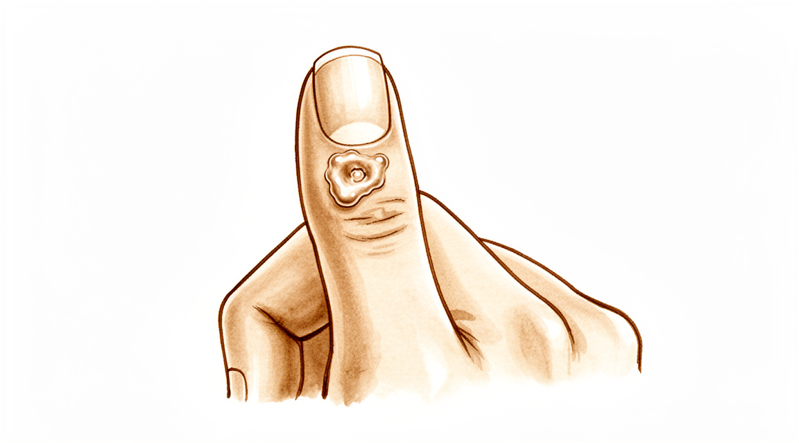
您可能会注意到手指顶部有一个小而硬的肿块,通常位于远端指间关节附近。这个肿块是一种黏液囊肿。它摸起来通常像皮下的一颗小豌豆。如果您的皮肤较薄,您可能能清楚地看到它。囊肿可能会使覆盖其上的皮肤看起来发亮或紧绷。在某些情况下,它可能导致指甲呈现波浪状或脊状生长模式。
囊肿周围的区域可能会感到压痛或酸痛。按压手指或弯曲手指时可能会感到疼痛。日常活动可能会变得困难。拿取物品、打字或抓握工具时可能会疼痛。您可能会发现扣纽扣或拉上夹克拉链很困难。有些人报告说,夜间或长时间使用手部后疼痛会加重。如果您将手放在身下休息,不适感可能会干扰睡眠。
您的外科医生会检查囊肿并评估其对手指活动的影响。疼痛通常源于潜在的关节改变,而不仅仅是肿块本身。如果骨赘(骨刺)仍然存在,仅切除囊肿可能无法缓解疼痛。您的外科医生可能会讨论切除骨赘,以帮助减轻对关节和皮肤的压力。这种方法有助于皮肤更好地愈合,并降低囊肿复发的可能性。
在许多情况下,治疗骨赘会导致囊肿完全消退。您可能不需要复杂的手术。单纯切除囊肿联合骨赘切除术的复发率极低。甚至更微创的选项,如仅切除骨赘,在大多数情况下也能实现完全消退。如果需要皮瓣,患者对疤痕的满意度很高。您很可能对外观感到满意,并愿意再次接受该手术。
您的外科医生会根据您的具体需求制定计划。目标是缓解疼痛并恢复功能。大多数患者发现,治疗后,他们的手指感觉更舒适,外观也更好。一旦主要问题得到解决,皮肤具有良好的恢复潜力。您可以预期,在仔细护理伤口的情况下,恢复过程将是顺利的。
实际发生了什么
黏液囊肿是一种形成于手指上的小型充满液体的囊袋。它通常出现在手指尖端附近,靠近甲床。囊肿位于关节囊的上方,关节囊是包裹在关节周围以保持其稳定的坚韧纤维性套筒。
其根本原因是关节的退行性关节炎。随着软骨——覆盖在骨头末端的光滑涂层——发生退化,身体会尝试修复损伤。这一过程通常会形成骨赘,也称为骨刺。这些是从关节表面向外生长的细小粗糙的额外骨性突起。
可以将关节囊想象成一个垫片或密封件。当骨刺摩擦这个密封件时,会刺激组织。这种刺激导致关节滑膜渗出滑液,滑液是保持关节顺畅运动的天然润滑剂。液体通过关节囊的薄弱部位渗出,形成你在皮肤上看到的可见隆起。
由于囊肿与关节相连,其中充满了相同的润滑液。液体的压力可能导致囊肿上方的皮肤变薄且脆弱。在某些情况下,囊肿可能会压迫附近的神经或影响甲母质,甲母质是指位于甲上皮下方、负责生长指甲的组织。这种压力会导致你在指甲上看到的沟槽或脊状突起。
仅切除囊肿往往会导致其复发,因为骨刺仍然存在。骨刺会继续刺激关节滑膜,导致更多液体渗出。要打破这一循环,必须处理潜在的骨刺。当骨刺被移除时,刺激停止,关节滑膜得以愈合。这就是为什么治疗骨骼与治疗囊肿本身同样重要。
我们能采取的措施
您的治疗过程通常从简单的自我护理和专业指导开始。您可以尝试让手指休息,并避免对囊肿施加压力的活动。物理治疗有助于维持关节的活动度,并保持周围肌肉的力量。这种方法旨在通过非侵入性手段减少刺激并改善功能。您应给予保守治疗充分的尝试时间,以观察症状是否缓解。许多患者发现,通过调整日常习惯并遵循温和的锻炼计划,即可获得足够的缓解,从而避免进一步干预。
如果自我护理效果不佳,您的外科医生可能会建议药物治疗以控制疼痛和炎症。皮质类固醇注射是治疗此类囊肿的常见选择。掌侧皮质类固醇注射便于针头准确且一致地置入关节。与其他技术相比,该方法最大限度地减少了潜在的软组织损伤和感染风险。注射有助于缓解驱动囊肿生长的炎症。虽然证据强调了这种方法的安全性和便捷性,但重要的是要理解,注射通常仅用于管理症状,而非永久去除囊肿。效果会持续一段时间,但潜在的退行性关节炎仍然存在。您和您的外科医生将决定这种暂时缓解是否足够,或者是否需要采取进一步措施。
当保守治疗达到极限,且囊肿引起持续性疼痛、畸形或功能障碍时,会考虑手术治疗。您的外科医生将讨论针对您具体病例的最佳手术方案。目标是去除囊肿并解决根本原因,这通常是关节炎引起的骨赘(骨刺)。去除这些骨赘至关重要,因为它能显著降低囊肿复发的几率。某些技术涉及连同小块皮瓣一起切除囊肿以确保适当愈合,而其他技术则主要关注骨骼。您的外科医生将选择一种方法,在低复发率和良好的手部美容效果之间取得最佳平衡。
预期情况
黏液囊肿是小的、充满液体的肿块,通常出现在手指或拇指的远端关节附近。它们与该关节的退行性关节炎密切相关。由于囊肿源于这种潜在的关节改变,如果未解决根本原因,囊肿本身可能会持续存在或复发。然而,经过适当治疗,预后通常非常良好。
您的外科医生可能会建议切除囊肿以及引起囊肿的任何骨赘(骨刺)。这种方法可以极罕见地消除黏液囊肿的复发。如果您的外科医生选择使用局部推进皮瓣的技术,复发率仅为1.4%。在某些情况下,您的外科医生可能仅切除骨赘而不取出囊肿。这种侵入性较小的方法在大多数情况下可实现完全缓解。无论软组织的具体计划如何,您的外科医生都会切除骨赘以防止囊肿复发。
您可以对结果感到高度满意,尤其是关于疤痕的外观。许多患者表示他们会再次接受该手术。所使用的外科技术旨在简单可靠。例如,某些方法允许在不增加指甲基质风险的情况下去除变薄的皮肤。其他方法使用皮肤移植,提供令人满意的美容效果,同时具有可接受的复发率。即使是复杂的病例,如囊肿生长在指神经内部,也能取得成功的结果。
如果不治疗,囊肿可能会持续存在或增大,可能导致皮肤变薄并增加感染风险。通过同时处理囊肿和潜在的关节炎,您的外科医生旨在提供持久的解决方案。大多数患者发现手术后手指的外观和感觉都更好。目标不仅是去除肿块,还要防止其复发。您可以期待一个直接的恢复过程,重点在于保护手术部位,同时让关节愈合。
何时就医
若发现手指靠近指甲处有小肿块,请咨询全科医生。若囊肿引起持续疼痛且休息后未缓解,请要求专科医生评估。若出现关节无力或不稳,请及时就医。注意是否有卡顿或关节不稳感。若症状影响睡眠或工作,请联系您的外科医生。该区域症状突然加重也需进行检查。早期评估有助于预防并发症。您的外科医生可讨论切除囊肿及骨赘等方案。该方法通常满意度高且复发率低。治疗根本原因有助于皮肤自然恢复。
Evidence & references
Overview
- Scientific data regarding mucous cysts consist almost entirely of retrospective studies [1].
- Much of the management or recommendations for mucous cysts is based on expert opinion [1].
- Total dorsal capsulectomy alone is a simple treatment for mucous cysts that does not lead to any recurrence [2].
- Excision of the cyst combined with complete removal of the marginal osteophyte eradicates mucous cysts with extremely rare recurrence [3].
- Osteophyte excision without cyst excision may be a good treatment choice for mucous cysts of the finger, providing a less invasive method with complete resolution in most cases [5].
- Osteophyte removal results in a low cyst recurrence rate, indicating it should be undertaken regardless of the surgeon's plan for the soft tissues [13].
- The Zitelli bilobed flap allows excision of the cyst and thinned skin with no added risk to the nail matrix [6].
- The use of a Wolfe graft for mucous cysts is simple, easy to perform, and provides satisfactory cosmesis with acceptable recurrence rates [7].
- Surgical excision with a local advancement skin flap is a reliable treatment for digital mucous cysts, demonstrating a low recurrence rate of 1.4% and high patient satisfaction regarding the scar and willingness to undergo the procedure again [9].
- A surgical technique involving excision of the cyst, synovectomy, and débridement of osteophytes with rotational flap closure resulted in no recurrences in thirty-six patients [10].
- Pathohistological analysis is useful in cases where doubts arise about the initial diagnosis of a benign tumorous lesion [4].
- Eccrine porocarcinomas have a substantial risk of metastasis, high risk of local recurrence, and are potentially fatal [8].
Anatomy & Pathophysiology
- Scientific data regarding mucous cysts consist almost entirely of retrospective studies [1].
- Much of the management or recommendations for mucous cysts is based on expert opinion [1].
- Mucous cysts are associated with marginal osteophytes at the distal interphalangeal joint [3].
- The primary pathology in mucous cysts involves osteophytes, and removal of these osteophytes allows for skin recovery potential [20].
- Ultrasound is a powerful modality for evaluating pathologic conditions in the hand and wrist [16].
- Ultrasound provides a cost-effective and expedient alternative or adjunct to MRI for hand and wrist evaluation [16].
- Ultrasound is best used when there is a specific clinical question regarding a well-localized abnormality [16].
- Pathohistological analysis is useful in cases where doubts arise about the initial diagnosis of a benign tumorous lesion [4].
- Subungual keratoacanthoma may show locally aggressive behaviour but does not metastasize [14].
Classification
- Scientific data regarding mucous cysts consist almost entirely of retrospective studies [1].
- Much of the management or recommendations for mucous cysts is based on expert opinion [1].
- Total dorsal capsulectomy alone is a simple treatment for mucous cysts that does not lead to any recurrence [2].
- Excision of the cyst combined with complete removal of the marginal osteophyte eradicates mucous cysts with extremely rare recurrence [3].
- Pathohistological analysis is useful in cases where doubts arise about the initial diagnosis of a benign tumorous lesion [4].
- Osteophyte excision without cyst excision may be a good treatment choice for mucous cyst of the finger, providing a less invasive method with complete resolution in most cases [5].
- The Zitelli bilobed flap allows excision of the cyst and thinned skin with no added risk to the nail matrix [6].
- The use of a Wolfe graft is simple, easy to perform, and provides satisfactory cosmesis with acceptable recurrence rates [7].
- Eccrine porocarcinomas have a substantial risk of metastasis, high risk of local recurrence, and are potentially fatal [8].
- Surgical excision with a local advancement skin flap is a reliable treatment for digital mucous cysts, demonstrating a low recurrence rate of 1.4% and high patient satisfaction regarding the scar and willingness to undergo the procedure again [9].
- A surgical technique involving excision of the cyst, synovectomy, and débridement of osteophytes with rotational flap closure resulted in no recurrences in thirty-six patients [10].
- There is a statistically significant difference in recurrence rates between Type I giant cell tumours of the tendon sheath (0%) and Type II tumours (38%) [11].
- Recurrence in Type II giant cell tumours of the tendon sheath is likely due to undetected satellite lesions or incomplete excision [11].
- Incomplete excision of a granular cell nerve tumor can lead to recurrence [12].
- Osteophyte removal results in a low cyst recurrence rate [13].
- Osteophyte removal should be undertaken regardless of the surgeon's plan for the soft tissues [13].
Clinical Presentation
- Scientific data regarding mucous cysts consist almost entirely of retrospective studies [1].
- Much of the management or recommendations for mucous cysts is based on expert opinion [1].
- Malignant natural-killer cell neoplasms can present as a mucous cyst on the distal interphalangeal joint of the finger [4].
- Eccrine porocarcinomas can present as a hand cyst [8].
- Subungual keratoacanthoma may present as a condition masquerading as flexor tenosynovitis in the finger [14].
- Ultrasound is a powerful modality for the evaluation of pathologic conditions in the hand and wrist [16].
- Ultrasound provides a cost-effective and expedient alternative and/or adjunct to MRI for hand and wrist evaluation [16].
- Ultrasound is best used when there is a specific clinical question regarding a well-localized abnormality in the hand or wrist [16].
Investigations
- Scientific data regarding mucous cysts consist almost entirely of retrospective studies [1].
- Much of the management of mucous cysts is based on expert opinion [1].
- Pathohistological analysis is useful when doubts arise about the initial diagnosis of a benign tumorous lesion [4].
- Eccrine porocarcinomas have a substantial risk of metastasis, high risk of local recurrence, and are potentially fatal [8].
- Ultrasound is a powerful modality for evaluation of pathologic conditions in the hand and wrist [16].
- Ultrasound provides a cost-effective and expedient alternative and/or adjunct to MRI [16].
- Ultrasound is best used when there is a specific clinical question regarding a well-localized abnormality [16].
Treatment
- Scientific data regarding mucous cysts consist almost entirely of retrospective studies, and much of what is done or recommended is based on expert opinion [1].
- Total dorsal capsulectomy alone is a simple treatment for mucous cysts that does not lead to any recurrence [2].
- Excision of the cyst and complete removal of the marginal osteophyte eradicates mucous cysts with extremely rare recurrence [3].
- Osteophyte excision without cyst excision may be a good treatment choice for mucous cyst of the finger, providing a less invasive method with complete resolution in most cases [5].
- Osteophyte removal results in a low cyst recurrence rate, indicating that it should be undertaken regardless of the surgeon's plan for the soft tissues [13].
- The Zitelli bilobed flap allows excision of the cyst and thinned skin with no added risk to the nail matrix [6].
- Use of Wolfe graft is simple, easy to perform, and provides satisfactory cosmesis with acceptable recurrence rates [7].
- Surgical excision with a local advancement skin flap is a reliable treatment for digital mucous cysts, demonstrating a low recurrence rate of 1.4% and high patient satisfaction regarding the scar and willingness to undergo the procedure again [9].
- A surgical technique involving excision of the cyst, synovectomy, and débridement of osteophytes with rotational flap closure resulted in no recurrences in thirty-six patients [10].
- Pathohistological analysis is useful in cases where doubts arise about the initial diagnosis of a benign tumorous lesion [4].
Complications
- Scientific data regarding mucous cysts consist almost entirely of retrospective studies, with many recommendations based on expert opinion [1].
- Total dorsal capsulectomy alone for mucous cysts did not lead to any recurrence [2].
- Excision of the cyst and complete removal of the marginal osteophyte eradicates mucous cysts with extremely rare recurrence [3].
- Osteophyte excision without cyst excision may provide complete resolution in most cases [5].
- The Zitelli bilobed flap allows excision of the cyst and thinned skin with no added risk to the nail matrix [6].
- Use of a Wolfe graft provides satisfactory cosmesis with acceptable recurrence rates [7].
- Surgical excision with a local advancement skin flap demonstrates a low recurrence rate of 1.4% and high patient satisfaction regarding the scar [9].
- A surgical technique involving excision of the cyst, synovectomy, and débridement of osteophytes with rotational flap closure resulted in no recurrences in thirty-six patients [10].
- Incomplete excision can lead to recurrence of granular cell nerve tumors [12].
- Type II giant cell tumors of the tendon sheath have a 38% recurrence rate, likely due to undetected satellite lesions or incomplete excision [11].
- Malignant natural-killer cell neoplasms can present as mucous cysts on the distal interphalangeal joint [4].
- Pathohistological analysis is useful in cases where doubts arise about the initial diagnosis of a benign tumorous lesion [4].
- Eccrine porocarcinomas have a substantial risk of metastasis, high risk of local recurrence, and are potentially fatal [8].
Recovery
- Scientific data regarding mucous cysts consist almost entirely of retrospective studies [1].
- Much of the treatment for mucous cysts is based on expert opinion [1].
- Total dorsal capsulectomy alone is a simple treatment for mucous cysts that does not lead to any recurrence [2].
- Excision of the cyst and complete removal of the marginal osteophyte eradicates mucous cysts with extremely rare recurrence [3].
- Osteophyte excision without cyst excision may be a good treatment choice for mucous cysts of the finger, providing a less invasive method with complete resolution in most cases [5].
- The Zitelli bilobed flap allows excision of the cyst and thinned skin with no added risk to the nail matrix [6].
- The use of a Wolfe graft is simple, easy to perform, and provides satisfactory cosmesis with acceptable recurrence rates [7].
- Surgical excision with a local advancement skin flap is a reliable treatment for digital mucous cysts, demonstrating a low recurrence rate of 1.4% and high patient satisfaction regarding the scar and willingness to undergo the procedure again [9].
- Pathohistological analysis is useful in cases where doubts arise about the initial diagnosis of a benign tumorous lesion [4].
- Eccrine porocarcinomas have a substantial risk of metastasis, high risk of local recurrence, and are potentially fatal [8].
- Incomplete excision can lead to recurrence in granular cell nerve tumors [12].
Key Evidence
- [L4] The scientific data regarding mucous cysts consist almost entirely of retrospective studies, and much of what is done or recommended is based on expert opinion. [1] (10.1016/j.jhsa.2010.01.029)
- [L4] A total dorsal capsulectomy alone was a simple treatment for mucous cysts and did not lead to any recurrence. [2] (10.1016/j.jhsa.2014.03.004)
- [L4] Excision of the cyst and complete removal of the marginal osteophyte eradicates mucous cysts with extremely rare recurrence. [3] (10.2106/00004623-197355030-00013)
- [L5] This case emphasizes the utility of a pathohistological analysis in cases where doubts arise about the initial diagnosis of a benign tumorous lesion. [4] (10.1007/s00402-008-0794-4)
- [L4] Osteophyte excision without cyst excision may be a good treatment choice for mucous cyst of the finger, providing a less invasive method with complete resolution in most cases. [5] (10.1177/1753193413478549)
- [L4] It allows excision of the cyst and thinned skin with no added risk to the nail matrix. [6] (10.1016/j.jhsa.2017.03.013)
- [L4] The technique is simple, easy to perform, and provides satisfactory cosmesis with acceptable recurrence rates. [7] (10.1177/1753193408103498)
- [L4] Prompt recognition and appropriate treatment are critical because eccrine porocarcinomas have a substantial risk of metastasis, high risk of local recurrence, and are potentially fatal. [8] (10.1016/j.jhsa.2016.07.112)
- [L4] Surgical excision with a local advancement skin flap is a reliable treatment for digital mucous cysts, demonstrating a low recurrence rate of 1.4% and high patient satisfaction regarding the scar and willingness to undergo the procedure again. [9] (10.1177/1753193413508540)
- [L4] A new surgical technique involving excision of the cyst, synovectomy, and débridement of osteophytes with rotational flap closure resulted in no recurrences in thirty-six patients. [10] (10.2106/00004623-197254070-00008)
- [L3] The study found a statistically significant difference in recurrence rates between Type I tumours (0%) and Type II tumours (38%), with recurrence in Type II likely due to undetected satellite lesions or incomplete excision. [11] (10.1054/jhsb.2000.0522)
- [Case_report] The author notes that while the true recurrence rate is unknown, incomplete excision can lead to recurrence. [12] (10.1016/j.jhsa.2009.05.011)
- [Commentary] The article shows that osteophyte removal results in a low cyst recurrence rate, indicating that it should be undertaken regardless of the surgeon's plan for the soft tissues. [13] (10.1177/1753193413510663)
- [L4] Subungual keratoacanthoma may show locally aggressive behaviour but does not metastasize. [14] (10.1177/1753193409360605)
- [L5] Ultrasound is a powerful modality for evaluation of pathologic conditions in the hand and wrist, providing a cost-effective and expedient alternative and/or adjunct to MRI, best used when there is a specific clinical question regarding a well-localized abnormality. [16] (10.1016/j.jhsa.2009.02.010)
- [L5] The authors of the original study believe that extensive damage to the skin is unnecessary and that the skin has recovery potential once the main problem (osteophytes) is removed, favoring a less invasive approach over techniques requiring skin flaps. [20] (10.1177/1753193414546443)
References
[1] Mucous Cysts. The Journal of Hand Surgery. 2010. DOI: 10.1016/j.jhsa.2010.01.029 [2] Total Dorsal Capsulectomy for the Treatment of Mucous Cysts. The Journal of Hand Surgery. 2014. DOI: 10.1016/j.jhsa.2014.03.004 [3] Marginal Osteophyte Excision in Treatment of Mucous Cysts. The Journal of Bone & Joint Surgery. 1973. DOI: 10.2106/00004623-197355030-00013 [4] Malignant Natural-Killer cell neoplasm presenting as a mucous cyst on the distal interphalangeal joint of the finger. Archives of Orthopaedic and Trauma Surgery. 2008. DOI: 10.1007/s00402-008-0794-4 [5] Osteophyte excision without cyst excision for a mucous cyst of the finger. Journal of Hand Surgery (European Volume). 2013. DOI: 10.1177/1753193413478549 [6] The Zitelli Bilobed Flap on Skin Coverage After Mucous Cyst Excision: A Retrospective Cohort of 33 Cases. The Journal of Hand Surgery. 2017. DOI: 10.1016/j.jhsa.2017.03.013 [7] Use of Wolfe Graft for the Treatment of Mucous Cysts. Journal of Hand Surgery (European Volume). 2009. DOI: 10.1177/1753193408103498 [8] Eccrine Porocarcinoma Presenting as a Hand Cyst. The Journal of Hand Surgery. 2016. DOI: 10.1016/j.jhsa.2016.07.112 [9] A reliable surgical treatment for digital mucous cysts. Journal of Hand Surgery (European Volume). 2013. DOI: 10.1177/1753193413508540 [10] Etiology and Treatment of the So-Called Mucous Cyst of the Finger. The Journal of Bone & Joint Surgery. 1972. DOI: 10.2106/00004623-197254070-00008 [11] Giant Cell Tumours of Tendon Sheath: Classification and Recurrence Rate. Journal of Hand Surgery. 2001. DOI: 10.1054/jhsb.2000.0522 [12] Granular Cell Nerve Tumor in the Hand: Case Report. The Journal of Hand Surgery. 2009. DOI: 10.1016/j.jhsa.2009.05.011 [13] Commentary on Lee et al. Osteophyte excision without cyst excision for a mucous cyst of the finger. Journal of Hand Surgery (European Volume). 2014. DOI: 10.1177/1753193413510663 [14] Metastases to the finger masquerading as flexor tenosynovitis. Journal of Hand Surgery (European Volume). 2010. DOI: 10.1177/1753193409360605 [16] Ultrasound of the Hand and Wrist. The Journal of Hand Surgery. 2009. DOI: 10.1016/j.jhsa.2009.02.010 [20] Re: Lee HJ, Kim PT, Jeon IH, et al. Osteophyte excision without cyst excision for a mucous cyst of the finger. J Hand Surg Eur. 2014, 39: 258–61. Journal of Hand Surgery (European Volume). 2014. DOI: 10.1177/1753193414546443


