Mucous Cyst Info Evidence
Last reviewed
Also on YouTube.
Video transcript
A small, firm bump can appear near the end of a finger, just below the nail, and it may catch or feel tender when you grip a jar or do up a button. This fluid-filled swelling sits on the top joint and is often linked to wear and tear arthritis. The skin above it may look thin or stretched, and you might feel a dull ache or tight sensation right where the cyst sits. While the bump itself is not dangerous, it tends to make simple movements uncomfortable. The main goal of treatment is to remove the pressure and the underlying bone spur causing the problem. You may notice that the discomfort grows worse when you use your finger for gripping or pinching tasks. Resting on that hand while sleeping on your side can also wake you up if the pressure builds overnight. Many people find the ache is most noticeable in the morning or after a long day of activity. Your surgeon will carefully look at how the cyst affects your daily life before deciding on a plan. These recognisable signs help determine whether conservative care or a procedure is the right path forward. A mucous cyst forms when a small fluid-filled sac pushes against the tough sleeve covering your finger joint. Over time, wear and tear arthritis can cause a bony bump to grow at the joint edge. This bump acts like a small rock that presses against the joint capsule until a weak spot develops. Fluid then leaks out from inside the joint and gets trapped under the skin, making the area look thin. Removing the bony bump is essential to stop the leak, as treating the bone rather than just the surface often leads to lasting relief. Many cysts do not require immediate surgery, so you can try simple self-care and see if the swelling settles. A physiotherapist will guide you through gentle exercises to reduce stiffness and improve how the joint moves. If the cyst remains painful or limits your range of motion, a cortisone injection may be offered. This shot is placed directly into the joint space to reduce swelling and lower the risk of skin damage. Surgery is considered only when conservative care reaches its limit, and most people tend to be very satisfied with the outcome. You should see your general practitioner if persistent pain does not improve with regular rest. Ask for a specialist review if symptoms begin to interfere with your sleep or work, or if you experience a sudden worsening of discomfort. Professional guidance helps you choose the right path toward lasting comfort. Early advice supports your joint and surrounding skin through every stage of recovery.
Mucous cysts – common bumps near finger joints, often linked to arthritis, and treatment options.
What you're feeling
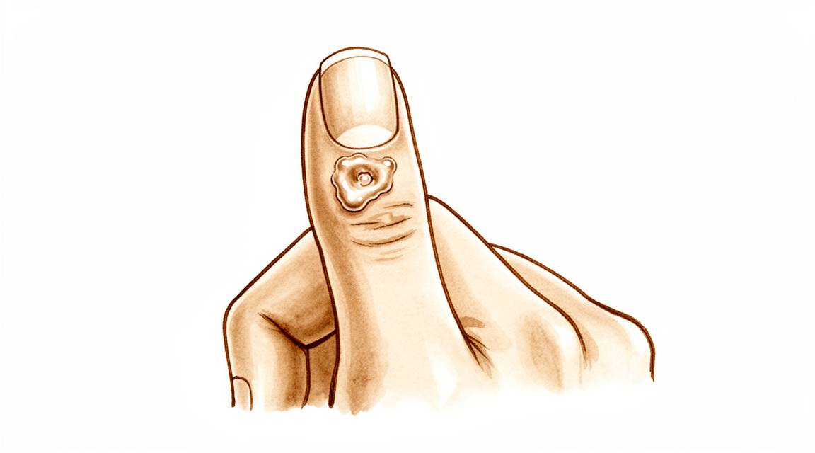
You may notice a small, firm bump on the top of your finger, usually near the last joint. This bump is a mucous cyst. It often feels like a small pea under the skin. You might see it clearly if your skin is thin there. The cyst can make the skin over it look shiny or stretched. In some cases, it may cause the fingernail to grow in a wavy or ridged pattern.
The area around the cyst can feel tender or achy. You might feel pain when you press on it or when you bend your finger. Daily tasks can become difficult. Reaching for objects, typing, or gripping tools may hurt. You might find it hard to fasten buttons or zip up a jacket. Some people report that the pain is worse at night or after using their hands for a long time. The discomfort can interfere with sleeping if you rest on that hand.
Your surgeon will look at the cyst and check how it affects your finger’s movement. The pain often comes from the underlying joint changes, not just the bump itself. Removing the cyst alone might not stop the pain if the bone spur (osteophyte) remains. Your surgeon may discuss removing the bone spur to help relieve pressure on the joint and skin. This approach can help the skin heal better and reduce the chance of the cyst coming back.
In many cases, treating the bone spur leads to complete resolution of the cyst. You may not need complex surgery. Simple removal of the cyst combined with bone spur removal has an extremely rare recurrence rate. Even less invasive options, like removing just the bone spur, can provide complete resolution in most cases. If a skin flap is needed, patient satisfaction regarding the scar is high. You are likely to be happy with the appearance and willing to have the procedure again.
Your surgeon will tailor the plan to your specific needs. The goal is to relieve your pain and restore function. Most patients find that after treatment, their finger feels more comfortable and looks better. The skin has good recovery potential once the main problem is addressed. You can expect a straightforward recovery with careful wound care.
What's actually happening
A mucous cyst is a small, fluid-filled sac that forms on your finger. It usually appears near the tip of the finger, close to the nail bed. The cyst sits on top of the joint capsule, which is the tough, fibrous sleeve that wraps around your joint to keep it stable.
The root cause is wear-and-tear arthritis in the joint. As the cartilage—the smooth coating on the ends of your bones—breaks down, your body tries to repair the damage. This process often creates bone spurs, called osteophytes. These are small, rough bumps of extra bone that grow out from the joint surface.
Think of the joint capsule like a gasket or a seal. When the bone spur rubs against this seal, it irritates the tissue. This irritation causes the joint lining to leak synovial fluid, which is the natural lubricant that keeps your joint moving smoothly. The fluid pushes through the weakened spot in the capsule, forming the visible bump you see on your skin.
Because the cyst is connected to the joint, it is filled with this same lubricating fluid. The pressure from the fluid can make the skin over the cyst thin and fragile. In some cases, the cyst can press on nearby nerves or affect the nail matrix, which is the tissue under your cuticle that grows your nail. This pressure is what leads to the grooves or ridges you might see in your nail.
Removing just the cyst often leads to it coming back because the bone spur remains. The spur continues to irritate the joint lining, causing more fluid to leak. To stop this cycle, the underlying bone spur must be addressed. When the spur is removed, the irritation stops, and the joint lining can heal. This is why treating the bone is just as important as treating the cyst itself.
What we can do about it
Your journey usually begins with simple self-care and professional guidance. You can try resting the finger and avoiding activities that put pressure on the cyst. Physiotherapy helps you maintain movement in the joint and keeps the surrounding muscles strong. This approach aims to reduce irritation and improve function without invasive steps. You should give this conservative care a fair trial to see if symptoms settle. Many patients find that managing daily habits and following a gentle exercise plan provides enough relief to avoid further intervention.
If self-care is not enough, your surgeon may suggest medical management to control pain and inflammation. Corticosteroid injections are a common option for these cysts. A volar corticosteroid injection allows for easy and consistent needle placement into the joint. This method minimizes potential soft tissue damage and infection risks compared to other techniques. The injection helps calm the inflammation that drives cyst growth. While the evidence highlights the safety and ease of this approach, it is important to understand that injections typically manage symptoms rather than permanently remove the cyst. The effect lasts for a period of time, but the underlying wear-and-tear arthritis remains. You and your surgeon will decide if this temporary relief is sufficient or if further steps are needed.
Surgery is considered when conservative care has reached its limit and the cyst causes persistent pain, deformity, or functional problems. Your surgeon will discuss the best surgical option for your specific case. The goal is to remove the cyst and address the root cause, which is often bone spurs (osteophytes) from arthritis. Removing these bone spurs is crucial because it significantly lowers the chance of the cyst coming back. Some techniques involve removing the cyst along with a small skin flap to ensure proper healing, while others focus primarily on the bone. Your surgeon will choose the method that offers the best balance of low recurrence rates and good cosmetic results for your hand.
What to expect
Mucous cysts are small, fluid-filled lumps that often appear near the end joint of your finger or thumb. They are closely linked to wear-and-tear arthritis in that joint. Because they stem from this underlying joint change, the cyst itself may persist or return if the root cause is not addressed. However, with proper treatment, the outlook is generally very positive.
Your surgeon will likely recommend removing the cyst along with any bone spurs (osteophytes) causing it. This approach eradicates mucous cysts with extremely rare recurrence. If your surgeon chooses a technique using a local advancement skin flap, the recurrence rate is low at 1.4%. In some cases, your surgeon may remove only the bone spur without taking out the cyst. This less invasive method provides complete resolution in most cases. Regardless of the specific plan for the soft tissues, your surgeon will remove the bone spur to prevent the cyst from coming back.
You can expect high satisfaction with the results, particularly regarding the appearance of the scar. Many patients report they would undergo the procedure again. The surgical techniques used are designed to be simple and reliable. For example, some methods allow for the removal of thinned skin without adding risk to your nail matrix. Others use skin grafts that provide satisfactory cosmetic results with acceptable recurrence rates. Even complex cases, such as cysts growing inside the digital nerve, can result in successful outcomes.
If left untreated, the cyst may remain or grow, potentially thinning the skin and increasing the risk of infection. By addressing both the cyst and the underlying arthritis, your surgeon aims for a lasting solution. Most patients find that their finger looks and feels better after the procedure. The goal is not just to remove the lump, but to stop it from returning. You can expect a straightforward recovery process focused on protecting the surgical site while allowing your joint to heal.
When to see someone
See your GP if you notice a small bump on your finger near the nail. Ask for a specialist review if the cyst causes persistent pain that does not improve with rest. Seek care if you experience weakness or instability in the joint. Watch for locking or giving way sensations. Contact your surgeon if symptoms interfere with sleep or work. Sudden worsening of the area also warrants a check-up. Early evaluation helps prevent complications. Your surgeon can discuss options like removing the cyst and any bone spurs. This approach often leads to high satisfaction and low recurrence rates. Treating the underlying cause helps the skin recover naturally.
Evidence & references
title: "Mucous Cyst" slug: mucous-cyst region: hand audience: patient mesh_terms: ["Ganglion Cysts", "Finger Joint", "Fingers", "Cysts", "Soft Tissue Neoplasms", "Sweat Gland Neoplasms", "Diagnosis, Differential", "Osteophyte"] article_count: 20 model_used: Qwen3.6-35B-A3B-Q8_0.gguf generated_at: '2026-06-16T19:30:57+00:00' key_articles: - title: "Mucous Cysts" ref_num: 1 evidence_tier: paper evidence_level: 4 doi: 10.1016/j.jhsa.2010.01.029 year: 2010 - title: "Total Dorsal Capsulectomy for the Treatment of Mucous Cysts" ref_num: 2 evidence_tier: paper evidence_level: 4 doi: 10.1016/j.jhsa.2014.03.004 year: 2014 - title: "Marginal Osteophyte Excision in Treatment of Mucous Cysts" ref_num: 3 evidence_tier: paper evidence_level: 4 doi: 10.2106/00004623-197355030-00013 year: 1973 - title: "Malignant Natural-Killer cell neoplasm presenting as a mucous cyst on the distal interphalangeal joint of the finger" ref_num: 4 evidence_tier: paper evidence_level: 5 doi: 10.1007/s00402-008-0794-4 year: 2008 - title: "Osteophyte excision without cyst excision for a mucous cyst of the finger" ref_num: 5 evidence_tier: paper evidence_level: 4 doi: 10.1177/1753193413478549 year: 2013 - title: "The Zitelli Bilobed Flap on Skin Coverage After Mucous Cyst Excision: A Retrospective Cohort of 33 Cases" ref_num: 6 evidence_tier: paper evidence_level: 4 doi: 10.1016/j.jhsa.2017.03.013 year: 2017 - title: "Use of Wolfe Graft for the Treatment of Mucous Cysts" ref_num: 7 evidence_tier: paper evidence_level: 4 doi: 10.1177/1753193408103498 year: 2009 - title: "Eccrine Porocarcinoma Presenting as a Hand Cyst" ref_num: 8 evidence_tier: paper evidence_level: 4 doi: 10.1016/j.jhsa.2016.07.112 year: 2016 - title: "A reliable surgical treatment for digital mucous cysts" ref_num: 9 evidence_tier: paper evidence_level: 4 doi: 10.1177/1753193413508540 year: 2013 - title: "Etiology and Treatment of the So-Called Mucous Cyst of the Finger" ref_num: 10 evidence_tier: paper evidence_level: 4 doi: 10.2106/00004623-197254070-00008 year: 1972 - title: "Giant Cell Tumours of Tendon Sheath: Classification and Recurrence Rate" ref_num: 11 evidence_tier: paper evidence_level: 3 doi: 10.1054/jhsb.2000.0522 year: 2001 - title: "Granular Cell Nerve Tumor in the Hand: Case Report" ref_num: 12 evidence_tier: case_report evidence_level: 4 doi: 10.1016/j.jhsa.2009.05.011 year: 2009 - title: "Commentary on Lee et al. Osteophyte excision without cyst excision for a mucous cyst of the finger" ref_num: 13 evidence_tier: commentary evidence_level: 5 doi: 10.1177/1753193413510663 year: 2014 - title: "Metastases to the finger masquerading as flexor tenosynovitis" ref_num: 14 evidence_tier: paper evidence_level: 4 doi: 10.1177/1753193409360605 year: 2010 - title: "Ultrasound of the Hand and Wrist" ref_num: 16 evidence_tier: paper evidence_level: 5 doi: 10.1016/j.jhsa.2009.02.010 year: 2009 - title: "Re: Lee HJ, Kim PT, Jeon IH, et al. Osteophyte excision without cyst excision for a mucous cyst of the finger. J Hand Surg Eur. 2014, 39: 258–61" ref_num: 20 evidence_tier: paper evidence_level: 5 doi: 10.1177/1753193414546443 year: 2014 synthesis_version: "v2" verifier_status: skipped
Overview
- Scientific data regarding mucous cysts consist almost entirely of retrospective studies [1].
- Much of the management or recommendations for mucous cysts is based on expert opinion [1].
- Total dorsal capsulectomy alone is a simple treatment for mucous cysts that does not lead to any recurrence [2].
- Excision of the cyst combined with complete removal of the marginal osteophyte eradicates mucous cysts with extremely rare recurrence [3].
- Osteophyte excision without cyst excision may be a good treatment choice for mucous cysts of the finger, providing a less invasive method with complete resolution in most cases [5].
- Osteophyte removal results in a low cyst recurrence rate, indicating it should be undertaken regardless of the surgeon's plan for the soft tissues [13].
- The Zitelli bilobed flap allows excision of the cyst and thinned skin with no added risk to the nail matrix [6].
- The use of a Wolfe graft for mucous cysts is simple, easy to perform, and provides satisfactory cosmesis with acceptable recurrence rates [7].
- Surgical excision with a local advancement skin flap is a reliable treatment for digital mucous cysts, demonstrating a low recurrence rate of 1.4% and high patient satisfaction regarding the scar and willingness to undergo the procedure again [9].
- A surgical technique involving excision of the cyst, synovectomy, and débridement of osteophytes with rotational flap closure resulted in no recurrences in thirty-six patients [10].
- Pathohistological analysis is useful in cases where doubts arise about the initial diagnosis of a benign tumorous lesion [4].
- Eccrine porocarcinomas have a substantial risk of metastasis, high risk of local recurrence, and are potentially fatal [8].
Anatomy & Pathophysiology
- Scientific data regarding mucous cysts consist almost entirely of retrospective studies [1].
- Much of the management or recommendations for mucous cysts is based on expert opinion [1].
- Mucous cysts are associated with marginal osteophytes at the distal interphalangeal joint [3].
- The primary pathology in mucous cysts involves osteophytes, and removal of these osteophytes allows for skin recovery potential [20].
- Ultrasound is a powerful modality for evaluating pathologic conditions in the hand and wrist [16].
- Ultrasound provides a cost-effective and expedient alternative or adjunct to MRI for hand and wrist evaluation [16].
- Ultrasound is best used when there is a specific clinical question regarding a well-localized abnormality [16].
- Pathohistological analysis is useful in cases where doubts arise about the initial diagnosis of a benign tumorous lesion [4].
- Subungual keratoacanthoma may show locally aggressive behaviour but does not metastasize [14].
Classification
- Scientific data regarding mucous cysts consist almost entirely of retrospective studies [1].
- Much of the management or recommendations for mucous cysts is based on expert opinion [1].
- Total dorsal capsulectomy alone is a simple treatment for mucous cysts that does not lead to any recurrence [2].
- Excision of the cyst combined with complete removal of the marginal osteophyte eradicates mucous cysts with extremely rare recurrence [3].
- Pathohistological analysis is useful in cases where doubts arise about the initial diagnosis of a benign tumorous lesion [4].
- Osteophyte excision without cyst excision may be a good treatment choice for mucous cyst of the finger, providing a less invasive method with complete resolution in most cases [5].
- The Zitelli bilobed flap allows excision of the cyst and thinned skin with no added risk to the nail matrix [6].
- The use of a Wolfe graft is simple, easy to perform, and provides satisfactory cosmesis with acceptable recurrence rates [7].
- Eccrine porocarcinomas have a substantial risk of metastasis, high risk of local recurrence, and are potentially fatal [8].
- Surgical excision with a local advancement skin flap is a reliable treatment for digital mucous cysts, demonstrating a low recurrence rate of 1.4% and high patient satisfaction regarding the scar and willingness to undergo the procedure again [9].
- A surgical technique involving excision of the cyst, synovectomy, and débridement of osteophytes with rotational flap closure resulted in no recurrences in thirty-six patients [10].
- There is a statistically significant difference in recurrence rates between Type I giant cell tumours of the tendon sheath (0%) and Type II tumours (38%) [11].
- Recurrence in Type II giant cell tumours of the tendon sheath is likely due to undetected satellite lesions or incomplete excision [11].
- Incomplete excision of a granular cell nerve tumor can lead to recurrence [12].
- Osteophyte removal results in a low cyst recurrence rate [13].
- Osteophyte removal should be undertaken regardless of the surgeon's plan for the soft tissues [13].
Clinical Presentation
- Scientific data regarding mucous cysts consist almost entirely of retrospective studies [1].
- Much of the management or recommendations for mucous cysts is based on expert opinion [1].
- Malignant natural-killer cell neoplasms can present as a mucous cyst on the distal interphalangeal joint of the finger [4].
- Eccrine porocarcinomas can present as a hand cyst [8].
- Subungual keratoacanthoma may present as a condition masquerading as flexor tenosynovitis in the finger [14].
- Ultrasound is a powerful modality for the evaluation of pathologic conditions in the hand and wrist [16].
- Ultrasound provides a cost-effective and expedient alternative and/or adjunct to MRI for hand and wrist evaluation [16].
- Ultrasound is best used when there is a specific clinical question regarding a well-localized abnormality in the hand or wrist [16].
Investigations
- Scientific data regarding mucous cysts consist almost entirely of retrospective studies [1].
- Much of the management of mucous cysts is based on expert opinion [1].
- Pathohistological analysis is useful when doubts arise about the initial diagnosis of a benign tumorous lesion [4].
- Eccrine porocarcinomas have a substantial risk of metastasis, high risk of local recurrence, and are potentially fatal [8].
- Ultrasound is a powerful modality for evaluation of pathologic conditions in the hand and wrist [16].
- Ultrasound provides a cost-effective and expedient alternative and/or adjunct to MRI [16].
- Ultrasound is best used when there is a specific clinical question regarding a well-localized abnormality [16].
Treatment
- Scientific data regarding mucous cysts consist almost entirely of retrospective studies, and much of what is done or recommended is based on expert opinion [1].
- Total dorsal capsulectomy alone is a simple treatment for mucous cysts that does not lead to any recurrence [2].
- Excision of the cyst and complete removal of the marginal osteophyte eradicates mucous cysts with extremely rare recurrence [3].
- Osteophyte excision without cyst excision may be a good treatment choice for mucous cyst of the finger, providing a less invasive method with complete resolution in most cases [5].
- Osteophyte removal results in a low cyst recurrence rate, indicating that it should be undertaken regardless of the surgeon's plan for the soft tissues [13].
- The Zitelli bilobed flap allows excision of the cyst and thinned skin with no added risk to the nail matrix [6].
- Use of Wolfe graft is simple, easy to perform, and provides satisfactory cosmesis with acceptable recurrence rates [7].
- Surgical excision with a local advancement skin flap is a reliable treatment for digital mucous cysts, demonstrating a low recurrence rate of 1.4% and high patient satisfaction regarding the scar and willingness to undergo the procedure again [9].
- A surgical technique involving excision of the cyst, synovectomy, and débridement of osteophytes with rotational flap closure resulted in no recurrences in thirty-six patients [10].
- Pathohistological analysis is useful in cases where doubts arise about the initial diagnosis of a benign tumorous lesion [4].
Complications
- Scientific data regarding mucous cysts consist almost entirely of retrospective studies, with many recommendations based on expert opinion [1].
- Total dorsal capsulectomy alone for mucous cysts did not lead to any recurrence [2].
- Excision of the cyst and complete removal of the marginal osteophyte eradicates mucous cysts with extremely rare recurrence [3].
- Osteophyte excision without cyst excision may provide complete resolution in most cases [5].
- The Zitelli bilobed flap allows excision of the cyst and thinned skin with no added risk to the nail matrix [6].
- Use of a Wolfe graft provides satisfactory cosmesis with acceptable recurrence rates [7].
- Surgical excision with a local advancement skin flap demonstrates a low recurrence rate of 1.4% and high patient satisfaction regarding the scar [9].
- A surgical technique involving excision of the cyst, synovectomy, and débridement of osteophytes with rotational flap closure resulted in no recurrences in thirty-six patients [10].
- Incomplete excision can lead to recurrence of granular cell nerve tumors [12].
- Type II giant cell tumors of the tendon sheath have a 38% recurrence rate, likely due to undetected satellite lesions or incomplete excision [11].
- Malignant natural-killer cell neoplasms can present as mucous cysts on the distal interphalangeal joint [4].
- Pathohistological analysis is useful in cases where doubts arise about the initial diagnosis of a benign tumorous lesion [4].
- Eccrine porocarcinomas have a substantial risk of metastasis, high risk of local recurrence, and are potentially fatal [8].
Recovery
- Scientific data regarding mucous cysts consist almost entirely of retrospective studies [1].
- Much of the treatment for mucous cysts is based on expert opinion [1].
- Total dorsal capsulectomy alone is a simple treatment for mucous cysts that does not lead to any recurrence [2].
- Excision of the cyst and complete removal of the marginal osteophyte eradicates mucous cysts with extremely rare recurrence [3].
- Osteophyte excision without cyst excision may be a good treatment choice for mucous cysts of the finger, providing a less invasive method with complete resolution in most cases [5].
- The Zitelli bilobed flap allows excision of the cyst and thinned skin with no added risk to the nail matrix [6].
- The use of a Wolfe graft is simple, easy to perform, and provides satisfactory cosmesis with acceptable recurrence rates [7].
- Surgical excision with a local advancement skin flap is a reliable treatment for digital mucous cysts, demonstrating a low recurrence rate of 1.4% and high patient satisfaction regarding the scar and willingness to undergo the procedure again [9].
- Pathohistological analysis is useful in cases where doubts arise about the initial diagnosis of a benign tumorous lesion [4].
- Eccrine porocarcinomas have a substantial risk of metastasis, high risk of local recurrence, and are potentially fatal [8].
- Incomplete excision can lead to recurrence in granular cell nerve tumors [12].
Key Evidence
- [L4] The scientific data regarding mucous cysts consist almost entirely of retrospective studies, and much of what is done or recommended is based on expert opinion. (10.1016/j.jhsa.2010.01.029)
- [L4] A total dorsal capsulectomy alone was a simple treatment for mucous cysts and did not lead to any recurrence. (10.1016/j.jhsa.2014.03.004)
- [L4] Excision of the cyst and complete removal of the marginal osteophyte eradicates mucous cysts with extremely rare recurrence. (10.2106/00004623-197355030-00013)
- [L5] This case emphasizes the utility of a pathohistological analysis in cases where doubts arise about the initial diagnosis of a benign tumorous lesion. (10.1007/s00402-008-0794-4)
- [L4] Osteophyte excision without cyst excision may be a good treatment choice for mucous cyst of the finger, providing a less invasive method with complete resolution in most cases. (10.1177/1753193413478549)
- [L4] It allows excision of the cyst and thinned skin with no added risk to the nail matrix. (10.1016/j.jhsa.2017.03.013)
- [L4] The technique is simple, easy to perform, and provides satisfactory cosmesis with acceptable recurrence rates. (10.1177/1753193408103498)
- [L4] Prompt recognition and appropriate treatment are critical because eccrine porocarcinomas have a substantial risk of metastasis, high risk of local recurrence, and are potentially fatal. (10.1016/j.jhsa.2016.07.112)
- [L4] Surgical excision with a local advancement skin flap is a reliable treatment for digital mucous cysts, demonstrating a low recurrence rate of 1.4% and high patient satisfaction regarding the scar and willingness to undergo the procedure again. (10.1177/1753193413508540)
- [L4] A new surgical technique involving excision of the cyst, synovectomy, and débridement of osteophytes with rotational flap closure resulted in no recurrences in thirty-six patients. (10.2106/00004623-197254070-00008)
- [L3] The study found a statistically significant difference in recurrence rates between Type I tumours (0%) and Type II tumours (38%), with recurrence in Type II likely due to undetected satellite lesions or incomplete excision. (10.1054/jhsb.2000.0522)
- [Case_report] The author notes that while the true recurrence rate is unknown, incomplete excision can lead to recurrence. (10.1016/j.jhsa.2009.05.011)
- [Commentary] The article shows that osteophyte removal results in a low cyst recurrence rate, indicating that it should be undertaken regardless of the surgeon's plan for the soft tissues. (10.1177/1753193413510663)
- [L4] Subungual keratoacanthoma may show locally aggressive behaviour but does not metastasize. (10.1177/1753193409360605)
- [L5] Ultrasound is a powerful modality for evaluation of pathologic conditions in the hand and wrist, providing a cost-effective and expedient alternative and/or adjunct to MRI, best used when there is a specific clinical question regarding a well-localized abnormality. (10.1016/j.jhsa.2009.02.010)
- [L5] The authors of the original study believe that extensive damage to the skin is unnecessary and that the skin has recovery potential once the main problem (osteophytes) is removed, favoring a less invasive approach over techniques requiring skin flaps. (10.1177/1753193414546443)
References
[1] Mucous Cysts. The Journal of Hand Surgery. 2010. DOI: 10.1016/j.jhsa.2010.01.029 [2] Total Dorsal Capsulectomy for the Treatment of Mucous Cysts. The Journal of Hand Surgery. 2014. DOI: 10.1016/j.jhsa.2014.03.004 [3] Marginal Osteophyte Excision in Treatment of Mucous Cysts. The Journal of Bone & Joint Surgery. 1973. DOI: 10.2106/00004623-197355030-00013 [4] Malignant Natural-Killer cell neoplasm presenting as a mucous cyst on the distal interphalangeal joint of the finger. Archives of Orthopaedic and Trauma Surgery. 2008. DOI: 10.1007/s00402-008-0794-4 [5] Osteophyte excision without cyst excision for a mucous cyst of the finger. Journal of Hand Surgery (European Volume). 2013. DOI: 10.1177/1753193413478549 [6] The Zitelli Bilobed Flap on Skin Coverage After Mucous Cyst Excision: A Retrospective Cohort of 33 Cases. The Journal of Hand Surgery. 2017. DOI: 10.1016/j.jhsa.2017.03.013 [7] Use of Wolfe Graft for the Treatment of Mucous Cysts. Journal of Hand Surgery (European Volume). 2009. DOI: 10.1177/1753193408103498 [8] Eccrine Porocarcinoma Presenting as a Hand Cyst. The Journal of Hand Surgery. 2016. DOI: 10.1016/j.jhsa.2016.07.112 [9] A reliable surgical treatment for digital mucous cysts. Journal of Hand Surgery (European Volume). 2013. DOI: 10.1177/1753193413508540 [10] Etiology and Treatment of the So-Called Mucous Cyst of the Finger. The Journal of Bone & Joint Surgery. 1972. DOI: 10.2106/00004623-197254070-00008 [11] Giant Cell Tumours of Tendon Sheath: Classification and Recurrence Rate. Journal of Hand Surgery. 2001. DOI: 10.1054/jhsb.2000.0522 [12] Granular Cell Nerve Tumor in the Hand: Case Report. The Journal of Hand Surgery. 2009. DOI: 10.1016/j.jhsa.2009.05.011 [13] Commentary on Lee et al. Osteophyte excision without cyst excision for a mucous cyst of the finger. Journal of Hand Surgery (European Volume). 2014. DOI: 10.1177/1753193413510663 [14] Metastases to the finger masquerading as flexor tenosynovitis. Journal of Hand Surgery (European Volume). 2010. DOI: 10.1177/1753193409360605 [16] Ultrasound of the Hand and Wrist. The Journal of Hand Surgery. 2009. DOI: 10.1016/j.jhsa.2009.02.010 [20] Re: Lee HJ, Kim PT, Jeon IH, et al. Osteophyte excision without cyst excision for a mucous cyst of the finger. J Hand Surg Eur. 2014, 39: 258–61. Journal of Hand Surgery (European Volume). 2014. DOI: 10.1177/1753193414546443


