Liberação do Túnel Radial
Patients › Rehabilitation
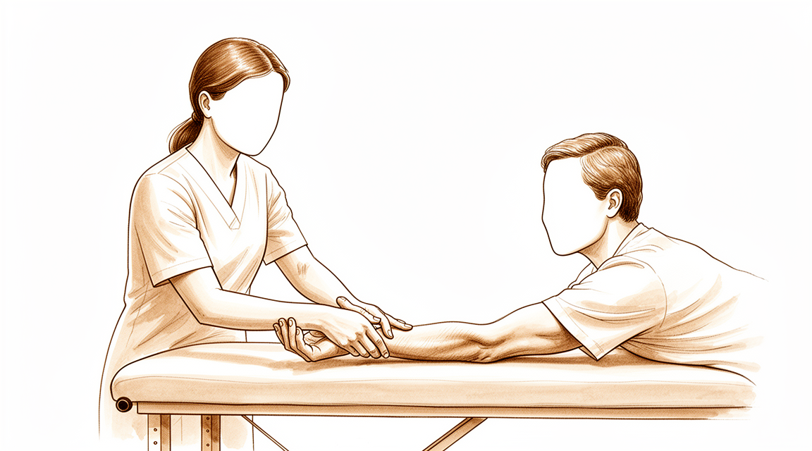
Post-operative exercises and precautions after radial tunnel release, including radial nerve glides.
Este protocolo orienta a sua recuperação após a liberação do túnel radial com o Dr. Kieran Hirpara no Mater Private Hospital Rockhampton. Ele explica o que esperar nas semanas seguintes à cirurgia e apresenta o programa de exercícios do material pós-operatório fornecido. Traga esta página ou seu PDF para a sua primeira consulta de fisioterapia ou terapia da mão, para que a reabilitação seja coordenada. Seu terapeuta pode ajustar o plano conforme a evolução da sua recuperação.
Se tiver alguma preocupação sobre sua ferida após a cirurgia, entre em contato com a clínica. Frequentemente, é útil tirar uma foto da ferida e enviá-la por e-mail para avaliação.
O que esperar
O cuidado da sua ferida é explicado nas orientações de cuidado de feridas da clínica. À medida que a ferida cicatriza, o nervo libertado pode aderir aos tecidos circundantes; os exercícios de deslizamento abaixo são muito importantes para o manter em movimento livre e evitar que fique preso.
Por vezes, a ferida pode tornar-se sensível. Isto é normal e pode ser prevenido ou minimizado ao iniciar a dessensibilização diária: bater suavemente e esfregar sobre a ferida (ou sobre a curadura), começando imediatamente após a sua cirurgia. Este tipo de "feedback sensorial" permite que o nervo normalize o toque e a textura.
Assim que a ferida estiver totalmente cicatrizada, inicie a massagem da cicatriz: círculos firmes sobre a incisão. Consulte as orientações de cuidado de feridas para mais informações sobre a gestão da cicatriz.
É importante ter expectativas realistas sobre a recuperação. O nervo radial tem de percorrer e esticar uma quantidade mensurável durante os movimentos normais do braço, por isso, mantê-lo em movimento precocemente é o que evita que ele adira aos tecidos em cicatrização [1]. Mesmo assim, o alívio da dor após a libertação do túnel radial é frequentemente gradual e não imediato, e para algumas pessoas é apenas parcial. Honestamente, esta operação é menos previsível do que alguns outros procedimentos de libertação nervosa: a síndrome do túnel radial pode ser difícil de diagnosticar com certeza e muitas vezes sobrepõe-se ao cotovelo de tenista, o que é parte da razão pela qual os resultados variam. Estudos publicados a longo prazo relatam bons resultados em cerca de dois terços dos pacientes em geral, com os melhores resultados naqueles que apresentam apenas sintomas do túnel radial [2][3]. A recuperação tende a ser mais lenta e menos completa quando há também cotovelo de tenista (epicondilite lateral), mais de uma compressão nervosa no mesmo braço, ou um pedido de compensação laboral [2][4]. O seu programa de deslizamento nervoso e a dessensibilização graduada são as partes da reabilitação mais sob o seu controlo, e a prática diária constante dá ao nervo a melhor hipótese de se estabilizar.
Precauções e limitações
O uso funcional leve da sua mão é incentivado para tarefas diárias, como cuidados pessoais, alimentação, vestir-se, escrever e digitar. Além disso, os limites são simples: não levantar pesos, não fazer força de preensão, não suportar peso e não utilizar máquinas vibratórias (por exemplo, ferramentas elétricas ou cortador de grama) por até 6 semanas após a cirurgia, e a direção de veículos é limitada nas primeiras 1–2 semanas.
Para o seu fisioterapeuta:
Objetivos
- Prevenir a aderência do nervo liberado à ferida em cicatrização (programa de deslizamento nervoso)
- Reduzir a sensibilidade da ferida por meio de dessensibilização gradual
- Manter a amplitude de movimento do punho, antebraço e cotovelo
- Apoiar o uso funcional leve da mão para atividades de vida diária
Conduta
- Dessensibilização diária: toques leves / fricção sobre a ferida (curativo), iniciando imediatamente no pós-operatório
- Massagem na cicatriz (círculos firmes sobre a incisão) após a cicatrização completa da ferida
- Programa de exercícios domiciliares conforme os cartões abaixo: alongamento de flexão / extensão do punho; alongamento de supinação / pronação do punho; flexão / extensão do cotovelo; deslizamentos do nervo radial
- Favorecer deslizamentos nervosos do tipo deslizante ("slider") em vez de tensão agressiva no final do arco de movimento: as técnicas deslizantes alcançam excursão nervosa substancialmente maior com muito menor tensão nervosa, o que é melhor tolerado em torno de um nervo recentemente descomprimido [1][5]
- A mobilização nervosa pode ser considerada como adjuvante ao programa; a base de evidências para a mobilização neural em condições relacionadas a nervos é favorável, mas de certeza variável, portanto a progressão deve ser guiada pelos sintomas [6]
Precauções
- Apenas uso funcional leve da mão (cuidados pessoais, alimentação, vestir-se, escrever, digitar)
- Não levantar pesos, não fazer força de preensão, não suportar peso e não utilizar máquinas vibratórias (por exemplo, ferramentas elétricas, cortador de grama) por até 6 semanas após a cirurgia
- A direção de veículos é limitada nas primeiras 1–2 semanas
- Os deslizamentos e alongamentos nervosos devem ser leves e essencialmente sem dor; evite forçar um arco de movimento que reproduza a dor nervosa pré-operatória
Estes são os exercícios do material pós-operatório fornecido, iniciados após a cirurgia e continuados em casa conforme orientação do seu fisioterapeuta ou terapeuta da mão. As repetições, tempos de manutenção e frequência estão listados em cada cartão.
Seus exercícios
Este programa de exercícios foi elaborado em parceria com Sarah Farrell, Bacharel em Terapia Ocupacional (BOccThy), Terapeuta de Mão Acreditada (AHT).
Após o seu protocolo
Este protocolo complementa as orientações gerais de recuperação da clínica; consulte o manejo da dor pós-operatória, o cuidado com a ferida e os fundamentos da terapia manual. Para informações sobre o procedimento cirúrgico e a condição que ele trata, consulte a liberação do túnel radial e a síndrome do túnel radial.
Referências
[1] Wright TW, Glowczewskie F, Cowin D, Wheeler DL. Excursão e tensão do nervo radial no cotovelo e punho associadas ao movimento do membro superior. J Hand Surg Am. 2005;30(5):990–996. https://pubmed.ncbi.nlm.nih.gov/16182056/ [2] Lee JT, Azari K, Jones NF. Resultados a longo prazo da liberação do túnel radial — o efeito do cotovelo de tenista coexistente, síndromes compressivas múltiplas e compensação ao trabalhador. J Plast Reconstr Aesthet Surg. 2008;61(9):1095–1099. https://www.sciencedirect.com/science/article/abs/pii/S1748681507004044 [3] Sotereanos DG, Varitimidis SE, Giannakopoulos PN, Westkaemper JG. Resultados do tratamento cirúrgico da síndrome do túnel radial. J Hand Surg Am. 1999;24(3):566–570. https://pubmed.ncbi.nlm.nih.gov/10357537/ [4] Naam NH, Nemani S. Síndrome do túnel radial. Orthop Clin North Am. 2012;43(4):529–536. (Síndrome do Túnel Radial, StatPearls.) https://www.ncbi.nlm.nih.gov/books/NBK555937/ [5] Coppieters MW, Butler DS. Os "deslizadores" deslizam e os "tensionadores" tensionam? Uma análise das técnicas neurodinâmicas e considerações sobre sua aplicação. Man Ther. 2008;13(3):213–221. https://pubmed.ncbi.nlm.nih.gov/17398140/ [6] Basson A, Olivier B, Ellis R, Coppieters M, Stewart A, Mudzi W. A eficácia da mobilização neural para condições neuromusculoesqueléticas: uma revisão sistemática e meta-análise. J Orthop Sports Phys Ther. 2017;47(9):593–615. https://pubmed.ncbi.nlm.nih.gov/28704626/
Evidence & references
Radial Tunnel Release — Evidence Brief & Post-operative Rehabilitation
Topic scope: post-operative rehabilitation after surgical decompression / neurolysis of the posterior interosseous nerve (deep branch of the radial nerve) in the radial tunnel of the proximal forearm, performed for radial tunnel syndrome (RTS). This is an elbow / proximal-forearm topic — anatomically and clinically distinct from carpal-tunnel and cubital-tunnel decompression. Like other nerve decompressions it is an early-motion pathway (early elbow/forearm/wrist motion, radial-nerve glides, oedema and scar care). The scope deliberately foregrounds the diagnostic controversy and the more variable, lower success rates that distinguish RTS release from the better-validated carpal-tunnel and cubital-tunnel operations.
Defining principle of the rehab here: a decompressed nerve does not create a healing construct that needs months of protection — it needs early, gentle movement to stop it adhering to the operative bed and to restore its glide. So the rehab is an early-motion programme: light functional hand use from day 1, radial-nerve sliders, graded desensitisation and (once healed) scar massage; heavier loading deferred to ~6 weeks. But two honesty caveats sit over the whole topic. First, RTS is a contested diagnosis — there is no confirmatory imaging or electrodiagnostic test, it is a diagnosis of exclusion, and a substantial body of opinion regards it as a variant of recalcitrant lateral epicondylitis. Second, outcomes after release are more variable and on average lower than carpal- or cubital-tunnel release — good results cluster around two-thirds overall, and fall further with co-existing tennis elbow, multiple compression sites, or a workers'-compensation context. Patient expectations should be set accordingly.
A. THE DIAGNOSTIC CONTROVERSY (read first — it frames everything)
RTS is among the most contested entities in upper-limb surgery, and the rehab brief is incomplete without it:
- No confirmatory test. Electromyography and nerve-conduction studies are characteristically normal in RTS (compression is intermittent/dynamic and predominantly of a motor nerve carrying few pain fibres), and MRI is frequently negative — denervation oedema in supinator/extensors is suggestive but inconsistent, and a normal scan does not exclude the diagnosis. RTS is therefore a clinical diagnosis of exclusion, resting on point tenderness ~4 cm distal to the lateral epicondyle (over the radial tunnel rather than the epicondyle), pain on resisted supination / resisted long-finger extension, and — for some surgeons — temporary relief from a diagnostic local-anaesthetic block at the radial tunnel.
- Overlap with lateral epicondylitis (tennis elbow). The two coexist frequently and share the lateral-elbow pain territory. A recognised school of thought holds that "RTS" is often severe, recalcitrant lateral epicondylitis rather than a discrete compression neuropathy. Importantly, routine PIN release added to lateral-epicondylitis surgery has not been shown to improve outcomes, so the diagnosis should be secure before a decompression is planned.
- Practical consequence. Surgery is a last resort after prolonged failed conservative care (activity modification, splinting, anti-inflammatories, sometimes a steroid injection), and is best reserved for patients with proximal-forearm pain and no better explanation. This uncertainty is the single most important reason post-operative expectations must be framed honestly.
B. RELEASE OUTCOMES (variable — and why)
- Headline success ~two-thirds. Across the older long-term series, roughly 67% good, 15% fair, 18% poor after radial tunnel decompression — markedly more variable than carpal- or cubital-tunnel release. A 2008 long-term series (Lee, Azari, Jones) and a 1999 series (Sotereanos et al.) both document this spread; the Sotereanos cohort reported good/excellent results in only ~39% by objective assessment (though ~64% by patient self-rating), underscoring how outcome depends on the metric used.
- Co-existing lateral epicondylitis lowers success. Success falls to roughly 40% when tennis elbow coexists, versus far higher with isolated RTS.
- Multiple compression sites and workers'-compensation context lower success — reported ~58% success in compensation cases vs ~73% without. These are the same modifiers named in the patient protocol.
- 2025 systematic review (Raymond et al., HAND). 11 studies, 401 limbs (381 patients). Outcomes were heterogeneous; a dorsal approach between ECRB and EDC was associated with the most favourable Roles-and-Maudsley scores and satisfaction. The review's central message is that the overall evidence is low-grade (observational), the diagnosis non-standardised, and the effectiveness of conservative treatment essentially untested — a "tendency" toward benefit rather than proof.
- Resorption-style "spontaneous improvement" does not apply here — unlike calcific tendinitis, RTS does not self-resolve through a biological cycle; conservative care manages symptoms rather than curing a deposit.
C. SURGICAL APPROACH (shapes the early rehab)
- What is done. Complete neurolysis of the radial nerve at its bifurcation, decompressing the deep branch (PIN) and superficial sensory branch, releasing the arcade of Frohse (the proximal supinator edge), the leash of Henry (radial recurrent vessels), the ECRB fascial edge, and the distal supinator border. Any constrictive bands or vessels are divided.
- Approaches. Dorsal (Thompson, between ECRB/EDC or the brachioradialis–ECRL interval), volar/anterior (Henry), or transmuscular. Anatomical studies map the trade-offs; the dorsal ECRB–EDC interval performed best in the 2025 review. The superficial radial branch matters — it is a recognised source of post-operative dysaesthesia if irritated.
- Rehab implication. A muscle-splitting/dorsal exposure through the extensor mass means early gentle forearm rotation and wrist motion are encouraged but heavy resisted supination/extension is deferred; the incision sits over a mobile, frequently sensitive area, so desensitisation and scar care carry real weight here.
D. POST-OP THERAPY ROLE (nerve/tendon glides, oedema, scar)
The decompressed nerve must glide, not adhere. The mechanical rationale is well quantified: the radial nerve translates and stretches a measurable amount across the elbow and wrist during ordinary arm motion (Wright et al. 2005), so early motion is what keeps it free of the healing bed.
- Early motion, immediately. Early active elbow, forearm and wrist movement within pain limits from the first post-op days; most protocols use no rigid splinting (or a removable splint for comfort/night only).
- Radial-nerve glides — favour "sliders" over "tensioners". Sliding (slider) neurodynamic techniques achieve substantially greater nerve excursion at much lower nerve strain than end-range tensioners — preferable around a freshly decompressed nerve. Neural-mobilisation evidence across neuromusculoskeletal conditions is supportive but of variable certainty, so progression is symptom-guided and essentially pain-free; mechanism work (e.g., the MONET protocol) is still maturing.
- Oedema and desensitisation. Graded desensitisation (tapping/rubbing over the dressing) from day 1 normalises touch and pre-empts a sensitive scar — particularly relevant given superficial- radial-branch proximity.
- Scar management once healed. Massage, pressure, and silicone are advocated to loosen skin–tissue adhesions and aid remodelling, started once the wound is closed/sutures out.
- Strengthening deferred. Light functional ADL use throughout; resisted strengthening of wrist/ elbow and fine-motor work introduced from ~6 weeks. Heavy work and vibration tools avoided to ~6–8 weeks.
Phased post-op timeline (maps to the patient protocol phases)
| Phase | Window | Splint | Motion / nerve work | Load / strengthening | Notes |
|---|---|---|---|---|---|
| I — Protect & glide | Day 0–2 wk | None, or removable for comfort/night | Early pain-free active elbow/forearm/wrist ROM; radial-nerve sliders; desensitisation from day 1 | Light functional ADL use only (self-care, feeding, dressing, writing, typing) | Stop the nerve adhering; settle the wound. No lifting/gripping/weight-bearing/vibration tools. Driving limited first 1–2 wk |
| II — Restore motion | 2–6 wk | Off | Progress full active + gentle assisted ROM; continue sliders; scar massage once healed | Still no resisted loading; ADL use continues | Sensitivity/dysaesthesia common and usually settles; keep glides gentle |
| III — Strengthen & return | ~6 wk onward | Off | Full ROM goal; sliders as needed | Begin graded wrist/elbow strengthening + fine-motor work from ~6 wk; advance work/heavy tasks thereafter | Vibration tools/heavy work resume ~6–8 wk. Pain relief is often gradual and may be partial — counsel accordingly |
E. COMPLICATIONS / DOWNSIDES
- Incomplete or no pain relief — the dominant "complication," tied directly to diagnostic uncertainty; relief is frequently gradual and sometimes partial.
- Superficial-radial-branch dysaesthesia / scar sensitivity — recognised; desensitisation and careful technique mitigate it.
- Transient PIN weakness (finger/thumb extension) from retraction — usually recovers.
- Adhesion/recurrence of symptoms if early glide is neglected.
- Standard wound risks (infection, haematoma) — uncommon.
F. KEY CONTROVERSIES / EVIDENCE QUALITY
- Does RTS exist as a discrete entity? Genuinely contested. No confirmatory test; substantial opinion equates much of it with recalcitrant lateral epicondylitis. This is the defining controversy and must shape consent and expectation-setting. Unresolved — expert opinion divided.
- Patient selection drives outcome more than technique. Isolated RTS does best; coexisting tennis elbow, multiple compressions, and compensation context predict worse results. Moderate (consistent across cohorts).
- Approach choice. A dorsal ECRB–EDC interval was favoured in the 2025 SR, but the evidence is observational and confounded by diagnostic heterogeneity. Weak–moderate.
- The rehab protocol itself is consensus/expert — drawn from surgeon and hand-therapy guidance (early motion, sliders, desensitisation, scar care), not from a rehab RCT. Phase timings are typical, not trial-derived. Weak / consensus.
- Conservative-treatment efficacy is essentially untested — the 2025 SR notes no usable trials of non-operative care, so "failed conservative management" before surgery rests on practice convention. Weak.
G. EVIDENCE STRENGTH FLAGS (summary)
- STRONG: the mechanical rationale for early nerve glide — quantified radial-nerve excursion/ strain across elbow and wrist (Wright et al. 2005); slider-vs-tensioner excursion/strain physiology.
- MODERATE: patient-selection modifiers of outcome (lateral epicondylitis, multiple compressions, workers' compensation lower success); ~two-thirds overall good-result rate from long-term cohorts; dorsal-approach signal from the 2025 systematic review (low-grade studies).
- WEAK / CONSENSUS: the existence and diagnostic criteria of RTS (no confirmatory test; overlap with lateral epicondylitis); the post-operative rehabilitation protocol (surgeon/ hand-therapy guidance, no rehab RCT); neural-mobilisation certainty (supportive but variable); efficacy of conservative care (essentially untested).
CITATIONS
RAG corpus (180,000+ Orthopaedic articles)
- Posterior Interosseous Nerve Compression in the Forearm, AKA Radial Tunnel Syndrome. HAND. 2022. DOI: 10.1177/15589447221122822
- Radial Tunnel Syndrome: Emphasis on the Superficial Branch of the Radial Nerve. J Hand Surg Eur. 2009. DOI: 10.1177/1753193408099832
- Anatomical Study of the Surgical Approaches to the Radial Tunnel. J Hand Surg Am. 2015. DOI: 10.1016/j.jhsa.2015.03.009
- MR Imaging Features of Radial Tunnel Syndrome: Initial Experience. Radiology. 2006. DOI: 10.1148/radiol.2401050028
- Management of Lateral Epicondylitis: Current Concepts. J Am Acad Orthop Surg (JAAOS). 2008. DOI: 10.5435/00124635-200801000-00004
- Uncommon Nerve Compression Syndromes of the Upper Extremity. J Am Acad Orthop Surg (JAAOS). 1998. DOI: 10.5435/00124635-199811000-00006
- Radial Nerve Excursion and Strain at the Elbow and Wrist Associated With Upper-Extremity Motion. J Hand Surg Am. 2005. DOI: 10.1016/j.jhsa.2005.06.008
- Evidence and Techniques in Rehabilitation Following Nerve Injuries. Hand Clin. 2013. DOI: 10.1016/j.hcl.2013.04.012
- Preventive Strategies, Exercises and Rehabilitation of Hand Compression Neuropathies. J Hand Ther. 2022. DOI: 10.1016/j.jht.2021.11.003
- Mechanisms of Neurodynamic Treatments (MONET): a protocol for a mechanistic study. BMC Musculoskelet Disord. 2024. DOI: 10.1186/s12891-024-07713-6
Radial-tunnel literature (URLs)
- Clinical Outcomes of Operative Management for Radial Tunnel Syndrome According to Surgical Approach: a Systematic Review. HAND. 2025. https://journals.sagepub.com/doi/10.1177/15589447251315761
- The Epidemiology of Radial Tunnel Syndrome and Its Overlap With Lateral Epicondylitis. J Hand Surg Am. 2023. https://www.jhandsurg.org/article/S0363-5023(23)00138-7/abstract
- Lee JT, Azari K, Jones NF. Long-term results of radial tunnel release — the effect of co-existing tennis elbow, multiple compression syndromes and workers' compensation. J Plast Reconstr Aesthet Surg. 2008. https://www.sciencedirect.com/science/article/abs/pii/S1748681507004044
- Sotereanos DG, et al. Results of surgical treatment for radial tunnel syndrome. J Hand Surg Am. 1999. https://pubmed.ncbi.nlm.nih.gov/10357537/
- Interventions for treating the radial tunnel syndrome: a systematic review of observational studies (DARE). https://www.ncbi.nlm.nih.gov/books/NBK75403/
- Radial Tunnel Syndrome (StatPearls). https://www.ncbi.nlm.nih.gov/books/NBK555937/
- Orthopedic Management of Radial Tunnel Syndrome: A Diagnostic and Treatment Dilemma. PMC. https://pmc.ncbi.nlm.nih.gov/articles/PMC10081130/
- Radial Tunnel Syndrome: Case Report and Comprehensive Critical Review of a Compression Neuropathy Surrounded by Controversy. PMC. https://pmc.ncbi.nlm.nih.gov/articles/PMC9896270/
Published rehab protocols (patient-guidance — basis for the phase structure)
- Radial Tunnel Release post-op protocol (Santa Barbara Orthopedic / Mencias). https://www.sbortho.com/wp-content/uploads/2023/09/radial-tunnel-release-new.pdf
- Radial Tunnel Syndrome — conservative and post-operative rehabilitation. Physiopedia. https://www.physio-pedia.com/Radial_Tunnel_Syndrome
- Basson A, et al. The effectiveness of neural mobilization for neuromusculoskeletal conditions: a systematic review and meta-analysis. J Orthop Sports Phys Ther. 2017. https://pubmed.ncbi.nlm.nih.gov/28704626/
- Coppieters MW, Butler DS. Do "sliders" slide and "tensioners" tension? Man Ther. 2008. https://pubmed.ncbi.nlm.nih.gov/17398140/


