

桡骨管松解术 资料
本方案指导您在 Mater Private Hospital Rockhampton 接受 Kieran Hirpara 医生进行的桡骨管松解术后的康复过程。它说明了术后数周内的预期情况,并制定了术后手册中的锻炼计划。请在首次物理治疗或手部治疗就诊时携带此页面或其 PDF 文件,以确保您的康复过程协调一致。您的治疗师可能会根据康复进展调整计划。
如果您对术后伤口有任何疑虑,请联系诊所。通常,拍摄伤口照片并通过电子邮件发送以供审阅会很有帮助。
预期情况
伤口护理说明详见本诊所的伤口护理建议。随着伤口愈合,被释放的神经可能会与周围组织粘连;以下滑动练习对于保持神经自由移动、防止其被牵拉固定至关重要。
有时伤口可能会变得敏感。这是正常现象,可通过立即开始每日脱敏训练来预防或减轻:在手术后立即开始,轻轻拍打和摩擦伤口(或敷料)。这种类型的“感觉反馈”有助于神经正常化触觉和纹理感知。
伤口完全愈合后,开始进行疤痕按摩:在切口处进行有力的环形按摩。有关疤痕管理的更多信息,请参阅伤口护理建议。
对康复抱持现实期望非常重要。桡神经在普通手臂运动中必须移动并拉伸一定的可测量距离,因此早期保持其活动是防止其粘附于愈合组织的关键 [1]。即便如此,桡骨管松解术后的疼痛缓解通常是渐进的,而非立即见效,且对某些患者而言可能仅为部分缓解。诚实地说,与其他一些神经松解手术相比,该手术的可预测性较低:桡骨管综合征难以确诊,且常与网球肘重叠,这也是结果存在差异的部分原因。已发表的长期研究报告显示,总体而言约三分之二的患者预后良好,其中仅表现为桡骨管症状的患者效果最佳 [2][3]。如果同时存在网球肘(肱骨外上髁炎)、同侧手臂存在多处神经卡压,或涉及工伤赔偿索赔,康复过程往往更慢且不完全 [2][4]。您的神经滑动计划和分级脱敏训练是康复过程中最由您掌控的部分,坚持每日练习能为神经提供最佳的稳定机会。
注意事项与限制
鼓励手部进行轻度功能性使用,以完成日常生活任务,如自我护理、进食、穿衣、书写和打字。除此之外,限制很简单:术后最长6周内禁止提重物、抓握、承重或使用振动机械(例如电动工具或割草机),且驾驶限制持续前2–3周。
致您的物理治疗师:
目标
- 防止已松解的神经与愈合中的伤口粘连(神经滑动计划)
- 通过分级脱敏来稳定伤口敏感性
- 维持腕部、前臂和肘部的活动范围
- 支持手部进行轻度功能性使用,以完成日常生活活动
管理
- 每日脱敏:立即在术后开始,对伤口(敷料)进行轻柔的拍打/摩擦
- 一旦伤口完全愈合,进行瘢痕按摩(在切口上进行有力的环形按摩)
- 按照以下卡片执行家庭锻炼计划:腕屈曲/伸展拉伸;腕旋后/旋前拉伸;肘屈曲/伸展;桡神经滑动
- 优先采用温和的滑动型(“slider”)神经滑动,而非剧烈的末端范围张力拉伸:滑动技术在产生显著更大的神经位移的同时,神经应变要小得多,这在近期减压后的神经周围耐受性更好 [1][5]
- 可考虑将神经动员作为该计划的辅助手段;神经动员在神经相关疾病中的证据基础是支持的,但确定性各异,因此进展应以症状为导向 [6]
注意事项
- 仅限手部轻度功能性使用(自我护理、进食、穿衣、书写、打字)
- 术后最长6周内禁止提重物、抓握、承重或使用振动机械(例如电动工具、割草机)
- 驾驶限制持续前2–3周
- 神经滑动和拉伸应轻柔且基本无痛;避免强行进入会重现术前神经疼痛的范围
这些是术后手册中的锻炼项目,术后开始并在物理治疗师或手部治疗师的指导下在家继续。重复次数、保持时间和频率列在每张卡片上。
您的锻炼
Kieran Hirpara 4.0
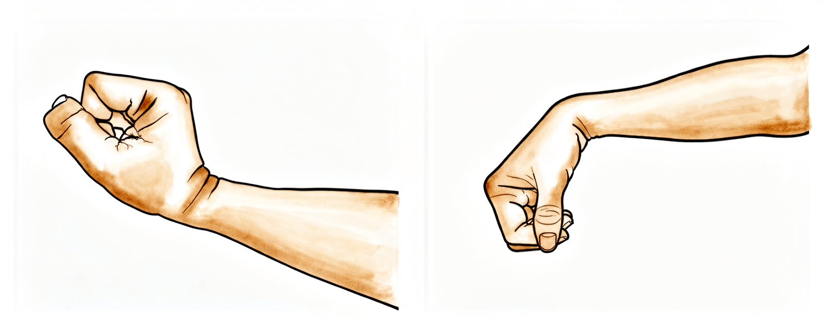
腕关节屈曲/伸展拉伸
将肘部支撑在桌面上,轻轻前后摆动手腕(或如图所示,将手腕悬空置于桌边或扶手椅边缘)。待感觉较为舒适后,用另一只手按压手掌,将手腕向后推,然后向前推,保持手指放松。每个方向保持拉伸姿势15秒;每个方向重复5次。
10次,每日4–5次
Kieran Hirpara 4.0
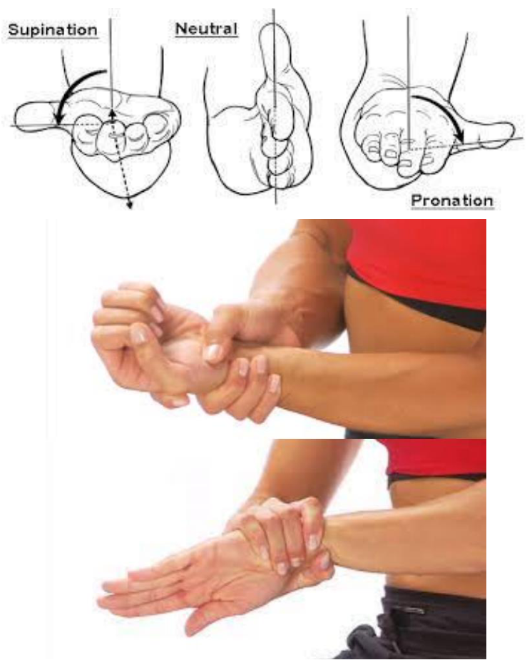
腕部旋后/旋前拉伸
将肘部置于身体一侧弯曲,掌心朝向天花板。将手旋至掌心向上,保持15秒;然后旋至掌心向下,保持15秒;重复5次。随着舒适度增加,用对侧手——握住您的手腕,而非手掌——轻轻将旋转进一步推向每个位置,直至感到拉伸感。
5 次重复,每天 4 次,每周 5–7 天
Kieran Hirpara 4.0
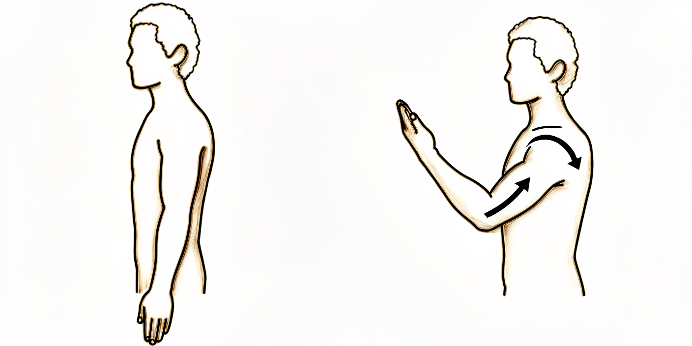
肘关节屈曲/伸展
手臂置于体侧,尽可能轻柔地伸直前臂和肘部。保持此极限拉伸姿势3–5秒。反向重复该动作,向上弯曲前臂,尝试用手触碰肩部。
每日2次,每次10次
Kieran Hirpara 4.0
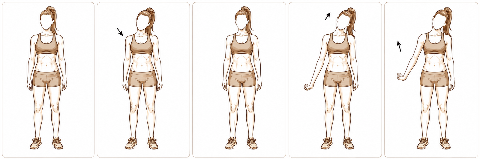
桡神经滑动
双臂自然下垂站立。将肩部下沉,手指向地面伸展。将手臂内旋(拇指朝向身体),并屈曲手腕,掌心向上。轻轻将头部向远离拉伸侧的方向倾斜,然后将手臂向上并远离身体抬起。每个滑动姿势保持3至5秒。
5–8 次,每天 2–4 次,每周 6–7 天
本锻炼方案由职业治疗学士(BOccThy)、认证手治疗师(AHT)Sarah Farrell 共同编写。
术后方案
本方案与诊所的通用康复建议配合使用;请参阅术后疼痛管理、伤口护理和手部治疗基础。关于手术本身及其治疗的病症,请参阅桡骨管松解术和桡骨管综合征。
参考文献
[1] Wright TW, Glowczewskie F, Cowin D, Wheeler DL. 上肢运动时肘部和腕部桡神经的位移和应变. J Hand Surg Am. 2005;30(5):990–996. https://pubmed.ncbi.nlm.nih.gov/16182056/ [2] Lee JT, Azari K, Jones NF. 桡管松解术的长期疗效——合并网球肘、多重压迫综合征及工伤赔偿的影响. J Plast Reconstr Aesthet Surg. 2008;61(9):1095–1099. https://www.sciencedirect.com/science/article/abs/pii/S1748681507004044 [3] Sotereanos DG, Varitimidis SE, Giannakopoulos PN, Westkaemper JG. 桡管综合征手术治疗的结果. J Hand Surg Am. 1999;24(3):566–570. https://pubmed.ncbi.nlm.nih.gov/10357537/ [4] Naam NH, Nemani S. 桡管综合征. Orthop Clin North Am. 2012;43(4):529–536. (桡管综合征,StatPearls.) https://www.ncbi.nlm.nih.gov/books/NBK555937/ [5] Coppieters MW, Butler DS. “滑动者”滑动,“张紧者”张紧吗?神经动力学技术及其应用的分析和考量. Man Ther. 2008;13(3):213–221. https://pubmed.ncbi.nlm.nih.gov/17398140/ [6] Basson A, Olivier B, Ellis R, Coppieters M, Stewart A, Mudzi W. 神经松动术治疗神经肌肉骨骼疾病的有效性:系统评价和荟萃分析. J Orthop Sports Phys Ther. 2017;47(9):593–615. https://pubmed.ncbi.nlm.nih.gov/28704626/
Evidence & references
This is the clinical evidence summary written for health professionals. It is technical, and it lists the research this page was built from. You do not need to read it to understand your treatment or to make a decision about it.
Topic scope: post-operative rehabilitation after surgical decompression / neurolysis of the posterior interosseous nerve (deep branch of the radial nerve) in the radial tunnel of the proximal forearm, performed for radial tunnel syndrome (RTS). This is an elbow / proximal-forearm topic — anatomically and clinically distinct from carpal-tunnel and cubital-tunnel decompression. Like other nerve decompressions it is an early-motion pathway (early elbow/forearm/wrist motion, radial-nerve glides, oedema and scar care). The scope deliberately foregrounds the diagnostic controversy and the more variable, lower success rates that distinguish RTS release from the better-validated carpal-tunnel and cubital-tunnel operations.
Defining principle of the rehab here: a decompressed nerve does not create a healing construct that needs months of protection — it needs early, gentle movement to stop it adhering to the operative bed and to restore its glide. So the rehab is an early-motion programme: light functional hand use from day 1, radial-nerve sliders, graded desensitisation and (once healed) scar massage; heavier loading deferred to ~6 weeks. But two honesty caveats sit over the whole topic. First, RTS is a contested diagnosis — there is no confirmatory imaging or electrodiagnostic test, it is a diagnosis of exclusion, and a substantial body of opinion regards it as a variant of recalcitrant lateral epicondylitis. Second, outcomes after release are more variable and on average lower than carpal- or cubital-tunnel release — good results cluster around two-thirds overall, and fall further with co-existing tennis elbow, multiple compression sites, or a workers'-compensation context. Patient expectations should be set accordingly.
A. THE DIAGNOSTIC CONTROVERSY (read first — it frames everything)
RTS is among the most contested entities in upper-limb surgery, and the rehab brief is incomplete without it:
- No confirmatory test. Electromyography and nerve-conduction studies are characteristically normal in RTS (compression is intermittent/dynamic and predominantly of a motor nerve carrying few pain fibres), and MRI is frequently negative — denervation oedema in supinator/extensors is suggestive but inconsistent, and a normal scan does not exclude the diagnosis. RTS is therefore a clinical diagnosis of exclusion, resting on point tenderness ~4 cm distal to the lateral epicondyle (over the radial tunnel rather than the epicondyle), pain on resisted supination / resisted long-finger extension, and — for some surgeons — temporary relief from a diagnostic local-anaesthetic block at the radial tunnel.
- Overlap with lateral epicondylitis (tennis elbow). The two coexist frequently and share the lateral-elbow pain territory. A recognised school of thought holds that "RTS" is often severe, recalcitrant lateral epicondylitis rather than a discrete compression neuropathy. Importantly, routine PIN release added to lateral-epicondylitis surgery has not been shown to improve outcomes, so the diagnosis should be secure before a decompression is planned.
- Practical consequence. Surgery is a last resort after prolonged failed conservative care (activity modification, splinting, anti-inflammatories, sometimes a steroid injection), and is best reserved for patients with proximal-forearm pain and no better explanation. This uncertainty is the single most important reason post-operative expectations must be framed honestly.
B. RELEASE OUTCOMES (variable — and why)
- Headline success ~two-thirds. Across the older long-term series, roughly 67% good, 15% fair, 18% poor after radial tunnel decompression — markedly more variable than carpal- or cubital-tunnel release. A 2008 long-term series (Lee, Azari, Jones) and a 1999 series (Sotereanos et al.) both document this spread; the Sotereanos cohort reported good/excellent results in only ~39% by objective assessment (though ~64% by patient self-rating), underscoring how outcome depends on the metric used.
- Co-existing lateral epicondylitis lowers success. Success falls to roughly 40% when tennis elbow coexists, versus far higher with isolated RTS.
- Multiple compression sites and workers'-compensation context lower success — reported ~58% success in compensation cases vs ~73% without. These are the same modifiers named in the patient protocol.
- 2025 systematic review (Raymond et al., HAND). 11 studies, 401 limbs (381 patients). Outcomes were heterogeneous; a dorsal approach between ECRB and EDC was associated with the most favourable Roles-and-Maudsley scores and satisfaction. The review's central message is that the overall evidence is low-grade (observational), the diagnosis non-standardised, and the effectiveness of conservative treatment essentially untested — a "tendency" toward benefit rather than proof.
- Resorption-style "spontaneous improvement" does not apply here — unlike calcific tendinitis, RTS does not self-resolve through a biological cycle; conservative care manages symptoms rather than curing a deposit.
C. SURGICAL APPROACH (shapes the early rehab)
- What is done. Complete neurolysis of the radial nerve at its bifurcation, decompressing the deep branch (PIN) and superficial sensory branch, releasing the arcade of Frohse (the proximal supinator edge), the leash of Henry (radial recurrent vessels), the ECRB fascial edge, and the distal supinator border. Any constrictive bands or vessels are divided.
- Approaches. Dorsal (Thompson, between ECRB/EDC or the brachioradialis–ECRL interval), volar/anterior (Henry), or transmuscular. Anatomical studies map the trade-offs; the dorsal ECRB–EDC interval performed best in the 2025 review. The superficial radial branch matters — it is a recognised source of post-operative dysaesthesia if irritated.
- Rehab implication. A muscle-splitting/dorsal exposure through the extensor mass means early gentle forearm rotation and wrist motion are encouraged but heavy resisted supination/extension is deferred; the incision sits over a mobile, frequently sensitive area, so desensitisation and scar care carry real weight here.
D. POST-OP THERAPY ROLE (nerve/tendon glides, oedema, scar)
The decompressed nerve must glide, not adhere. The mechanical rationale is well quantified: the radial nerve translates and stretches a measurable amount across the elbow and wrist during ordinary arm motion (Wright et al. 2005), so early motion is what keeps it free of the healing bed.
- Early motion, immediately. Early active elbow, forearm and wrist movement within pain limits from the first post-op days; most protocols use no rigid splinting (or a removable splint for comfort/night only).
- Radial-nerve glides — favour "sliders" over "tensioners". Sliding (slider) neurodynamic techniques achieve substantially greater nerve excursion at much lower nerve strain than end-range tensioners — preferable around a freshly decompressed nerve. Neural-mobilisation evidence across neuromusculoskeletal conditions is supportive but of variable certainty, so progression is symptom-guided and essentially pain-free; mechanism work (e.g., the MONET protocol) is still maturing.
- Oedema and desensitisation. Graded desensitisation (tapping/rubbing over the dressing) from day 1 normalises touch and pre-empts a sensitive scar — particularly relevant given superficial- radial-branch proximity.
- Scar management once healed. Massage, pressure, and silicone are advocated to loosen skin–tissue adhesions and aid remodelling, started once the wound is closed/sutures out.
- Strengthening deferred. Light functional ADL use throughout; resisted strengthening of wrist/ elbow and fine-motor work introduced from ~6 weeks. Heavy work and vibration tools avoided to ~6–8 weeks.
Phased post-op timeline (maps to the patient protocol phases)
| Phase | Window | Splint | Motion / nerve work | Load / strengthening | Notes |
|---|---|---|---|---|---|
| I — Protect & glide | Day 0–2 wk | None, or removable for comfort/night | Early pain-free active elbow/forearm/wrist ROM; radial-nerve sliders; desensitisation from day 1 | Light functional ADL use only (self-care, feeding, dressing, writing, typing) | Stop the nerve adhering; settle the wound. No lifting/gripping/weight-bearing/vibration tools. Driving limited first 1–2 wk |
| II — Restore motion | 2–6 wk | Off | Progress full active + gentle assisted ROM; continue sliders; scar massage once healed | Still no resisted loading; ADL use continues | Sensitivity/dysaesthesia common and usually settles; keep glides gentle |
| III — Strengthen & return | ~6 wk onward | Off | Full ROM goal; sliders as needed | Begin graded wrist/elbow strengthening + fine-motor work from ~6 wk; advance work/heavy tasks thereafter | Vibration tools/heavy work resume ~6–8 wk. Pain relief is often gradual and may be partial — counsel accordingly |
E. COMPLICATIONS / DOWNSIDES
- Incomplete or no pain relief — the dominant "complication," tied directly to diagnostic uncertainty; relief is frequently gradual and sometimes partial.
- Superficial-radial-branch dysaesthesia / scar sensitivity — recognised; desensitisation and careful technique mitigate it.
- Transient PIN weakness (finger/thumb extension) from retraction — usually recovers.
- Adhesion/recurrence of symptoms if early glide is neglected.
- Standard wound risks (infection, haematoma) — uncommon.
F. KEY CONTROVERSIES / EVIDENCE QUALITY
- Does RTS exist as a discrete entity? Genuinely contested. No confirmatory test; substantial opinion equates much of it with recalcitrant lateral epicondylitis. This is the defining controversy and must shape consent and expectation-setting. Unresolved — expert opinion divided.
- Patient selection drives outcome more than technique. Isolated RTS does best; coexisting tennis elbow, multiple compressions, and compensation context predict worse results. Moderate (consistent across cohorts).
- Approach choice. A dorsal ECRB–EDC interval was favoured in the 2025 SR, but the evidence is observational and confounded by diagnostic heterogeneity. Weak–moderate.
- The rehab protocol itself is consensus/expert — drawn from surgeon and hand-therapy guidance (early motion, sliders, desensitisation, scar care), not from a rehab RCT. Phase timings are typical, not trial-derived. Weak / consensus.
- Conservative-treatment efficacy is essentially untested — the 2025 SR notes no usable trials of non-operative care, so "failed conservative management" before surgery rests on practice convention. Weak.
G. EVIDENCE STRENGTH FLAGS (summary)
- STRONG: the mechanical rationale for early nerve glide — quantified radial-nerve excursion/ strain across elbow and wrist (Wright et al. 2005); slider-vs-tensioner excursion/strain physiology.
- MODERATE: patient-selection modifiers of outcome (lateral epicondylitis, multiple compressions, workers' compensation lower success); ~two-thirds overall good-result rate from long-term cohorts; dorsal-approach signal from the 2025 systematic review (low-grade studies).
- WEAK / CONSENSUS: the existence and diagnostic criteria of RTS (no confirmatory test; overlap with lateral epicondylitis); the post-operative rehabilitation protocol (surgeon/ hand-therapy guidance, no rehab RCT); neural-mobilisation certainty (supportive but variable); efficacy of conservative care (essentially untested).
CITATIONS
RAG corpus (180,000+ Orthopaedic articles)
- Posterior Interosseous Nerve Compression in the Forearm, AKA Radial Tunnel Syndrome. HAND. 2022. DOI: 10.1177/15589447221122822
- Radial Tunnel Syndrome: Emphasis on the Superficial Branch of the Radial Nerve. J Hand Surg Eur. 2009. DOI: 10.1177/1753193408099832
- Anatomical Study of the Surgical Approaches to the Radial Tunnel. J Hand Surg Am. 2015. DOI: 10.1016/j.jhsa.2015.03.009
- MR Imaging Features of Radial Tunnel Syndrome: Initial Experience. Radiology. 2006. DOI: 10.1148/radiol.2401050028
- Management of Lateral Epicondylitis: Current Concepts. J Am Acad Orthop Surg (JAAOS). 2008. DOI: 10.5435/00124635-200801000-00004
- Uncommon Nerve Compression Syndromes of the Upper Extremity. J Am Acad Orthop Surg (JAAOS). 1998. DOI: 10.5435/00124635-199811000-00006
- Radial Nerve Excursion and Strain at the Elbow and Wrist Associated With Upper-Extremity Motion. J Hand Surg Am. 2005. DOI: 10.1016/j.jhsa.2005.06.008
- Evidence and Techniques in Rehabilitation Following Nerve Injuries. Hand Clin. 2013. DOI: 10.1016/j.hcl.2013.04.012
- Preventive Strategies, Exercises and Rehabilitation of Hand Compression Neuropathies. J Hand Ther. 2022. DOI: 10.1016/j.jht.2021.11.003
- Mechanisms of Neurodynamic Treatments (MONET): a protocol for a mechanistic study. BMC Musculoskelet Disord. 2024. DOI: 10.1186/s12891-024-07713-6
Radial-tunnel literature (URLs)
- Clinical Outcomes of Operative Management for Radial Tunnel Syndrome According to Surgical Approach: a Systematic Review. HAND. 2025. https://journals.sagepub.com/doi/10.1177/15589447251315761
- The Epidemiology of Radial Tunnel Syndrome and Its Overlap With Lateral Epicondylitis. J Hand Surg Am. 2023. https://www.jhandsurg.org/article/S0363-5023(23)00138-7/abstract
- Lee JT, Azari K, Jones NF. Long-term results of radial tunnel release — the effect of co-existing tennis elbow, multiple compression syndromes and workers' compensation. J Plast Reconstr Aesthet Surg. 2008. https://www.sciencedirect.com/science/article/abs/pii/S1748681507004044
- Sotereanos DG, et al. Results of surgical treatment for radial tunnel syndrome. J Hand Surg Am. 1999. https://pubmed.ncbi.nlm.nih.gov/10357537/
- Interventions for treating the radial tunnel syndrome: a systematic review of observational studies (DARE). https://www.ncbi.nlm.nih.gov/books/NBK75403/
- Radial Tunnel Syndrome (StatPearls). https://www.ncbi.nlm.nih.gov/books/NBK555937/
- Orthopedic Management of Radial Tunnel Syndrome: A Diagnostic and Treatment Dilemma. PMC. https://pmc.ncbi.nlm.nih.gov/articles/PMC10081130/
- Radial Tunnel Syndrome: Case Report and Comprehensive Critical Review of a Compression Neuropathy Surrounded by Controversy. PMC. https://pmc.ncbi.nlm.nih.gov/articles/PMC9896270/
Published rehab protocols (patient-guidance — basis for the phase structure)
- Radial Tunnel Release post-op protocol (Santa Barbara Orthopedic / Mencias). https://www.sbortho.com/wp-content/uploads/2023/09/radial-tunnel-release-new.pdf
- Radial Tunnel Syndrome — conservative and post-operative rehabilitation. Physiopedia. https://www.physio-pedia.com/Radial_Tunnel_Syndrome
- Basson A, et al. The effectiveness of neural mobilization for neuromusculoskeletal conditions: a systematic review and meta-analysis. J Orthop Sports Phys Ther. 2017. https://pubmed.ncbi.nlm.nih.gov/28704626/
- Coppieters MW, Butler DS. Do "sliders" slide and "tensioners" tension? Man Ther. 2008. https://pubmed.ncbi.nlm.nih.gov/17398140/





