Radial Tunnel Release Info Evidence
Last reviewed
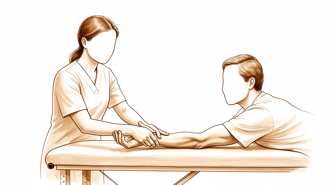
Post-operative exercises and precautions after radial tunnel release, including radial nerve glides.
This protocol guides your recovery after radial tunnel release with Dr Kieran Hirpara at Mater Private Hospital Rockhampton. It explains what to expect in the weeks after surgery and sets out the exercise program from your post-operative handout — bring this page or its PDF to your first physiotherapy or hand therapy visit so your rehabilitation stays coordinated. Your therapist may adjust the plan depending on how your recovery progresses.
If you have any concerns about your wound after surgery, get in touch with the rooms. It is often helpful to take a photo of the wound and email it for review.
What to expect
Care of your wound is explained in the practice's wound care advice. As your wound heals, the released nerve can stick to the surrounding tissue — the gliding exercises below are very important to keep it moving freely and stop it from becoming tethered.
Sometimes the wound can become sensitive. This is normal, and can be prevented or minimised by commencing daily desensitisation: gently tapping and rubbing over the wound (or the dressing), starting immediately following your surgery. This type of "sensory feedback" allows the nerve to normalise touch and texture.
Once the wound is fully healed, commence scar massage — firm circles over the incision. Refer to the wound care advice for more information on scar management.
It is important to have realistic expectations of recovery. The radial nerve has to travel and stretch a measurable amount during ordinary arm movements, so keeping it moving early is what stops it adhering to the healing tissues [1]. Even so, the relief of pain after radial tunnel release is often gradual rather than immediate, and for some people it is only partial. This operation is, honestly, less predictable than some other nerve-release procedures: radial tunnel syndrome can be hard to diagnose with certainty and it often overlaps with tennis elbow, which is part of why results vary. Published long-term studies report good outcomes in around two-thirds of patients overall, with the best results in those who have radial tunnel symptoms alone [2][3]. Recovery tends to be slower and less complete when there is also tennis elbow (lateral epicondylitis), more than one nerve compression in the same arm, or a workers' compensation claim [2][4]. Your nerve-gliding program and graded desensitisation are the parts of rehabilitation most within your control, and steady daily practice gives the nerve its best chance to settle.
Precautions and limitations
Light functional use of your hand is encouraged for daily living tasks such as self-care, feeding, dressing, writing and typing. Beyond that, the limits are simple: no lifting, gripping, weight-bearing or use of vibration machinery (for example, power tools or a lawn mower) for up to 6 weeks after surgery, and driving is limited for the first 1–2 weeks.
For your physiotherapist:
Goals
- Prevent the released nerve adhering to the healing wound (nerve gliding program)
- Settle wound sensitivity through graded desensitisation
- Maintain wrist, forearm and elbow range of motion
- Support light functional use of the hand for activities of daily living
Management
- Daily desensitisation — gentle tapping / rubbing over the wound (dressing) — commencing immediately post-operatively
- Scar massage (firm circles over the incision) once the wound is fully healed
- Home exercise program as per the cards below: wrist flexion / extension stretch; wrist supination / pronation stretch; elbow flexion / extension; radial nerve glides
- Favour gentle sliding-type ("slider") nerve glides over aggressive end-range tensioning: sliding techniques achieve substantially greater nerve excursion at much lower nerve strain, which is better tolerated around a recently decompressed nerve [1][5]
- Nerve mobilisation may be considered as an adjunct to the program; the evidence base for neural mobilisation in nerve-related conditions is supportive but of variable certainty, so progression should be symptom-guided [6]
Precautions
- Light functional use of the hand only (self-care, feeding, dressing, writing, typing)
- No lifting, gripping, weight-bearing or use of vibration machinery (e.g. power tools, lawn mower) for up to 6 weeks post-op
- Driving is limited for the first 1–2 weeks
- Nerve glides and stretches should be gentle and essentially pain-free — avoid forcing into a range that reproduces the pre-operative nerve pain
These are the exercises from your post-operative handout, started after surgery and continued at home as guided by your physiotherapist or hand therapist. The repetitions, hold times and frequency are listed on each card.
Your exercises
Kieran Hirpara 4.0
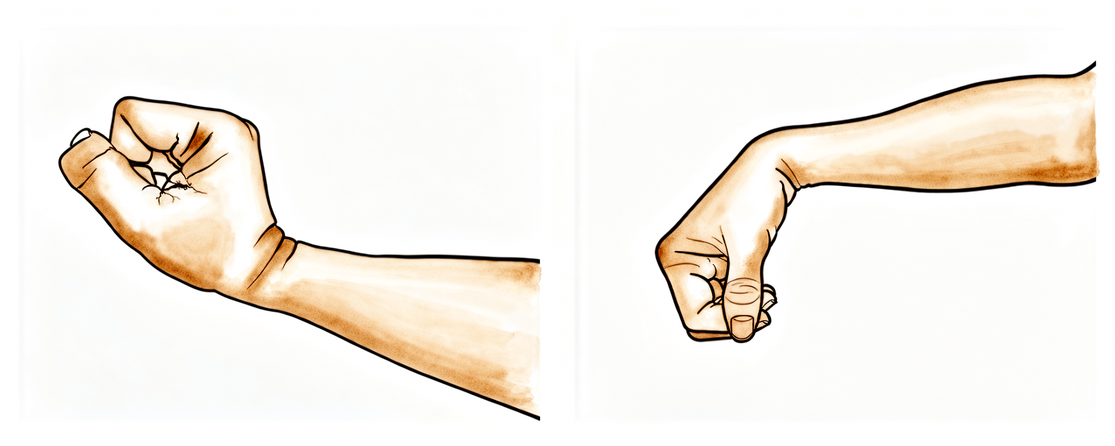
Wrist flexion / extension stretch
Rest your elbow on a table and gently rock your wrist back and forth (or rest it over the edge of a table or armchair, as pictured). Once more comfortable, use your other hand on your palm to push the wrist backwards, then forwards, keeping your fingers loose. Hold each stretch for 15 seconds; repeat 5 times in each direction.
10 repetitions, 4–5 times daily
Kieran Hirpara 4.0
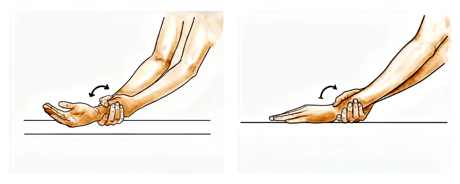
Wrist supination / pronation stretch
Bend your elbow at the side of your body, palm facing the ceiling. Turn your hand palm up and hold for 15 seconds, then palm down and hold for 15 seconds; repeat 5 times. As comfort increases, use your opposite hand — holding at your WRIST, not your hand — to gently push the rotation further into each position until you feel a stretch.
5 repetitions, 4 times a day, 5–7 days per week
Kieran Hirpara 4.0
Elbow flexion / extension
With your arm by your side, gently straighten your forearm and elbow as far as you can. Hold this end-range stretch for 3–5 seconds. Repeat in the opposite direction, bending your forearm up and attempting to touch your shoulder with your hand.
10 repetitions, 2 times a day, daily
Kieran Hirpara 4.0
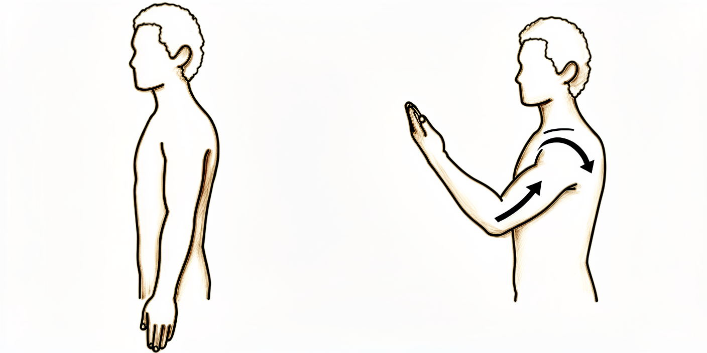
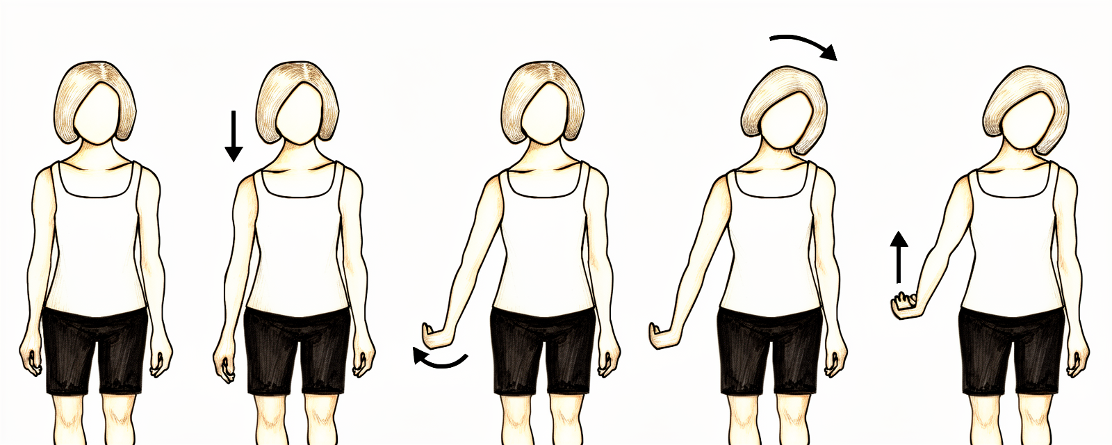
Radial nerve glides
Stand with your arms loose at your sides. Drop your shoulder down and reach your fingers toward the floor. Internally rotate your arm (thumb toward your body) and flex your wrist, palm up. Gently tilt your head away from the side you are stretching, then raise your arm up and away from your body. Hold each position of the glide for 3 to 5 seconds.
5–8 repetitions, 2–4 times a day, 6–7 days per week
This exercise program was written in association with Sarah Farrell, Bachelor of Occupational Therapy (BOccThy), Accredited Hand Therapist (AHT).
After your protocol
This protocol works alongside the practice's general recovery advice — see managing post-operative pain, wound care and hand therapy basics. For the operation itself and the condition it treats, see radial tunnel release and radial tunnel syndrome.
References
- Wright TW, Glowczewskie F, Cowin D, Wheeler DL. Radial nerve excursion and strain at the elbow and wrist associated with upper-extremity motion. J Hand Surg Am. 2005;30(5):990–996.
- Lee JT, Azari K, Jones NF. Long term results of radial tunnel release — the effect of co-existing tennis elbow, multiple compression syndromes and workers' compensation. J Plast Reconstr Aesthet Surg. 2008;61(9):1095–1099.
- Sotereanos DG, Varitimidis SE, Giannakopoulos PN, Westkaemper JG. Results of surgical treatment for radial tunnel syndrome. J Hand Surg Am. 1999;24(3):566–570.
- Naam NH, Nemani S. Radial tunnel syndrome. Orthop Clin North Am. 2012;43(4):529–536. (Radial Tunnel Syndrome, StatPearls.)
- Coppieters MW, Butler DS. Do "sliders" slide and "tensioners" tension? An analysis of neurodynamic techniques and considerations regarding their application. Man Ther. 2008;13(3):213–221.
- Basson A, Olivier B, Ellis R, Coppieters M, Stewart A, Mudzi W. The effectiveness of neural mobilization for neuromusculoskeletal conditions: a systematic review and meta-analysis. J Orthop Sports Phys Ther. 2017;47(9):593–615.
Evidence & references
Radial Tunnel Release — Evidence Brief & Post-operative Rehabilitation
Topic scope: post-operative rehabilitation after surgical decompression / neurolysis of the posterior interosseous nerve (deep branch of the radial nerve) in the radial tunnel of the proximal forearm, performed for radial tunnel syndrome (RTS). This is an elbow / proximal-forearm topic — anatomically and clinically distinct from carpal-tunnel and cubital-tunnel decompression. Like other nerve decompressions it is an early-motion pathway (early elbow/forearm/wrist motion, radial-nerve glides, oedema and scar care). The scope deliberately foregrounds the diagnostic controversy and the more variable, lower success rates that distinguish RTS release from the better-validated carpal-tunnel and cubital-tunnel operations.
Defining principle of the rehab here: a decompressed nerve does not create a healing construct that needs months of protection — it needs early, gentle movement to stop it adhering to the operative bed and to restore its glide. So the rehab is an early-motion programme: light functional hand use from day 1, radial-nerve sliders, graded desensitisation and (once healed) scar massage; heavier loading deferred to ~6 weeks. But two honesty caveats sit over the whole topic. First, RTS is a contested diagnosis — there is no confirmatory imaging or electrodiagnostic test, it is a diagnosis of exclusion, and a substantial body of opinion regards it as a variant of recalcitrant lateral epicondylitis. Second, outcomes after release are more variable and on average lower than carpal- or cubital-tunnel release — good results cluster around two-thirds overall, and fall further with co-existing tennis elbow, multiple compression sites, or a workers'-compensation context. Patient expectations should be set accordingly.
A. THE DIAGNOSTIC CONTROVERSY (read first — it frames everything)
RTS is among the most contested entities in upper-limb surgery, and the rehab brief is incomplete without it:
- No confirmatory test. Electromyography and nerve-conduction studies are characteristically normal in RTS (compression is intermittent/dynamic and predominantly of a motor nerve carrying few pain fibres), and MRI is frequently negative — denervation oedema in supinator/extensors is suggestive but inconsistent, and a normal scan does not exclude the diagnosis. RTS is therefore a clinical diagnosis of exclusion, resting on point tenderness ~4 cm distal to the lateral epicondyle (over the radial tunnel rather than the epicondyle), pain on resisted supination / resisted long-finger extension, and — for some surgeons — temporary relief from a diagnostic local-anaesthetic block at the radial tunnel.
- Overlap with lateral epicondylitis (tennis elbow). The two coexist frequently and share the lateral-elbow pain territory. A recognised school of thought holds that "RTS" is often severe, recalcitrant lateral epicondylitis rather than a discrete compression neuropathy. Importantly, routine PIN release added to lateral-epicondylitis surgery has not been shown to improve outcomes, so the diagnosis should be secure before a decompression is planned.
- Practical consequence. Surgery is a last resort after prolonged failed conservative care (activity modification, splinting, anti-inflammatories, sometimes a steroid injection), and is best reserved for patients with proximal-forearm pain and no better explanation. This uncertainty is the single most important reason post-operative expectations must be framed honestly.
B. RELEASE OUTCOMES (variable — and why)
- Headline success ~two-thirds. Across the older long-term series, roughly 67% good, 15% fair, 18% poor after radial tunnel decompression — markedly more variable than carpal- or cubital-tunnel release. A 2008 long-term series (Lee, Azari, Jones) and a 1999 series (Sotereanos et al.) both document this spread; the Sotereanos cohort reported good/excellent results in only ~39% by objective assessment (though ~64% by patient self-rating), underscoring how outcome depends on the metric used.
- Co-existing lateral epicondylitis lowers success. Success falls to roughly 40% when tennis elbow coexists, versus far higher with isolated RTS.
- Multiple compression sites and workers'-compensation context lower success — reported ~58% success in compensation cases vs ~73% without. These are the same modifiers named in the patient protocol.
- 2025 systematic review (Raymond et al., HAND). 11 studies, 401 limbs (381 patients). Outcomes were heterogeneous; a dorsal approach between ECRB and EDC was associated with the most favourable Roles-and-Maudsley scores and satisfaction. The review's central message is that the overall evidence is low-grade (observational), the diagnosis non-standardised, and the effectiveness of conservative treatment essentially untested — a "tendency" toward benefit rather than proof.
- Resorption-style "spontaneous improvement" does not apply here — unlike calcific tendinitis, RTS does not self-resolve through a biological cycle; conservative care manages symptoms rather than curing a deposit.
C. SURGICAL APPROACH (shapes the early rehab)
- What is done. Complete neurolysis of the radial nerve at its bifurcation, decompressing the deep branch (PIN) and superficial sensory branch, releasing the arcade of Frohse (the proximal supinator edge), the leash of Henry (radial recurrent vessels), the ECRB fascial edge, and the distal supinator border. Any constrictive bands or vessels are divided.
- Approaches. Dorsal (Thompson, between ECRB/EDC or the brachioradialis–ECRL interval), volar/anterior (Henry), or transmuscular. Anatomical studies map the trade-offs; the dorsal ECRB–EDC interval performed best in the 2025 review. The superficial radial branch matters — it is a recognised source of post-operative dysaesthesia if irritated.
- Rehab implication. A muscle-splitting/dorsal exposure through the extensor mass means early gentle forearm rotation and wrist motion are encouraged but heavy resisted supination/extension is deferred; the incision sits over a mobile, frequently sensitive area, so desensitisation and scar care carry real weight here.
D. POST-OP THERAPY ROLE (nerve/tendon glides, oedema, scar)
The decompressed nerve must glide, not adhere. The mechanical rationale is well quantified: the radial nerve translates and stretches a measurable amount across the elbow and wrist during ordinary arm motion (Wright et al. 2005), so early motion is what keeps it free of the healing bed.
- Early motion, immediately. Early active elbow, forearm and wrist movement within pain limits from the first post-op days; most protocols use no rigid splinting (or a removable splint for comfort/night only).
- Radial-nerve glides — favour "sliders" over "tensioners". Sliding (slider) neurodynamic techniques achieve substantially greater nerve excursion at much lower nerve strain than end-range tensioners — preferable around a freshly decompressed nerve. Neural-mobilisation evidence across neuromusculoskeletal conditions is supportive but of variable certainty, so progression is symptom-guided and essentially pain-free; mechanism work (e.g., the MONET protocol) is still maturing.
- Oedema and desensitisation. Graded desensitisation (tapping/rubbing over the dressing) from day 1 normalises touch and pre-empts a sensitive scar — particularly relevant given superficial- radial-branch proximity.
- Scar management once healed. Massage, pressure, and silicone are advocated to loosen skin–tissue adhesions and aid remodelling, started once the wound is closed/sutures out.
- Strengthening deferred. Light functional ADL use throughout; resisted strengthening of wrist/ elbow and fine-motor work introduced from ~6 weeks. Heavy work and vibration tools avoided to ~6–8 weeks.
Phased post-op timeline (maps to the patient protocol phases)
| Phase | Window | Splint | Motion / nerve work | Load / strengthening | Notes |
|---|---|---|---|---|---|
| I — Protect & glide | Day 0–2 wk | None, or removable for comfort/night | Early pain-free active elbow/forearm/wrist ROM; radial-nerve sliders; desensitisation from day 1 | Light functional ADL use only (self-care, feeding, dressing, writing, typing) | Stop the nerve adhering; settle the wound. No lifting/gripping/weight-bearing/vibration tools. Driving limited first 1–2 wk |
| II — Restore motion | 2–6 wk | Off | Progress full active + gentle assisted ROM; continue sliders; scar massage once healed | Still no resisted loading; ADL use continues | Sensitivity/dysaesthesia common and usually settles; keep glides gentle |
| III — Strengthen & return | ~6 wk onward | Off | Full ROM goal; sliders as needed | Begin graded wrist/elbow strengthening + fine-motor work from ~6 wk; advance work/heavy tasks thereafter | Vibration tools/heavy work resume ~6–8 wk. Pain relief is often gradual and may be partial — counsel accordingly |
E. COMPLICATIONS / DOWNSIDES
- Incomplete or no pain relief — the dominant "complication," tied directly to diagnostic uncertainty; relief is frequently gradual and sometimes partial.
- Superficial-radial-branch dysaesthesia / scar sensitivity — recognised; desensitisation and careful technique mitigate it.
- Transient PIN weakness (finger/thumb extension) from retraction — usually recovers.
- Adhesion/recurrence of symptoms if early glide is neglected.
- Standard wound risks (infection, haematoma) — uncommon.
F. KEY CONTROVERSIES / EVIDENCE QUALITY
- Does RTS exist as a discrete entity? Genuinely contested. No confirmatory test; substantial opinion equates much of it with recalcitrant lateral epicondylitis. This is the defining controversy and must shape consent and expectation-setting. Unresolved — expert opinion divided.
- Patient selection drives outcome more than technique. Isolated RTS does best; coexisting tennis elbow, multiple compressions, and compensation context predict worse results. Moderate (consistent across cohorts).
- Approach choice. A dorsal ECRB–EDC interval was favoured in the 2025 SR, but the evidence is observational and confounded by diagnostic heterogeneity. Weak–moderate.
- The rehab protocol itself is consensus/expert — drawn from surgeon and hand-therapy guidance (early motion, sliders, desensitisation, scar care), not from a rehab RCT. Phase timings are typical, not trial-derived. Weak / consensus.
- Conservative-treatment efficacy is essentially untested — the 2025 SR notes no usable trials of non-operative care, so "failed conservative management" before surgery rests on practice convention. Weak.
G. EVIDENCE STRENGTH FLAGS (summary)
- STRONG: the mechanical rationale for early nerve glide — quantified radial-nerve excursion/ strain across elbow and wrist (Wright et al. 2005); slider-vs-tensioner excursion/strain physiology.
- MODERATE: patient-selection modifiers of outcome (lateral epicondylitis, multiple compressions, workers' compensation lower success); ~two-thirds overall good-result rate from long-term cohorts; dorsal-approach signal from the 2025 systematic review (low-grade studies).
- WEAK / CONSENSUS: the existence and diagnostic criteria of RTS (no confirmatory test; overlap with lateral epicondylitis); the post-operative rehabilitation protocol (surgeon/ hand-therapy guidance, no rehab RCT); neural-mobilisation certainty (supportive but variable); efficacy of conservative care (essentially untested).
CITATIONS
RAG corpus (180,000+ Orthopaedic articles)
- Posterior Interosseous Nerve Compression in the Forearm, AKA Radial Tunnel Syndrome. HAND. 2022. DOI: 10.1177/15589447221122822
- Radial Tunnel Syndrome: Emphasis on the Superficial Branch of the Radial Nerve. J Hand Surg Eur. 2009. DOI: 10.1177/1753193408099832
- Anatomical Study of the Surgical Approaches to the Radial Tunnel. J Hand Surg Am. 2015. DOI: 10.1016/j.jhsa.2015.03.009
- MR Imaging Features of Radial Tunnel Syndrome: Initial Experience. Radiology. 2006. DOI: 10.1148/radiol.2401050028
- Management of Lateral Epicondylitis: Current Concepts. J Am Acad Orthop Surg (JAAOS). 2008. DOI: 10.5435/00124635-200801000-00004
- Uncommon Nerve Compression Syndromes of the Upper Extremity. J Am Acad Orthop Surg (JAAOS). 1998. DOI: 10.5435/00124635-199811000-00006
- Radial Nerve Excursion and Strain at the Elbow and Wrist Associated With Upper-Extremity Motion. J Hand Surg Am. 2005. DOI: 10.1016/j.jhsa.2005.06.008
- Evidence and Techniques in Rehabilitation Following Nerve Injuries. Hand Clin. 2013. DOI: 10.1016/j.hcl.2013.04.012
- Preventive Strategies, Exercises and Rehabilitation of Hand Compression Neuropathies. J Hand Ther. 2022. DOI: 10.1016/j.jht.2021.11.003
- Mechanisms of Neurodynamic Treatments (MONET): a protocol for a mechanistic study. BMC Musculoskelet Disord. 2024. DOI: 10.1186/s12891-024-07713-6
Radial-tunnel literature (URLs)
- Clinical Outcomes of Operative Management for Radial Tunnel Syndrome According to Surgical Approach: a Systematic Review. HAND. 2025. https://journals.sagepub.com/doi/10.1177/15589447251315761
- The Epidemiology of Radial Tunnel Syndrome and Its Overlap With Lateral Epicondylitis. J Hand Surg Am. 2023. https://www.jhandsurg.org/article/S0363-5023(23)00138-7/abstract
- Lee JT, Azari K, Jones NF. Long-term results of radial tunnel release — the effect of co-existing tennis elbow, multiple compression syndromes and workers' compensation. J Plast Reconstr Aesthet Surg. 2008. https://www.sciencedirect.com/science/article/abs/pii/S1748681507004044
- Sotereanos DG, et al. Results of surgical treatment for radial tunnel syndrome. J Hand Surg Am. 1999. https://pubmed.ncbi.nlm.nih.gov/10357537/
- Interventions for treating the radial tunnel syndrome: a systematic review of observational studies (DARE). https://www.ncbi.nlm.nih.gov/books/NBK75403/
- Radial Tunnel Syndrome (StatPearls). https://www.ncbi.nlm.nih.gov/books/NBK555937/
- Orthopedic Management of Radial Tunnel Syndrome: A Diagnostic and Treatment Dilemma. PMC. https://pmc.ncbi.nlm.nih.gov/articles/PMC10081130/
- Radial Tunnel Syndrome: Case Report and Comprehensive Critical Review of a Compression Neuropathy Surrounded by Controversy. PMC. https://pmc.ncbi.nlm.nih.gov/articles/PMC9896270/
Published rehab protocols (patient-guidance — basis for the phase structure)
- Radial Tunnel Release post-op protocol (Santa Barbara Orthopedic / Mencias). https://www.sbortho.com/wp-content/uploads/2023/09/radial-tunnel-release-new.pdf
- Radial Tunnel Syndrome — conservative and post-operative rehabilitation. Physiopedia. https://www.physio-pedia.com/Radial_Tunnel_Syndrome
- Basson A, et al. The effectiveness of neural mobilization for neuromusculoskeletal conditions: a systematic review and meta-analysis. J Orthop Sports Phys Ther. 2017. https://pubmed.ncbi.nlm.nih.gov/28704626/
- Coppieters MW, Butler DS. Do "sliders" slide and "tensioners" tension? Man Ther. 2008. https://pubmed.ncbi.nlm.nih.gov/17398140/


