Giải phóng Hố Quay
Patients › Rehabilitation
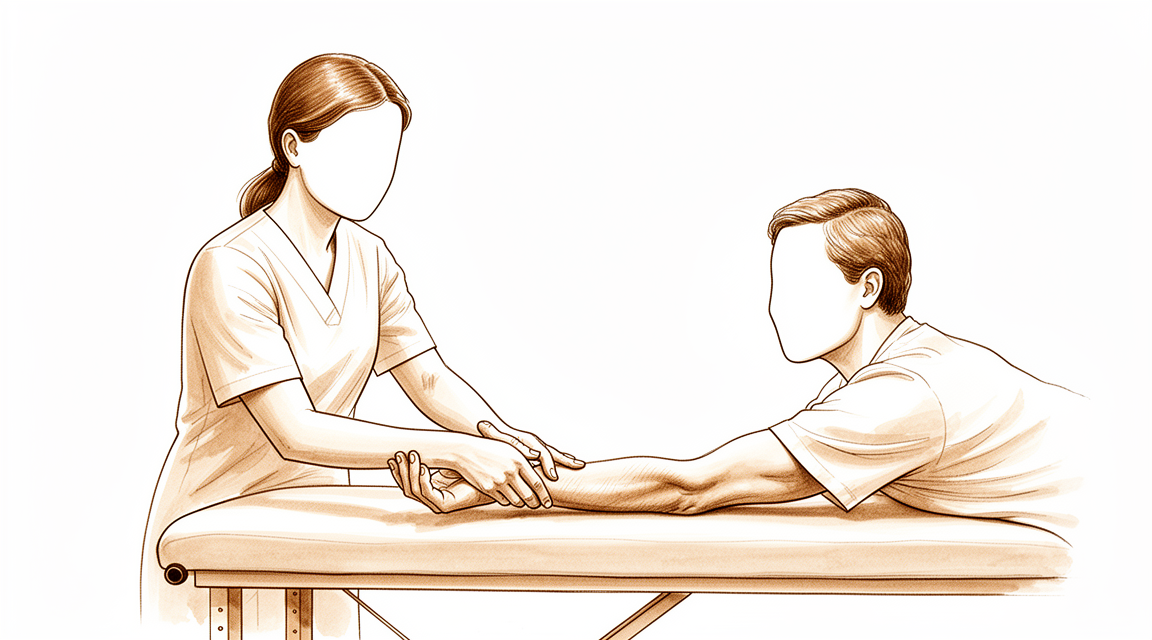
Post-operative exercises and precautions after radial tunnel release, including radial nerve glides.
Hướng dẫn này hỗ trợ quá trình hồi phục của bạn sau phẫu thuật giải phóng hố thần kinh trụ dưới sự chỉ định của bác sĩ Kieran Hirpara tại Bệnh viện tư nhân Mater Rockhampton. Hướng dẫn giải thích những điều bạn có thể mong đợi trong những tuần sau phẫu thuật và trình bày chương trình bài tập từ tài liệu hướng dẫn sau phẫu thuật. Hãy mang theo trang này hoặc file PDF của nó đến buổi trị liệu vật lý hoặc trị liệu tay đầu tiên để quá trình phục hồi chức năng được phối hợp nhịp nhàng. Chuyên viên trị liệu của bạn có thể điều chỉnh kế hoạch dựa trên tiến triển của quá trình hồi phục.
Nếu bạn có bất kỳ lo ngại nào về vết mổ sau phẫu thuật, hãy liên hệ với phòng khám. Việc chụp ảnh vết mổ và gửi qua email để xem xét thường rất hữu ích.
Những điều cần biết
Việc chăm sóc vết mổ của bạn được giải thích trong hướng dẫn chăm sóc vết mổ của phòng khám. Khi vết mổ lành lại, dây thần kinh được giải phóng có thể dính vào các mô xung quanh; các bài tập trượt dưới đây rất quan trọng để giữ cho nó di chuyển tự do và ngăn ngừa tình trạng bị mắc kẹt.
Đôi khi vết mổ có thể trở nên nhạy cảm. Đây là hiện tượng bình thường và có thể được ngăn ngừa hoặc giảm thiểu bằng cách bắt đầu quá trình giảm nhạy cảm hàng ngày: nhẹ nhàng vỗ và xoa lên vết mổ (hoặc băng gạc), bắt đầu ngay sau khi phẫu thuật. Loại "phản hồi cảm giác" này cho phép dây thần kinh bình thường hóa khả năng cảm nhận chạm và kết cấu.
Sau khi vết mổ lành hoàn toàn, hãy bắt đầu mát-xa sẹo: thực hiện các vòng tròn mạnh lên đường rạch. Tham khảo hướng dẫn chăm sóc vết mổ để biết thêm thông tin về quản lý sẹo.
Điều quan trọng là phải có kỳ vọng thực tế về quá trình hồi phục. Dây thần kinh quay phải di chuyển và kéo dãn một khoảng đo được trong các cử động bình thường của cánh tay, vì vậy việc giữ cho nó di chuyển sớm là yếu tố ngăn ngừa tình trạng dính vào các mô đang lành [1]. Dù vậy, việc giảm đau sau khi giải phóng hội chứng ống thần kinh quay thường diễn ra từ từ chứ không phải ngay lập tức, và ở một số người, kết quả chỉ là một phần. Phẫu thuật này, một cách trung thực, ít dự đoán được hơn so với một số thủ thuật giải phóng thần kinh khác: hội chứng ống thần kinh quay có thể khó chẩn đoán chắc chắn và thường chồng chéo với bệnh khuỷu tay quần vợt, đây là một phần lý do khiến kết quả điều trị khác nhau. Các nghiên cứu dài hạn được công bố báo cáo kết quả khả quan ở khoảng hai phần ba bệnh nhân nói chung, với kết quả tốt nhất ở những người chỉ có triệu chứng ống thần kinh quay [2][3]. Quá trình hồi phục có xu hướng chậm hơn và ít hoàn toàn hơn khi có kèm theo bệnh khuỷu tay quần vợt (viêm gai ngoài khuỷu tay), nhiều hơn một dây thần kinh bị chèn ép ở cùng một cánh tay, hoặc có khiếu nại bồi thường người lao động [2][4]. Chương trình trượt thần kinh và giảm nhạy cảm theo cấp độ của bạn là những phần của quá trình phục hồi chức năng nằm trong tầm kiểm soát của bạn nhiều nhất, và việc thực hành đều đặn hàng ngày sẽ mang lại cơ hội tốt nhất cho dây thần kinh ổn định.
Các biện pháp phòng ngừa và hạn chế
Việc sử dụng tay ở mức độ chức năng nhẹ được khuyến khích cho các nhiệm vụ sinh hoạt hàng ngày như tự chăm sóc bản thân, ăn uống, mặc quần áo, viết và đánh máy. Ngoài ra, các giới hạn rất đơn giản: không nâng vật, nắm chặt, chịu tải trọng hoặc sử dụng máy rung (ví dụ: dụng cụ điện hoặc máy cắt cỏ) trong tối đa 6 tuần sau phẫu thuật, và việc lái xe bị hạn chế trong 1–2 tuần đầu tiên.
Dành cho nhà vật lý trị liệu của bạn:
Mục tiêu
- Ngăn ngừa dây thần kinh đã được giải phóng dính vào vết thương đang lành (chương trình trượt dây thần kinh)
- Giảm độ nhạy cảm của vết thương thông qua quá trình giảm nhạy cảm có kiểm soát
- Duy trì tầm vận động của cổ tay, cẳng tay và khuỷu tay
- Hỗ trợ việc sử dụng tay ở mức độ chức năng nhẹ cho các hoạt động sinh hoạt hàng ngày
Quản lý điều trị
- Giảm nhạy cảm hàng ngày: vỗ nhẹ / xoa nhẹ lên vết thương (băng gạc), bắt đầu ngay sau phẫu thuật
- Xoa sẹo (vỗ tròn với lực vừa phải lên đường rạch) sau khi vết thương đã lành hoàn toàn
- Chương trình bài tập tại nhà theo các thẻ hướng dẫn bên dưới: căng duỗi gấp / duỗi cổ tay; căng duỗi xoay ngoài / xoay trong cổ tay; gấp / duỗi khuỷu tay; trượt dây thần kinh quay
- Ưu tiên các kỹ thuật trượt dây thần kinh nhẹ nhàng ("trượt") thay vì căng kéo mạnh ở tầm vận động tối đa: kỹ thuật trượt đạt được độ di chuyển của dây thần kinh lớn hơn đáng kể với mức độ căng dây thần kinh thấp hơn nhiều, điều này được dung nạp tốt hơn xung quanh dây thần kinh vừa được giải phóng chèn ép [1][5]
- Vận động dây thần kinh có thể được xem xét như một biện pháp bổ trợ cho chương trình; cơ sở bằng chứng về vận động dây thần kinh trong các tình trạng liên quan đến dây thần kinh là hỗ trợ nhưng có độ chắc chắn khác nhau, do đó việc tiến triển nên dựa trên triệu chứng [6]
Biện pháp phòng ngừa
- Chỉ sử dụng tay ở mức độ chức năng nhẹ (tự chăm sóc bản thân, ăn uống, mặc quần áo, viết, đánh máy)
- Không nâng vật, nắm chặt, chịu tải trọng hoặc sử dụng máy rung (ví dụ: dụng cụ điện, máy cắt cỏ) trong tối đa 6 tuần sau phẫu thuật
- Việc lái xe bị hạn chế trong 1–2 tuần đầu tiên
- Các bài tập trượt dây thần kinh và căng duỗi phải thực hiện nhẹ nhàng và về cơ bản không gây đau; tránh cố gắng ép vào tầm vận động làm tái hiện cơn đau dây thần kinh trước phẫu thuật
Đây là các bài tập từ tài liệu hướng dẫn sau phẫu thuật của bạn, bắt đầu sau phẫu thuật và tiếp tục tại nhà theo hướng dẫn của nhà vật lý trị liệu hoặc chuyên gia trị liệu tay. Số lần lặp lại, thời gian giữ và tần suất được liệt kê trên mỗi thẻ.
Các bài tập của bạn
Chương trình bài tập này được xây dựng với sự hợp tác của Sarah Farrell, Cử nhân Điều trị nghề nghiệp (BOccThy), Chuyên trị liệu tay được công nhận (AHT).
Sau khi thực hiện phác đồ
Phác đồ này được áp dụng song song với các lời khuyên chung về phục hồi chức năng của phòng khám; xem quản lý đau sau phẫu thuật, chăm sóc vết thương và cơ bản trị liệu tay. Về ca phẫu thuật và tình trạng bệnh mà nó điều trị, xem giải phóng ống thần kinh trụ ngoài và hội chứng ống thần kinh trụ ngoài.
Tài liệu tham khảo
[1] Wright TW, Glowczewskie F, Cowin D, Wheeler DL. Sự di chuyển và căng của dây thần kinh trụ tại khuỷu tay và cổ tay liên quan đến vận động chi trên. J Hand Surg Am. 2005;30(5):990–996. https://pubmed.ncbi.nlm.nih.gov/16182056/ [2] Lee JT, Azari K, Jones NF. Kết quả dài hạn của phẫu thuật giải phóng hố dây thần kinh trụ — ảnh hưởng của bệnh tennis elbow đồng thời, hội chứng chèn ép đa vị trí và bồi thường tai nạn lao động. J Plast Reconstr Aesthet Surg. 2008;61(9):1095–1099. https://www.sciencedirect.com/science/article/abs/pii/S1748681507004044 [3] Sotereanos DG, Varitimidis SE, Giannakopoulos PN, Westkaemper JG. Kết quả điều trị phẫu thuật hội chứng hố dây thần kinh trụ. J Hand Surg Am. 1999;24(3):566–570. https://pubmed.ncbi.nlm.nih.gov/10357537/ [4] Naam NH, Nemani S. Hội chứng hố dây thần kinh trụ. Orthop Clin North Am. 2012;43(4):529–536. (Hội chứng hố dây thần kinh trụ, StatPearls.) https://www.ncbi.nlm.nih.gov/books/NBK555937/ [5] Coppieters MW, Butler DS. Các "chất trượt" có trượt và các "chất căng" có căng không? Phân tích các kỹ thuật động học thần kinh và các cân nhắc về ứng dụng của chúng. Man Ther. 2008;13(3):213–221. https://pubmed.ncbi.nlm.nih.gov/17398140/ [6] Basson A, Olivier B, Ellis R, Coppieters M, Stewart A, Mudzi W. Hiệu quả của di chuyển thần kinh đối với các tình trạng cơ xương khớp thần kinh: một đánh giá hệ thống và phân tích tổng hợp. J Orthop Sports Phys Ther. 2017;47(9):593–615. https://pubmed.ncbi.nlm.nih.gov/28704626/
Evidence & references
Radial Tunnel Release — Evidence Brief & Post-operative Rehabilitation
Topic scope: post-operative rehabilitation after surgical decompression / neurolysis of the posterior interosseous nerve (deep branch of the radial nerve) in the radial tunnel of the proximal forearm, performed for radial tunnel syndrome (RTS). This is an elbow / proximal-forearm topic — anatomically and clinically distinct from carpal-tunnel and cubital-tunnel decompression. Like other nerve decompressions it is an early-motion pathway (early elbow/forearm/wrist motion, radial-nerve glides, oedema and scar care). The scope deliberately foregrounds the diagnostic controversy and the more variable, lower success rates that distinguish RTS release from the better-validated carpal-tunnel and cubital-tunnel operations.
Defining principle of the rehab here: a decompressed nerve does not create a healing construct that needs months of protection — it needs early, gentle movement to stop it adhering to the operative bed and to restore its glide. So the rehab is an early-motion programme: light functional hand use from day 1, radial-nerve sliders, graded desensitisation and (once healed) scar massage; heavier loading deferred to ~6 weeks. But two honesty caveats sit over the whole topic. First, RTS is a contested diagnosis — there is no confirmatory imaging or electrodiagnostic test, it is a diagnosis of exclusion, and a substantial body of opinion regards it as a variant of recalcitrant lateral epicondylitis. Second, outcomes after release are more variable and on average lower than carpal- or cubital-tunnel release — good results cluster around two-thirds overall, and fall further with co-existing tennis elbow, multiple compression sites, or a workers'-compensation context. Patient expectations should be set accordingly.
A. THE DIAGNOSTIC CONTROVERSY (read first — it frames everything)
RTS is among the most contested entities in upper-limb surgery, and the rehab brief is incomplete without it:
- No confirmatory test. Electromyography and nerve-conduction studies are characteristically normal in RTS (compression is intermittent/dynamic and predominantly of a motor nerve carrying few pain fibres), and MRI is frequently negative — denervation oedema in supinator/extensors is suggestive but inconsistent, and a normal scan does not exclude the diagnosis. RTS is therefore a clinical diagnosis of exclusion, resting on point tenderness ~4 cm distal to the lateral epicondyle (over the radial tunnel rather than the epicondyle), pain on resisted supination / resisted long-finger extension, and — for some surgeons — temporary relief from a diagnostic local-anaesthetic block at the radial tunnel.
- Overlap with lateral epicondylitis (tennis elbow). The two coexist frequently and share the lateral-elbow pain territory. A recognised school of thought holds that "RTS" is often severe, recalcitrant lateral epicondylitis rather than a discrete compression neuropathy. Importantly, routine PIN release added to lateral-epicondylitis surgery has not been shown to improve outcomes, so the diagnosis should be secure before a decompression is planned.
- Practical consequence. Surgery is a last resort after prolonged failed conservative care (activity modification, splinting, anti-inflammatories, sometimes a steroid injection), and is best reserved for patients with proximal-forearm pain and no better explanation. This uncertainty is the single most important reason post-operative expectations must be framed honestly.
B. RELEASE OUTCOMES (variable — and why)
- Headline success ~two-thirds. Across the older long-term series, roughly 67% good, 15% fair, 18% poor after radial tunnel decompression — markedly more variable than carpal- or cubital-tunnel release. A 2008 long-term series (Lee, Azari, Jones) and a 1999 series (Sotereanos et al.) both document this spread; the Sotereanos cohort reported good/excellent results in only ~39% by objective assessment (though ~64% by patient self-rating), underscoring how outcome depends on the metric used.
- Co-existing lateral epicondylitis lowers success. Success falls to roughly 40% when tennis elbow coexists, versus far higher with isolated RTS.
- Multiple compression sites and workers'-compensation context lower success — reported ~58% success in compensation cases vs ~73% without. These are the same modifiers named in the patient protocol.
- 2025 systematic review (Raymond et al., HAND). 11 studies, 401 limbs (381 patients). Outcomes were heterogeneous; a dorsal approach between ECRB and EDC was associated with the most favourable Roles-and-Maudsley scores and satisfaction. The review's central message is that the overall evidence is low-grade (observational), the diagnosis non-standardised, and the effectiveness of conservative treatment essentially untested — a "tendency" toward benefit rather than proof.
- Resorption-style "spontaneous improvement" does not apply here — unlike calcific tendinitis, RTS does not self-resolve through a biological cycle; conservative care manages symptoms rather than curing a deposit.
C. SURGICAL APPROACH (shapes the early rehab)
- What is done. Complete neurolysis of the radial nerve at its bifurcation, decompressing the deep branch (PIN) and superficial sensory branch, releasing the arcade of Frohse (the proximal supinator edge), the leash of Henry (radial recurrent vessels), the ECRB fascial edge, and the distal supinator border. Any constrictive bands or vessels are divided.
- Approaches. Dorsal (Thompson, between ECRB/EDC or the brachioradialis–ECRL interval), volar/anterior (Henry), or transmuscular. Anatomical studies map the trade-offs; the dorsal ECRB–EDC interval performed best in the 2025 review. The superficial radial branch matters — it is a recognised source of post-operative dysaesthesia if irritated.
- Rehab implication. A muscle-splitting/dorsal exposure through the extensor mass means early gentle forearm rotation and wrist motion are encouraged but heavy resisted supination/extension is deferred; the incision sits over a mobile, frequently sensitive area, so desensitisation and scar care carry real weight here.
D. POST-OP THERAPY ROLE (nerve/tendon glides, oedema, scar)
The decompressed nerve must glide, not adhere. The mechanical rationale is well quantified: the radial nerve translates and stretches a measurable amount across the elbow and wrist during ordinary arm motion (Wright et al. 2005), so early motion is what keeps it free of the healing bed.
- Early motion, immediately. Early active elbow, forearm and wrist movement within pain limits from the first post-op days; most protocols use no rigid splinting (or a removable splint for comfort/night only).
- Radial-nerve glides — favour "sliders" over "tensioners". Sliding (slider) neurodynamic techniques achieve substantially greater nerve excursion at much lower nerve strain than end-range tensioners — preferable around a freshly decompressed nerve. Neural-mobilisation evidence across neuromusculoskeletal conditions is supportive but of variable certainty, so progression is symptom-guided and essentially pain-free; mechanism work (e.g., the MONET protocol) is still maturing.
- Oedema and desensitisation. Graded desensitisation (tapping/rubbing over the dressing) from day 1 normalises touch and pre-empts a sensitive scar — particularly relevant given superficial- radial-branch proximity.
- Scar management once healed. Massage, pressure, and silicone are advocated to loosen skin–tissue adhesions and aid remodelling, started once the wound is closed/sutures out.
- Strengthening deferred. Light functional ADL use throughout; resisted strengthening of wrist/ elbow and fine-motor work introduced from ~6 weeks. Heavy work and vibration tools avoided to ~6–8 weeks.
Phased post-op timeline (maps to the patient protocol phases)
| Phase | Window | Splint | Motion / nerve work | Load / strengthening | Notes |
|---|---|---|---|---|---|
| I — Protect & glide | Day 0–2 wk | None, or removable for comfort/night | Early pain-free active elbow/forearm/wrist ROM; radial-nerve sliders; desensitisation from day 1 | Light functional ADL use only (self-care, feeding, dressing, writing, typing) | Stop the nerve adhering; settle the wound. No lifting/gripping/weight-bearing/vibration tools. Driving limited first 1–2 wk |
| II — Restore motion | 2–6 wk | Off | Progress full active + gentle assisted ROM; continue sliders; scar massage once healed | Still no resisted loading; ADL use continues | Sensitivity/dysaesthesia common and usually settles; keep glides gentle |
| III — Strengthen & return | ~6 wk onward | Off | Full ROM goal; sliders as needed | Begin graded wrist/elbow strengthening + fine-motor work from ~6 wk; advance work/heavy tasks thereafter | Vibration tools/heavy work resume ~6–8 wk. Pain relief is often gradual and may be partial — counsel accordingly |
E. COMPLICATIONS / DOWNSIDES
- Incomplete or no pain relief — the dominant "complication," tied directly to diagnostic uncertainty; relief is frequently gradual and sometimes partial.
- Superficial-radial-branch dysaesthesia / scar sensitivity — recognised; desensitisation and careful technique mitigate it.
- Transient PIN weakness (finger/thumb extension) from retraction — usually recovers.
- Adhesion/recurrence of symptoms if early glide is neglected.
- Standard wound risks (infection, haematoma) — uncommon.
F. KEY CONTROVERSIES / EVIDENCE QUALITY
- Does RTS exist as a discrete entity? Genuinely contested. No confirmatory test; substantial opinion equates much of it with recalcitrant lateral epicondylitis. This is the defining controversy and must shape consent and expectation-setting. Unresolved — expert opinion divided.
- Patient selection drives outcome more than technique. Isolated RTS does best; coexisting tennis elbow, multiple compressions, and compensation context predict worse results. Moderate (consistent across cohorts).
- Approach choice. A dorsal ECRB–EDC interval was favoured in the 2025 SR, but the evidence is observational and confounded by diagnostic heterogeneity. Weak–moderate.
- The rehab protocol itself is consensus/expert — drawn from surgeon and hand-therapy guidance (early motion, sliders, desensitisation, scar care), not from a rehab RCT. Phase timings are typical, not trial-derived. Weak / consensus.
- Conservative-treatment efficacy is essentially untested — the 2025 SR notes no usable trials of non-operative care, so "failed conservative management" before surgery rests on practice convention. Weak.
G. EVIDENCE STRENGTH FLAGS (summary)
- STRONG: the mechanical rationale for early nerve glide — quantified radial-nerve excursion/ strain across elbow and wrist (Wright et al. 2005); slider-vs-tensioner excursion/strain physiology.
- MODERATE: patient-selection modifiers of outcome (lateral epicondylitis, multiple compressions, workers' compensation lower success); ~two-thirds overall good-result rate from long-term cohorts; dorsal-approach signal from the 2025 systematic review (low-grade studies).
- WEAK / CONSENSUS: the existence and diagnostic criteria of RTS (no confirmatory test; overlap with lateral epicondylitis); the post-operative rehabilitation protocol (surgeon/ hand-therapy guidance, no rehab RCT); neural-mobilisation certainty (supportive but variable); efficacy of conservative care (essentially untested).
CITATIONS
RAG corpus (180,000+ Orthopaedic articles)
- Posterior Interosseous Nerve Compression in the Forearm, AKA Radial Tunnel Syndrome. HAND. 2022. DOI: 10.1177/15589447221122822
- Radial Tunnel Syndrome: Emphasis on the Superficial Branch of the Radial Nerve. J Hand Surg Eur. 2009. DOI: 10.1177/1753193408099832
- Anatomical Study of the Surgical Approaches to the Radial Tunnel. J Hand Surg Am. 2015. DOI: 10.1016/j.jhsa.2015.03.009
- MR Imaging Features of Radial Tunnel Syndrome: Initial Experience. Radiology. 2006. DOI: 10.1148/radiol.2401050028
- Management of Lateral Epicondylitis: Current Concepts. J Am Acad Orthop Surg (JAAOS). 2008. DOI: 10.5435/00124635-200801000-00004
- Uncommon Nerve Compression Syndromes of the Upper Extremity. J Am Acad Orthop Surg (JAAOS). 1998. DOI: 10.5435/00124635-199811000-00006
- Radial Nerve Excursion and Strain at the Elbow and Wrist Associated With Upper-Extremity Motion. J Hand Surg Am. 2005. DOI: 10.1016/j.jhsa.2005.06.008
- Evidence and Techniques in Rehabilitation Following Nerve Injuries. Hand Clin. 2013. DOI: 10.1016/j.hcl.2013.04.012
- Preventive Strategies, Exercises and Rehabilitation of Hand Compression Neuropathies. J Hand Ther. 2022. DOI: 10.1016/j.jht.2021.11.003
- Mechanisms of Neurodynamic Treatments (MONET): a protocol for a mechanistic study. BMC Musculoskelet Disord. 2024. DOI: 10.1186/s12891-024-07713-6
Radial-tunnel literature (URLs)
- Clinical Outcomes of Operative Management for Radial Tunnel Syndrome According to Surgical Approach: a Systematic Review. HAND. 2025. https://journals.sagepub.com/doi/10.1177/15589447251315761
- The Epidemiology of Radial Tunnel Syndrome and Its Overlap With Lateral Epicondylitis. J Hand Surg Am. 2023. https://www.jhandsurg.org/article/S0363-5023(23)00138-7/abstract
- Lee JT, Azari K, Jones NF. Long-term results of radial tunnel release — the effect of co-existing tennis elbow, multiple compression syndromes and workers' compensation. J Plast Reconstr Aesthet Surg. 2008. https://www.sciencedirect.com/science/article/abs/pii/S1748681507004044
- Sotereanos DG, et al. Results of surgical treatment for radial tunnel syndrome. J Hand Surg Am. 1999. https://pubmed.ncbi.nlm.nih.gov/10357537/
- Interventions for treating the radial tunnel syndrome: a systematic review of observational studies (DARE). https://www.ncbi.nlm.nih.gov/books/NBK75403/
- Radial Tunnel Syndrome (StatPearls). https://www.ncbi.nlm.nih.gov/books/NBK555937/
- Orthopedic Management of Radial Tunnel Syndrome: A Diagnostic and Treatment Dilemma. PMC. https://pmc.ncbi.nlm.nih.gov/articles/PMC10081130/
- Radial Tunnel Syndrome: Case Report and Comprehensive Critical Review of a Compression Neuropathy Surrounded by Controversy. PMC. https://pmc.ncbi.nlm.nih.gov/articles/PMC9896270/
Published rehab protocols (patient-guidance — basis for the phase structure)
- Radial Tunnel Release post-op protocol (Santa Barbara Orthopedic / Mencias). https://www.sbortho.com/wp-content/uploads/2023/09/radial-tunnel-release-new.pdf
- Radial Tunnel Syndrome — conservative and post-operative rehabilitation. Physiopedia. https://www.physio-pedia.com/Radial_Tunnel_Syndrome
- Basson A, et al. The effectiveness of neural mobilization for neuromusculoskeletal conditions: a systematic review and meta-analysis. J Orthop Sports Phys Ther. 2017. https://pubmed.ncbi.nlm.nih.gov/28704626/
- Coppieters MW, Butler DS. Do "sliders" slide and "tensioners" tension? Man Ther. 2008. https://pubmed.ncbi.nlm.nih.gov/17398140/


