Nerve Tests and Conduction Studies Info In-depth
Last reviewed
Patients › General-Health
What nerve conduction studies and EMG are, why they are done for carpal tunnel and other nerve problems, what to expect, and how accurate they are.
What it is
Nerve conduction studies measure how well your nerves are working. They show if nerve function is impaired and indicate the overall severity of conditions like carpal tunnel syndrome. These tests also help predict how well you might do after surgery.
Your doctor may order these tests, or an ultrasound, to confirm a diagnosis. Ultrasound is a valid alternative for checking ulnar nerve issues at the elbow. It confirms nerve integrity and shows indirect effects of surgery. Combining these tools helps identify actual nerve damage that may benefit from treatment.
These tests are especially useful when your signs and symptoms suggest mild-to-moderate nerve compression. They increase the chance of finding real median neuropathy that surgery can fix. Even if tests are negative, your doctor may still offer carpal tunnel release if symptoms fit.
The tests look at specific nerve signals. For example, they measure how fast signals travel in the ulnar nerve at the upper arm, elbow, and forearm. This is crucial for severe cases considering surgery. They also check the size of the median nerve. A larger nerve size often matches worse test results and stronger symptoms.
In some cases, like shoulder surgery, these insights predict functional outcomes. Your team uses this data to protect nerves during and after procedures. For ulnar nerve decompression, one specific signal amplitude predicts your recovery. Other standard signals do not.
You might have unique test patterns if you have double-crush syndrome, where nerves are compressed in more than one spot. This shows up as different timing in your nerve signals compared to simple carpal tunnel syndrome. The severity of your ulnar neuropathy at the elbow also matches the swelling size seen on ultrasound.
Overall, these tests give your doctor a clear picture of your nerve health. They guide decisions on whether surgery is right for you and help plan the best path to recovery.
Does it work?
Nerve conduction studies measure how well your nerves send signals. They are the best available indicator of how severe carpal tunnel syndrome is. Your doctor uses these results to understand the overall health of your nerves. These tests also have some value in predicting how well you might do after surgery.
Ultrasound is a valid alternative to these electrical tests for checking ulnar nerve issues at the elbow. It can confirm that your nerve is intact and show indirect effects of surgery. For median neuropathy, using electrodiagnostic studies or ultrasound can increase the chance of confirming the problem when your signs and symptoms suggest mild to moderate disease.
There is a big difference between what you feel and what tests show. Clinical signs and symptoms suggest a 73% prevalence of mild to moderate carpal tunnel syndrome. However, electrodiagnostic studies and ultrasound show a 51% prevalence. This means tests may find fewer cases than you experience.
For cubital tunnel syndrome, the evidence is mixed. Electrodiagnostic severity does not predict short- to midterm outcomes of surgery. Patient-reported preoperative disease severity may predict your expected postoperative change in functional improvement, while electrodiagnostic studies may not have prognostic value for these patients. However, one specific measure, compound muscle action potential amplitude, predicts functional outcomes after in situ decompression of the ulnar nerve.
The overall use of electrodiagnostic studies for carpal tunnel syndrome has been decreasing since at least 2014. This shift reflects changing practices and the availability of alternatives like ultrasound. For other conditions, such as suprascapular nerve dysfunction, insights from these tests are important for perioperative nerve preservation strategies and postoperative neurological assessments.
In some cases, non-surgical treatments show promise. Chitosan phonophoresis demonstrated significant improvements in nerve conduction, pain reduction, and enhancement of hand function for mild to moderate cubital tunnel syndrome. EMG-driven robotic treatment with 15 sessions of rehabilitation suggests that improvement in hand recovery may be possible for chronic stroke patients nine years after stroke. Your doctor will decide which test or approach is right for your specific situation.
Is it right for you?
Nerve conduction studies measure how well your nerves send signals. They are the best available indicator of overall disease severity in carpal tunnel syndrome. These tests also help predict your surgical outcome. If your signs and symptoms suggest mild to moderate median neuropathy, these studies may increase the chance of identifying actual nerve damage that surgery can fix. Ultrasound is a valid alternative for detecting ulnar neuropathy at the elbow. It can confirm nerve integrity and show indirect effects of surgery.
You might not need these tests if your case is straightforward. There is severe discordance between estimated prevalence based on symptoms (73%) versus test results (51%). This means many people with symptoms do not show up on tests. The overall use of these studies for carpal tunnel syndrome has been decreasing since at least 2014. For shoulder issues, electromyography does not show adequate effectiveness in diagnosing nerve lesions from rotator cuff tears. However, your doctor should still carefully evaluate nerve integrity in these cases.
These tests help your doctor plan the best path forward. Experts in electrodiagnostic studies and ultrasound should be part of your orthopaedic team. This integration helps with both diagnosis and rehabilitation. In some cases, like cubital tunnel syndrome, other treatments like chitosan phonophoresis can improve nerve conduction and reduce pain. Your doctor will decide if testing is necessary based on your specific symptoms and severity. It is a shared decision between you and your doctor. The goal is to ensure you receive the right care without unnecessary procedures.
The bottom line
Nerve tests help your doctor measure how well your nerves work and predict surgery results. These tests are especially useful if your symptoms suggest mild to moderate nerve pressure. They confirm whether surgery will likely help you. However, symptoms alone often overestimate the problem. Only about 51% of people with mild symptoms actually have nerve damage on testing. Your doctor uses these results to plan the best care for you.
Advanced reading: the deeper science (optional)
If you would like to understand your report rather than just receive it, this section explains what the equipment is actually measuring and how the pattern of results points to one kind of nerve problem rather than another. None of this is essential reading, but many people find it reassuring to know there is real, measurable physics behind the numbers.
The two halves of the study
A nerve test is really two examinations done in one sitting:
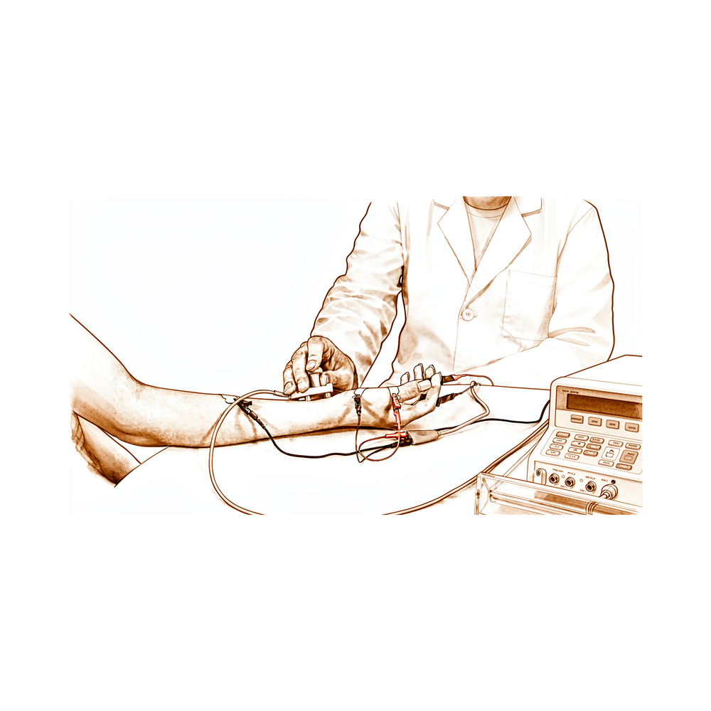
- Nerve conduction studies (NCS): small electrical pulses are delivered to a nerve through the skin, and the resulting signal is recorded a short distance away. This measures how well the nerve carries a signal.
- Needle electromyography (EMG): a fine needle electrode is placed into selected muscles to listen to their electrical activity, both at rest and while you gently contract. This measures the consequences of any nerve problem on the muscle itself.
The two halves answer different questions, and they are most powerful read together. The conduction studies localise and characterise the problem in the nerve; the needle exam shows whether muscle has been affected and whether it is recovering.
Motor versus sensory testing
Conduction studies come in two flavours, because a nerve has two jobs:
- Sensory studies stimulate the nerve and record the nerve's own electrical response a little further along. This response is called the sensory nerve action potential (SNAP). It reflects the health of the sensory fibres directly.
- Motor studies stimulate the nerve but record from a muscle that the nerve supplies. The muscle's response is called the compound muscle action potential (CMAP). So a motor study tests the whole chain: nerve, the junction with the muscle, and the muscle's ability to respond.
Both are done because some conditions affect sensory fibres first (many of the common compression problems begin this way), while others affect motor fibres, and the comparison helps pinpoint where the trouble lies.
Amplitude versus conduction velocity — the key idea
This is the single most useful concept for reading a nerve report. Each recorded signal is described by two broad properties, and they mean genuinely different things:
- Amplitude: the size (height) of the response. It reflects roughly how many working nerve fibres (axons) are contributing. When axons are lost, fewer fibres fire, and the amplitude falls. So a reduced amplitude points towards axon loss.
- Conduction velocity and latency: how fast the signal travels (velocity) and how long it takes to arrive (latency). These depend on the myelin, the fatty insulation wrapped around the axon that lets the signal jump along quickly. When myelin is damaged (demyelination), the signal slows down: velocity drops and latency lengthens, even if plenty of fibres are still present.
So, simplifying only slightly:
- Small signals, normal speed → the wiring is thinning out (axon loss).
- Normal-sized signals, slow speed → the insulation is damaged but the wires are largely intact (demyelination).
This is why the team does not just ask "is the nerve abnormal?" but "in what way is it abnormal?", because the pattern guides what is likely going on and how it tends to recover.
Conduction block
There is one more pattern worth knowing. Sometimes a nerve conducts normally below a problem area but the signal is much smaller when stimulated above it, as if part of the signal is being stopped at a specific point. This is called conduction block, and it is a feature of focal demyelination (a discrete spot of damaged insulation, often where a nerve is compressed). It is useful because it localises the problem to one segment of the nerve rather than the whole length.
The needle exam (EMG) and recruitment
The needle electrode does not deliver shocks; it listens. It picks up a few distinct things:
- At rest, a healthy muscle is electrically silent. If a muscle has lost its nerve supply, the muscle fibres become irritable and fire on their own. This shows up as fibrillations and positive sharp waves, small spontaneous signals that indicate the muscle has been recently denervated. They typically appear a couple of weeks after a nerve injury, not immediately.
- On gentle contraction, the examiner watches recruitment. Recruitment is how the muscle calls in its motor units (each motor unit being one nerve fibre and the muscle fibres it controls) as you push harder. If axons have been lost, there are fewer units to call on, so recruitment is reduced; the remaining units fire faster to compensate, but there are simply fewer of them.
- As a nerve recovers, surviving fibres sprout to "adopt" orphaned muscle fibres. This reinnervation produces larger, longer, more complex motor unit signals than normal. Their presence is actually an encouraging sign; it is the electrical signature of repair in progress.
Read together, these findings tell the team not only whether a muscle has been affected, but roughly when the problem started and whether recovery is already under way.
A note on tone
None of these findings is, by itself, alarming. They are descriptions, a vocabulary the team uses to translate your symptoms into something measurable. A reduced amplitude or a slowed velocity is information, not a verdict, and it is always interpreted alongside your history, your examination, and how things are changing over time.
To understand the underlying biology these tests are probing (how nerves carry signals, why myelin matters, and how nerves recover after injury), see the companion page on how nerves work and heal.


