Viêm xương khớp khuỷu tay
Patients › Elbow
Osteoarthritis of the elbow — primary and post-traumatic, conservative and surgical options.
Những gì bạn đang cảm thấy
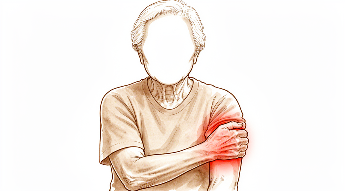
Bạn có thể nhận thấy khuỷu tay của mình đau âm ỉ, đặc biệt nếu bạn là nam giới trên 40 tuổi làm công việc nặng nhọc bằng tay. Viêm xương khớp do hao mòn này phổ biến hơn ở người lớn tuổi và nam giới. Một chấn thương trước đây ở khuỷu tay cũng làm tăng nguy cơ của bạn. Bạn có thể cảm thấy đau khi cố gắng duỗi thẳng hoặc gập cánh tay của bạn hoàn toàn. Khớp có thể cảm thấy cứng, khiến việc di chuyển qua toàn bộ phạm vi chuyển động trở nên khó khăn.
Các nhiệm vụ đơn giản có thể trở nên gây khó chịu. Bạn có thể gặp khó khăn khi với tay ra sau lưng để cài áo ngực hoặc nhét áo vào quần. Nâng vật có thể kích hoạt cơn đau nhói. Một số người cũng cảm thấy tê hoặc ngứa ran ở ngón áp út và ngón út. Điều này xảy ra vì dây thần kinh trụ, chạy gần khuỷu tay, có thể bị kích thích bởi cấu trúc khớp thay đổi.
Các triệu chứng của bạn có thể bùng phát sau khi bạn đã sử dụng cánh tay của mình trong một thời gian. Nghỉ ngơi thường mang lại sự giảm nhẹ. Tuy nhiên, bạn cũng có thể thức dậy vào ban đêm với cảm giác khó chịu. Cơn đau này có thể làm gián đoạn giấc ngủ của bạn nếu bạn nằm nghiêng về phía đó. Bác sĩ phẫu thuật của bạn sẽ xem xét các triệu chứng cụ thể, tuổi tác và nhu cầu hàng ngày của bạn để quyết định hướng đi tốt nhất.
Trong giai đoạn đầu, các phương pháp điều trị không phẫu thuật thường là bước đầu tiên. Những biện pháp bảo tồn này có thể mang lại sự giảm nhẹ cho nhiều người. Nếu cơn đau dai dẳng ở các cực của chuyển động, bác sĩ phẫu thuật của bạn có thể thảo luận về phẫu thuật nội soi. Thủ thuật xâm lấn tối thiểu này có thể giúp làm sạch chất thải và cải thiện chuyển động. Đối với các trường hợp nghiêm trọng nơi cơn đau xảy ra trong suốt tất cả các chuyển động, thay thế khuỷu tay toàn phần có thể được xem xét. Đây là một lựa chọn lớn được dành riêng cho các tình huống cụ thể do các biến chứng tiềm ẩn. Kế hoạch điều trị của bạn sẽ được cá nhân hóa theo tình huống độc đáo và nhu cầu chức năng của bạn.
Những gì thực sự đang xảy ra
Trong khuỷu tay của bạn, lớp phủ trơn tru trên đầu xương bị mòn đi. Đây là viêm xương khớp do hao mòn. Trong hầu hết các trường hợp, điều này xảy ra đầu tiên ở phần khớp bản lề chính. Khoảng cách giữa các xương thu hẹp, đặc biệt là nơi các xương cẳng tay gặp xương cánh tay. Khi khớp thay đổi, các khối xương nhỏ hình thành. Những khối này được gọi là gai xương. Chúng có thể cản trở chuyển động của bạn. Ví dụ, một gai xương có thể va vào một xương khác khi bạn duỗi thẳng cánh tay.
Khuỷu tay của bạn dựa vào các dây chằng chắc khỏe để duy trì sự ổn định. Các dây chằng này hoạt động như những sợi dây giữ các xương lại với nhau. Nếu một dây chằng bị yếu hoặc rách, các xương sẽ cọ xát vào nhau theo cách sai lệch. Căng thẳng bổ sung này làm tăng tốc độ hao mòn. Nó cũng có thể gây đau và mất ổn định. Bác sĩ phẫu thuật của bạn xem xét cả những thay đổi về xương và tình trạng sức khỏe của dây chằng để hiểu rõ tình huống cụ thể của bạn.
Đôi khi, xương dư thừa hình thành trong các mô mềm xung quanh khớp sau chấn thương hoặc phẫu thuật. Điều này được gọi là cốt hóa dị vị. Nó có thể tạo ra một khối cứng ngăn cản bạn cử động khuỷu tay hoàn toàn. Điều này khác với quá trình hao mòn thông thường. Đây là một vấn đề riêng biệt mà bác sĩ phẫu thuật sẽ kiểm tra nếu bạn có tình trạng cản trở cơ học đối với chuyển động.
Những thay đổi trong khớp của bạn giải thích lý do tại sao bạn cảm thấy đau và cứng khớp. Sự mất đi lớp sụn trơn tru có nghĩa là các xương cọ xát vào nhau. Các gai xương ngăn cản vật lý cánh tay của bạn di chuyển tự do. Các vấn đề về dây chằng khiến khớp cảm thấy lỏng lẻo hoặc không ổn định. Hiểu rõ những thay đổi cụ thể này giúp bác sĩ phẫu thuật của bạn lựa chọn phương pháp điều trị phù hợp cho bạn.
Những gì chúng tôi có thể làm về vấn đề này
Điều trị không phẫu thuật vẫn là bước đầu tiên trong quản lý sớm viêm xương khớp khuỷu tay. Quản lý không phẫu thuật có thể giúp giảm bớt triệu chứng ở các giai đoạn đầu của viêm khớp khuỷu tay. Bác sĩ phẫu thuật của bạn có thể sẽ khuyên dùng tự chăm sóc và vật lý trị liệu trước khi xem xét các lựa chọn khác. Các biện pháp này nhằm mục đích giảm đau và duy trì tầm vận động hiện tại của bạn. Bạn có thể sử dụng chườm đá hoặc chườm nóng để làm dịu tình trạng cứng khớp. Các bài tập nhẹ nhàng giúp duy trì sự vận động của khớp mà không gây thêm tổn thương. Hãy dành đủ thời gian cho phương pháp này phát huy tác dụng trước khi chuyển sang các phương pháp điều trị mạnh hơn.
Nếu các biện pháp đơn giản không đủ hiệu quả, bác sĩ phẫu thuật của bạn có thể thảo luận về quản lý bằng thuốc. Điều này thường bao gồm thuốc giảm đau và thuốc chống viêm để giúp bạn kiểm soát sự khó chịu hàng ngày. Trong một số trường hợp, tiêm thuốc có thể được khuyến nghị. Tiêm cortisone có thể giảm viêm và đau trong một thời gian hạn chế. Tiêm axit hyaluronic nhằm mục đích bôi trơn khớp, mặc dù bằng chứng về lợi ích lâu dài còn khác nhau. Tiêm huyết tương giàu tiểu cầu (PRP) sử dụng các thành phần máu của chính bạn để thúc đẩy quá trình lành thương, nhưng kết quả không được đảm bảo. Bác sĩ phẫu thuật của bạn sẽ tư vấn lựa chọn nào phù hợp với tình trạng cụ thể và mức độ đau của bạn.
Phẫu thuật được xem xét khi chăm sóc bảo tồn đã đạt đến giới hạn và các triệu chứng vẫn gây tàn tật. Phương pháp điều trị thích hợp cho viêm khớp khuỷu tay phụ thuộc vào nguyên nhân, mức độ nghiêm trọng, tuổi tác của bệnh nhân và các yêu cầu về chức năng. Đối với nhiều bệnh nhân, nạo vét phẫu thuật là một phương pháp điều trị hiệu quả cho các triệu chứng gây tàn tật của viêm xương khớp khuỷu tay nguyên phát với tỷ lệ biến chứng chấp nhận được. Thủ thuật này liên quan đến việc loại bỏ gai xương và mô viêm để cải thiện chuyển động và giảm đau. Nó an toàn và hiệu quả trong điều trị viêm xương khớp khuỷu tay. Nạo vét nội soi cung cấp giảm đau thỏa đáng, cải thiện tầm vận động khuỷu tay và kết quả chức năng tốt. Trong các trường hợp nghiêm trọng hơn khi khớp bị tổn thương đáng kể, thay khớp khuỷu tay toàn phần có thể là một lựa chọn. Điều này liên quan đến việc thay thế các bề mặt khớp bị tổn thương bằng các thành phần nhân tạo. Phẫu thuật tạo hình khớp xen kẽ là một lựa chọn khác cho một số bệnh nhân, sử dụng ghép mô để đệm cho khớp. Bác sĩ phẫu thuật của bạn sẽ xác định con đường phẫu thuật tốt nhất dựa trên nhu cầu cá nhân của bạn và giai đoạn bệnh của bạn.
Những gì cần mong đợi
Hành trình của bạn với bệnh viêm xương khớp do hao mòn khuỷu tay thường bắt đầu bằng điều trị không phẫu thuật. Đây là bước đầu tiên tiêu chuẩn cho các triệu chứng ở giai đoạn sớm. Bác sĩ phẫu thuật của bạn sẽ cá nhân hóa kế hoạch dựa trên độ tuổi, mức độ nghiêm trọng của tổn thương và nhu cầu sử dụng cánh tay của bạn. Ở các giai đoạn đầu, nghỉ ngơi, vật lý trị liệu và các phương pháp không phẫu thuật khác thường mang lại sự giảm đau đáng kể. Nhiều người nhận thấy các triệu chứng của họ giảm đủ để quản lý cuộc sống hàng ngày mà không cần phẫu thuật.
Nếu các biện pháp bảo tồn không làm giảm đau hoặc cứng khớp, bác sĩ phẫu thuật của bạn có thể thảo luận về các lựa chọn phẫu thuật. Lựa chọn phù hợp phụ thuộc vào việc bệnh viêm xương khớp có do chấn thương cũ hoặc phát triển tự nhiên, và mức độ vận động mà bạn vẫn còn. Đối với các trường hợp nhẹ đến trung bình, phẫu thuật nội soi cắt bỏ xương và bao khớp (một thủ thuật qua lỗ khóa để loại bỏ gai xương và làm chặt bao khớp) là một phương pháp an toàn và hiệu quả. Nó thường cải thiện đau và chức năng với nguy cơ biến chứng thấp. Phẫu thuật mở mang lại kết quả tương tự trong tay các bác sĩ giàu kinh nghiệm, mặc dù một số bệnh nhân có thể trải qua tình trạng cứng khớp trở lại theo thời gian.
Đối với bệnh ở giai đoạn tiến triển hơn, thay khớp là một lựa chọn cho một số bệnh nhân nhất định. Tuy nhiên, thay khớp khuỷu tay toàn phần thường không được khuyến nghị cho những người năng động. Nó có tỷ lệ biến chứng cao hơn và không bền vững bằng dưới mức sử dụng nặng. Nếu bạn trẻ tuổi hoặc rất năng động với bệnh viêm xương khớp sau chấn thương nghiêm trọng, thay khớp một phần (hemiarthroplasty) có thể được xem xét. Hãy lưu ý rằng phương pháp này có tỷ lệ cao cần phẫu thuật chỉnh sửa sau này.
Nhìn chung, hầu hết bệnh nhân thực hiện nạo khớp (làm sạch khớp) báo cáo chức năng và giảm đau thỏa đáng trong ngắn hạn đến trung hạn. Mặc dù tầm vận động có thể giảm nhẹ giữa thời gian theo dõi ngắn hạn và trung hạn sau một số thủ thuật nội soi, nhưng sự đánh đổi này thường đáng giá để giảm đau. Nếu không được điều trị, bệnh viêm xương khớp có xu hướng tiến triển, dẫn đến cứng khớp và đau tăng dần. Với quản lý thích hợp, bạn có thể mong đợi sự cải thiện đáng kể về chất lượng cuộc sống, mặc dù bạn phải chấp nhận rằng một số mất mát vận động hoặc nhu cầu về các thủ thuật trong tương lai là có thể tùy thuộc vào mức độ nghiêm trọng của tình trạng của bạn.
Khi nào cần gặp bác sĩ
Hãy gặp bác sĩ đa khoa nếu bạn có đau khuỷu tay dai dẳng không cải thiện khi nghỉ ngơi. Hãy yêu cầu đánh giá bởi bác sĩ chuyên khoa nếu bạn gặp phải tình trạng yếu cơ, mất ổn định hoặc kẹt khớp. Các triệu chứng ảnh hưởng đến giấc ngủ hoặc công việc của bạn cũng cần được chú ý. Sự gia tăng đột ngột của cơn đau là một lý do khác để tìm kiếm sự chăm sóc y tế. Thoái hóa khớp khuỷu tay là phổ biến, ảnh hưởng đến 55,0% những người từ 40 tuổi trở lên. Bệnh thường gây đau và hạn chế vận động. Điều trị bảo tồn sớm có thể giúp ích trong các giai đoạn đầu. Bác sĩ phẫu thuật của bạn sẽ điều chỉnh phương pháp điều trị dựa trên độ tuổi, mức độ nghiêm trọng và nhu cầu hàng ngày của bạn. Đừng bỏ qua các triệu chứng ảnh hưởng đến cuộc sống hàng ngày của bạn.
Evidence & references
Overview
- Nonoperative treatment is the first step in the early management of elbow osteoarthritis [2].
- Elbow arthroscopic osteocapsular arthroplasty is a safe and efficacious treatment for patients with mild to moderate osteoarthritis [1].
- Arthroscopic osteocapsular arthroplasty can be recommended for its favorable overall treatment outcomes for elbow osteoarthritis [29].
- Arthroscopic debridement for primary degenerative osteoarthritis of the elbow results in statistically significant and clinically relevant improvement in elbow range of motion and clinical outcomes with low complication and reoperation rates [16].
- Computer simulation studies recommend arthroscopic debridement in the surgical management of patients with osteoarthritis of the elbow [5].
- Open and arthroscopic debridement procedures are safe and effective in the treatment of elbow osteoarthritis [27].
- Capsulectomy and debridement for primary osteoarthritis of the elbow through a medial trans-flexor approach is associated with a low rate of complications and is safe and effective [3].
- Shoulder arthroplasty is a reasonable option for select patients with osteoarthritis [7].
- Elbow arthroplasty remains a poor option for active patients due to high rates of complications and limited durability [7].
- Total elbow replacement is a viable option for severe arthropathy in patients with congenital afibrinogenemia, achieving satisfactory results with no complications [11].
- The indications for total elbow arthroplasty are broadening, with use for acute trauma and osteoarthritis becoming increasingly common [14].
- Elbow hemiarthroplasty is an option for young or active patients with end-stage posttraumatic arthritis who are unwilling to accept activity limitations, though high rates of revision surgery and revision to total elbow arthroplasty occur [22].
- Interposition elbow arthroplasty using fascia lata graft for post-traumatic elbow osteoarthritis without ligament reconstruction provides favorable surgical outcomes with high satisfaction rates among young patients [26].
Anatomy & Pathophysiology
- Osteophytic change occurs predominantly in the ulnohumeral compartment of the elbow [33].
- Joint space narrowing more frequently affects the radiocapitellar articulation [33].
- Normal kinematics was preserved in the osteoarthritic elbow with a normal radiocapitellar joint [13].
- Three-dimensional computational models identified unique regions of impingement, such as between the radial head and a posterior capitellar osteophyte in extension [34].
- An increase in carrying angle is associated with radial deviation of stress [23].
- Deficiency of the ulnar collateral ligament (UCL) increased contact pressure within the posteromedial compartment of the elbow with an associated decrease in the contact area [46].
- Elbow ligament reconstruction effectively restores stability and limits progression to osteoarthritis in the long term [32].
- Both posterolateral and posteromedial rotatory instability must be addressed surgically to restore elbow stability [40].
- Heterotopic ossification should be considered in patients with a mechanical block to function after injury to the elbow or surgery [39].
- Flexion contracture is not primarily related to bony changes of the elbow [44].
Classification
- The prevalence of elbow osteoarthritis is 55.0% in respondents aged 40 years or older [8].
- The symptomatic prevalence of elbow osteoarthritis is 22.6% in respondents aged 40 years or older [8].
- Older age is a significant risk factor for elbow osteoarthritis [8].
- Male sex is a significant risk factor for elbow osteoarthritis [8].
- A history of elbow trauma is a significant risk factor for elbow osteoarthritis [8].
- Primary osteoarthritis of the elbow is characterized by relative preservation of articular cartilage [10].
- Primary osteoarthritis of the elbow is characterized by maintenance of joint space [10].
- Primary osteoarthritis of the elbow is characterized by hypertrophic osteophyte formation [10].
- Normal kinematics is preserved in the osteoarthritic elbow with a normal radiocapitellar joint (OAN group) [13].
- Both the BM and HR classification systems demonstrated substantial intraobserver and interobserver reliability for evaluating radiographic severity of post-traumatic arthritis and primary osteoarthritis of the elbow [25].
- The bony landmarks classification system effectively delineated osteophyte distribution in elbow patients [36].
Clinical Presentation
- Nonoperative treatment remains the first step in the early management of elbow osteoarthritis [2].
- Nonsurgical management may provide relief in early stages of elbow arthritis [15].
- The appropriate treatment for elbow arthritis depends on the etiology, severity, patient age, and functional demands [4].
- Surgical treatment for elbow arthritis is based on disease etiology, severity of degeneration, and patient age [6].
- Treatment of elbow arthritis must be individualized based on etiology, severity, patient age, and functional demands [15].
- The prevalence of elbow osteoarthritis was 55.0% in respondents aged 40 years or older [8].
- The symptomatic prevalence of elbow osteoarthritis was 22.6% in respondents aged 40 years or older [8].
- Older age is a significant risk factor for elbow osteoarthritis [8].
- Male sex is a significant risk factor for elbow osteoarthritis [8].
- A history of elbow trauma is a significant risk factor for elbow osteoarthritis [8].
- Primary osteoarthritis of the elbow is characterized by relative preservation of articular cartilage and maintenance of joint space with hypertrophic osteophyte formation [10].
- Primary elbow osteoarthritis predominantly affects middle-aged men undertaking heavy manual work [28].
- Primary elbow osteoarthritis presents with pain, limited movement, and potential ulnar nerve symptoms [28].
- Arthroscopic treatment of elbow osteoarthritis addresses pathologic processes associated with arthritis of the elbow [20].
- Arthroscopic treatment of elbow osteoarthritis is indicated for pain at motion extremes [15].
- Total elbow arthroplasty is indicated for pain throughout the arc of motion in elbow arthritis [15].
Investigations
- Nonoperative treatment remains the first step in the early management of elbow osteoarthritis [2].
- Primary osteoarthritis of the elbow is unique due to relative preservation of articular cartilage and maintenance of joint space with hypertrophic osteophyte formation [10].
- Osteophytic change occurs predominantly in the ulnohumeral compartment of the elbow, whereas joint space narrowing more frequently affects the radiocapitellar articulation [33].
- An increase in carrying angle is associated with radial deviation of stress [23].
- Both the BM and HR classification systems demonstrated substantial intraobserver and interobserver reliability for evaluating radiographic severity of post-traumatic arthritis and primary osteoarthritis of the elbow [25].
- CT has greater sensitivity than radiographs for the detection of osteophytes and loose bodies in primary elbow osteoarthritis [51].
- Three-dimensional computational models identified the locations and volumes of bony impingement in patients with osteoarthritis of the elbow and highlighted unique regions of impingement, such as between the radial head and a posterior capitellar osteophyte in extension [34].
- Normal kinematics was preserved in the osteoarthritic elbow with a normal radiocapitellar joint (OAN group) [13].
- The authors recommend performing an MRI if healing does not occur by a reasonable time despite successful bony healing to assess potential cartilage damage [50].
Treatment
Non-Operative Management
- Nonoperative treatment remains the first step in the early management of elbow osteoarthritis [2].
- Nonsurgical management may provide relief in early stages of elbow arthritis [15].
Surgical Decision-Making and Indications
- The appropriate treatment for elbow arthritis depends on the etiology, severity, patient age, and functional demands [4].
- Surgical treatment for elbow arthritis is based on disease etiology, severity of degeneration, and patient age [6].
- Treatment of elbow arthritis must be individualized based on etiology, severity, patient age, and functional demands [15].
- Primary elbow osteoarthritis predominantly affects middle-aged men undertaking heavy manual work, presenting with pain, limited movement, and potential ulnar nerve symptoms [28].
Arthroscopic and Open Debridement
- Elbow arthroscopic osteocapsular arthroplasty (AOA) is a safe, efficacious treatment for patients with mild to moderate osteoarthritis [1].
- Capsulectomy and debridement for primary osteoarthritis of the elbow through a medial trans-flexor approach is associated with a low rate of complications and is safe and effective [3].
- Arthroscopic debridement is recommended in the surgical management of patients with osteoarthritis of the elbow based on computer simulation [5].
- Elbow arthroscopic debridement for primary degenerative osteoarthritis results in statistically significant and clinically relevant improvement in elbow range of motion and clinical outcomes with low complication and reoperation rates [16].
- Open and arthroscopic debridement procedures are safe and effective in the treatment of elbow OA [27].
- Arthroscopic osteocapsular arthroplasty can be recommended for its favorable overall treatment outcomes for elbow osteoarthritis [29].
- Osteocapsular debridement is an effective surgical treatment option for patients with symptomatic primary elbow osteoarthritis who have failed conservative management [30].
- Osteocapsular debridement is an effective surgical treatment option for patients with symptomatic primary elbow osteoarthritis who have failed conservative management [31].
- Surgical debridement is an effective treatment for the disabling symptoms of primary elbow OA with an acceptable complication rate [37].
- Arthroscopic debridement for elbow osteoarthritis provides satisfactory pain relief, improvement of elbow motion, and good functional outcome [38].
- Osteocapsular debridement, a non-arthroplasty option, proves to be safe and effective in treating patients with elbow arthritis [47].
Total Elbow Arthroplasty
- Shoulder arthroplasty is a reasonable option for select patients with osteoarthritis, whereas elbow arthroplasty remains a poor option for active patients due to high rates of complications and limited durability [7].
- Total elbow replacement is a viable option for severe arthropathy in patients with congenital afibrinogenemia, achieving satisfactory results with no complications [11].
- The range of indications for total elbow arthroplasty is broadening, with use for acute trauma and osteoarthritis becoming increasingly more common [14].
- Total elbow arthroplasty is a surgical option for pain throughout the arc of motion in elbow arthritis [15].
Interposition Arthroplasty
- Interposition elbow arthroplasty using fascia lata graft for post-traumatic elbow osteoarthritis without ligament reconstruction provides favorable surgical outcomes with high satisfaction rates among young patients [26].
Complications
- Elbow arthroplasty remains a poor option for active patients due to high rates of complications and limited durability [7].
- Total elbow arthroplasty has an acceptable complication rate [42].
- Total elbow replacement is a viable option for severe arthropathy in patients with congenital afibrinogenemia, with no complications reported in the case [11].
Recovery
- Nonoperative treatment is the first step in the early management of elbow osteoarthritis [2].
- Treatment of elbow arthritis must be individualized based on etiology, severity, patient age, and functional demands [15].
- Nonsurgical management may provide relief in early stages of elbow arthritis [15].
- Surgical options for elbow arthritis range from arthroscopic debridement for pain at motion extremes to total elbow arthroplasty for pain throughout the arc of motion [15].
- Elbow arthroplasty remains a poor option for active patients due to high rates of complications and limited durability [7].
- Elbow arthroscopic osteocapsular arthroplasty (AOA) is a safe and efficacious treatment for patients with mild to moderate osteoarthritis [1].
- Arthroscopic debridement provides satisfactory elbow function and improvement in pain with little chance of reoperation at midterm follow-up for both posttraumatic and primary degenerative osteoarthritis [9].
- Clinical outcomes for primary elbow osteoarthritis patients undergoing arthroscopic OCA improve from preoperative assessment to short- and medium-term follow-up [18].
- Range of motion (ROM) decreases between short- and medium-term follow-up after arthroscopic OCA for primary elbow osteoarthritis [18].
- Open and arthroscopic treatment yields similar results in experienced hands, though patients may experience some degree of recurrence or motion loss [56].
- Capsulectomy and debridement through a medial trans-flexor approach is associated with a low rate of complications and is safe and effective for primary osteoarthritis of the elbow [3].
- Most patients undergoing open debridement and radiocapitellar replacement have an uneventful postoperative course, painless elbow joint, and satisfactory functional recovery at short-term follow-up [19].
- Radiocapitellar prosthetic arthroplasty largely preserves elbow kinematics and stability [24].
- Revision radiocapitellar arthroplasty by mismatched implant components is a salvage option that improved Mayo elbow performance scores from poor to good/excellent with no signs of implant failure at a minimum three-year follow-up [12].
- Elbow hemiarthroplasty is an option for young or active patients with end-stage posttraumatic arthritis who are unwilling to accept activity limitations, though high rates of revision surgery and revision to total elbow arthroplasty occur [22].
- Elbow ligament reconstruction effectively restores stability and limits progression to osteoarthritis in the long term [32].
Key Evidence
- [L4] Elbow AOA is a safe, efficacious treatment for patients with mild to moderate osteoarthritis. [1] (10.1016/j.jhsa.2015.11.018)
- [L5] Nonoperative treatment remains the first step in the early management of elbow osteoarthritis. [2] (10.2106/jbjs.e.00568)
- [L4] This approach is associated with a low rate of complications and is safe and effective for the treatment of primary osteoarthritis of the elbow. [3] (10.1016/j.jhsa.2011.07.018)
- [L5] The appropriate treatment for elbow arthritis depends on the etiology, severity, patient age, and functional demands. [4] (10.1016/j.jhsa.2009.02.019)
- [L4] The study recommends this technique in the surgical management of patients with osteoarthritis of the elbow. [5] (10.1302/0301-620x.96b2.30714)
- [L5] Surgical treatment for elbow arthritis is based on disease etiology, severity of degeneration, and patient age. [6] (10.1016/j.jhsa.2007.12.022)
- [L5] Shoulder arthroplasty is a reasonable option for select patients with osteoarthritis, whereas elbow arthroplasty remains a poor option for active patients due to high rates of complications and limited durability. [7] (10.1016/j.jht.2022.02.002)
- [L3] The prevalence of elbow OA was 55.0% in respondents aged 40 years or older, with a symptomatic prevalence of 22.6%; older age, male sex, and a history of elbow trauma were identified as significant risk factors. [8] (10.1016/j.jse.2018.02.049)
- [L3] Patients with either pathology can expect satisfactory elbow function and an improvement in pain with little chance of reoperation at the midterm of the follow-up duration. [9] (10.1016/j.jseint.2021.07.018)
- [L4] Primary osteoarthritis of the elbow is unique due to relative preservation of articular cartilage and maintenance of joint space with hypertrophic osteophyte formation. [10] (10.5435/00124635-200802000-00005)
- [L4] The patient achieved satisfactory results with no complications, demonstrating that total elbow replacement is a viable option for severe arthropathy in this rare condition. [11] (10.2106/jbjs.i.00149)
- [L4] At a minimum of three-year follow-up, both cases improved from poor to good and excellent Mayo elbow performance scores with no signs of implant failure on standard radiographs. [12] (10.1177/17585732241297152)
- [L4] Normal kinematics was preserved in the osteoarthritic elbow with a normal radiocapitellar joint (OAN group). [13] (10.1016/j.jhsa.2013.02.006)
- [L2] The range of indications for total elbow arthroplasty is broadening; total elbow arthroplasty for acute trauma and osteoarthritis is becoming increasingly more common. [14] (10.1302/2058-5241.5.190036)
- [L5] Treatment of elbow arthritis must be individualized based on etiology, severity, patient age, and functional demands; nonsurgical management may provide relief in early stages, while surgical options range from arthroscopic debridement for pain at motion extremes to total elbow arthroplasty for pain throughout the arc of motion. [15] (10.1016/j.jhsa.2012.12.037)
- [L1] Elbow arthroscopic debridement for primary degenerative osteoarthritis results in statistically significant and clinically relevant improvement in elbow range of motion and clinical outcomes with low complication and reoperation rates. [16] (10.1016/j.arthro.2017.08.247)
- [L4] Serial assessment of patients with primary elbow OA who underwent arthroscopic OCA showed that the clinical outcomes improved from preoperative assessment to short- and medium-term follow-up, although ROM decreased between short- and medium-term follow-up. [18] (10.1177/23259671231162398)
- [L4] Most patients had an uneventful postoperative course and have shown a painless elbow joint, with satisfactory functional recovery at short-term follow-up. [19] (10.1016/j.jse.2011.08.071)
- [L4] This procedure addresses the pathologic processes associated with arthritis of the elbow and was safe and effective in this series. [20] (10.1016/j.jse.2007.04.005)
- [L4] Elbow hemiarthroplasty is an option for young or active patients with end-stage posttraumatic arthritis who are unwilling to accept activity limitations, though high rates of revision surgery and revision to total elbow arthroplasty occur. [22] (10.5435/jaaos-d-18-00055)
- [L3] An increase in carrying angle is associated with radial deviation of stress. [23] (10.1097/corr.0000000000002921)
- [L4] The procedure largely preserves elbow kinematics and stability. [24] (10.1016/j.jse.2014.01.042)
- [L3] Both the BM and HR classification systems demonstrated substantial intraobserver and interobserver reliability for evaluating radiographic severity of post-traumatic arthritis and primary osteoarthritis of the elbow. [25] (10.1016/j.jse.2014.10.015)
- [L4] It provides favorable surgical outcomes with high satisfaction rates among young patients with elbow osteoarthritis. [26] (10.1016/j.jhsg.2024.05.002)
- [L5] However, from the data we obtained the open and arthroscopic debridement procedures seem to be safe and effective in the treatment of elbow OA. [27] (10.1186/s12891-018-2318-x)
- [L4] Primary elbow osteoarthritis predominantly affects middle-aged men undertaking heavy manual work, presenting with pain, limited movement, and potential ulnar nerve symptoms. [28] (10.1111/j.1758-5740.2010.00089.x)
- [L4] Arthroscopic osteocapsular arthroplasty can be recommended for its favorable overall treatment outcomes for elbow osteoarthritis. [29] (10.1016/j.jse.2019.09.036)
- [L1] Osteocapsular debridement is an effective surgical treatment option for patients with symptomatic primary elbow osteoarthritis who have failed conservative management. [30] (10.1016/j.jse.2020.01.061)
- [L2] Osteocapsular debridement is an effective surgical treatment option for patients with symptomatic primary elbow osteoarthritis who have failed conservative management. [31] (10.1016/j.jse.2020.01.060)
- [L4] Elbow ligament reconstruction by the technique of O'Driscoll et al effectively restores stability and limits progression to osteoarthritis in the long term. [32] (10.1016/j.jseint.2022.12.009)
- [L3] Osteophytic change occurs predominantly in the ulnohumeral compartment of the elbow, whereas joint space narrowing more frequently affects the radiocapitellar articulation. [33] (10.1016/j.jse.2006.08.005)
- [L4] Three-dimensional computational models identified the locations and volumes of bony impingement in patients with osteoarthritis of the elbow and highlighted unique regions of impingement, such as between the radial head and a posterior capitellar osteophyte in extension. [34] (10.1016/j.jhsa.2013.03.035)
- [L3] The bony landmarks classification system effectively delineated osteophyte distribution in elbow patients. [36] (10.1186/s13018-025-06145-9)
- [L1] Surgical debridement is an effective treatment for the disabling symptoms of primary elbow OA with an acceptable complication rate. [37] (10.1302/2058-5241.5.190095)
- [L4] Arthroscopic debridement for elbow osteoarthritis provides satisfactory pain relief, improvement of elbow motion, and good functional outcome. [38] (10.1016/j.jse.2014.01.009)
- [L4] Its presence should be considered in patients in whom there is a mechanical block to function after injury to the elbow or surgery. [39] (10.1016/j.jse.2007.06.018)
- [L4] Both directions of instability must be addressed surgically to restore elbow stability. [40] (10.1016/j.injury.2007.01.039)
- [L5] Total elbow arthroplasty has become a successful reconstruction for painful destructive arthritis about the elbow with an acceptable complication rate. [42] (10.1016/j.jhsa.2009.02.021)
- [L4] This indicates that flexion contracture is not primarily related to bony changes of the elbow. [44] (10.5435/jaaos-d-17-00110)
- [L5] Under the conditions tested, deficiency of the UCL increased contact pressure within the posteromedial compartment of the elbow with associated decrease in the contact area. [46] (10.1016/j.arthro.2013.03.046)
- [Commentary] Osteocapsular debridement, a non-arthroplasty option, proves to be safe and effective in treating patients with elbow arthritis, making the elbow more forgiving than previously thought. [47] (10.1016/j.arthro.2020.10.029)
- [Case_report] The authors recommend performing an MRI if healing does not occur by a reasonable time despite successful bony healing to assess potential cartilage damage. [50] (10.1007/s00402-005-0018-0)
- [L1] CT has greater sensitivity than radiographs for the detection of osteophytes and loose bodies in primary elbow osteoarthritis. [51] (10.1016/j.jse.2021.04.001)
- [Commentary] Open and arthroscopic treatment yields similar results in experienced hands, but patients may have some degree of recurrence or motion loss. [56] (10.1016/j.arthro.2019.02.004)
References
[1] Outcomes of Elbow Arthroscopic Osteocapsular Arthroplasty. The Journal of Hand Surgery. 2016. DOI: 10.1016/j.jhsa.2015.11.018 [2] Management of Elbow Osteoarthritis. The Journal of Bone & Joint Surgery. 2006. DOI: 10.2106/jbjs.e.00568 [3] Capsulectomy and Debridement for Primary Osteoarthritis of the Elbow Through a Medial Trans-Flexor Approach. The Journal of Hand Surgery. 2011. DOI: 10.1016/j.jhsa.2011.07.018 [4] Elbow Arthritis: Current Concepts. The Journal of Hand Surgery. 2009. DOI: 10.1016/j.jhsa.2009.02.019 [5] Arthroscopic debridement in the treatment of patients with osteoarthritis of the elbow, based on computer simulation. The Bone & Joint Journal. 2014. DOI: 10.1302/0301-620x.96b2.30714 [6] Surgical Options for the Arthritic Elbow. The Journal of Hand Surgery. 2008. DOI: 10.1016/j.jhsa.2007.12.022 [7] Surgical management of osteoarthritis in the shoulder and elbow. Journal of Hand Therapy. 2022. DOI: 10.1016/j.jht.2022.02.002 [8] The prevalence of elbow osteoarthritis in Japanese middle-aged and elderly populations: the relationship between risk factors and function. Journal of Shoulder and Elbow Surgery. 2018. DOI: 10.1016/j.jse.2018.02.049 [9] Midterm outcomes and survivorship of arthroscopic elbow debridement: a comparison of posttraumatic versus primary degenerative osteoarthritis. JSES International. 2022. DOI: 10.1016/j.jseint.2021.07.018 [10] Primary Osteoarthritis of the Elbow: Current Treatment Options. Journal of the American Academy of Orthopaedic Surgeons. 2008. DOI: 10.5435/00124635-200802000-00005 [11] Severe Elbow Arthropathy in a Patient with Congenital Afibrinogenemia. The Journal of Bone & Joint Surgery. 2010. DOI: 10.2106/jbjs.i.00149 [12] Revision radiocapitellar arthroplasty by mismatched implant components – A salvage option: A report of two cases with a minimum three-year follow-up. Shoulder & Elbow. 2024. DOI: 10.1177/17585732241297152 [13] Kinematic Changes in Elbow Osteoarthritis: In Vivo and 3-Dimensional Analysis Using Computed Tomographic Data. The Journal of Hand Surgery. 2013. DOI: 10.1016/j.jhsa.2013.02.006 [14] Global trends in indications for total elbow arthroplasty: a systematic review of national registries. EFORT Open Reviews. 2020. DOI: 10.1302/2058-5241.5.190036 [15] Elbow Arthritis: Current Concepts. The Journal of Hand Surgery. 2013. DOI: 10.1016/j.jhsa.2012.12.037 [16] Arthroscopic Debridement for Primary Degenerative Osteoarthritis of the Elbow Leads to Significant Improvement in Range of Motion and Clinical Outcomes: A Systematic Review. Arthroscopy. 2017. DOI: 10.1016/j.arthro.2017.08.247 [18] Serial Changes in Clinical Outcomes After Arthroscopic Osteocapsular Arthroplasty for Primary Elbow Osteoarthritis: A Medium-term Follow-up Study. Orthopaedic Journal of Sports Medicine. 2023. DOI: 10.1177/23259671231162398 [19] Open debridement and radiocapitellar replacement in primary and post-traumatic arthritis of the elbow: a multicenter study. Journal of Shoulder and Elbow Surgery. 2012. DOI: 10.1016/j.jse.2011.08.071 [20] Osteoarthritis of the elbow: Results of arthroscopic osteophyte resection and capsulectomy. Journal of Shoulder and Elbow Surgery. 2008. DOI: 10.1016/j.jse.2007.04.005 [22] Outcomes After Hemiarthroplasty of the Elbow for the Management of Posttraumatic Arthritis: Minimum 2-Year Follow-up. Journal of the American Academy of Orthopaedic Surgeons. 2019. DOI: 10.5435/jaaos-d-18-00055 [23] How Does the Subchondral Bone Density Distribution of the Distal Humerus Change Between Early and Advanced Stages of Osteoarthritis?. Clinical Orthopaedics & Related Research. 2023. DOI: 10.1097/corr.0000000000002921 [24] Radiocapitellar prosthetic arthroplasty: a report of 6 cases and review of the literature. Journal of Shoulder and Elbow Surgery. 2014. DOI: 10.1016/j.jse.2014.01.042 [25] Reliability testing of two classification systems for osteoarthritis and post-traumatic arthritis of the elbow. Journal of Shoulder and Elbow Surgery. 2015. DOI: 10.1016/j.jse.2014.10.015 [26] Functional Outcomes Following Interposition Elbow Arthroplasty Using Fascia Lata Graft for Post-Traumatic Elbow Osteoarthritis Without Ligament Reconstruction: A Minimum 3-Year Follow-Up Study. Journal of Hand Surgery Global Online. 2024. DOI: 10.1016/j.jhsg.2024.05.002 [27] Treatment of osteoarthritis of the elbow with open or arthroscopic debridement: a narrative review. BMC Musculoskeletal Disorders. 2018. DOI: 10.1186/s12891-018-2318-x [28] Primary Elbow Osteoarthritis: An Updated Review. Shoulder & Elbow. 2011. DOI: 10.1111/j.1758-5740.2010.00089.x [29] Arthroscopic osteocapsular arthroplasty for advanced-stage primary osteoarthritis of the elbow using a computed tomography–based classification. Journal of Shoulder and Elbow Surgery. 2020. DOI: 10.1016/j.jse.2019.09.036 [30] Patient and Procedure Specific Variables Associated with Revision or Removal of Radial Head Arthroplasty. Journal of Shoulder and Elbow Surgery. 2020. DOI: 10.1016/j.jse.2020.01.061 [31] The Clinical Impact of Different Approaches to Osteocapsular Debridement for Primary Osteoarthritis of the Elbow: A Systematic Review. Journal of Shoulder and Elbow Surgery. 2020. DOI: 10.1016/j.jse.2020.01.060 [32] Lateral elbow ligament reconstruction for posterolateral rotatory instability: 10 years follow-up in 32 patients. JSES International. 2023. DOI: 10.1016/j.jseint.2022.12.009 [33] Radiographic changes at the elbow in primary osteoarthritis: A comparison with normal aging of the elbow joint. Journal of Shoulder and Elbow Surgery. 2007. DOI: 10.1016/j.jse.2006.08.005 [34] Identifying the Location and Volume of Bony Impingement in Elbow Osteoarthritis by 3-Dimensional Computational Modeling. The Journal of Hand Surgery. 2013. DOI: 10.1016/j.jhsa.2013.03.035 [36] Bony landmarks guided mapping of the osteophytes of the elbow osteoarthritis patients: a three dimensional computed tomograph based study. Journal of Orthopaedic Surgery and Research. 2025. DOI: 10.1186/s13018-025-06145-9 [37] Arthroscopic and open debridement in primary elbow osteoarthritis: a systematic review and meta-analysis. EFORT Open Reviews. 2020. DOI: 10.1302/2058-5241.5.190095 [38] Arthroscopic débridement for primary osteoarthritis of the elbow: analysis of preoperative factors affecting outcome. Journal of Shoulder and Elbow Surgery. 2014. DOI: 10.1016/j.jse.2014.01.009 [39] Heterotopic ossification after the Outerbridge-Kashiwagi procedure in the elbow. Journal of Shoulder and Elbow Surgery. 2008. DOI: 10.1016/j.jse.2007.06.018 [40] Combined posterolateral and posteromedial rotatory instability of the elbow. Injury Extra. 2007. DOI: 10.1016/j.injury.2007.01.039 [42] Total Elbow Arthroplasty: Surgical Technique. The Journal of Hand Surgery. 2009. DOI: 10.1016/j.jhsa.2009.02.021 [44] Natural History of the Elbow Bony Architecture in Patients With Obstetric Brachial Plexus Injury and the Association With Flexion Contractures. Journal of the American Academy of Orthopaedic Surgeons. 2018. DOI: 10.5435/jaaos-d-17-00110 [46] Impact of Ulnar Collateral Ligament Tear on Contact Pressure and Contact Area in the Posteromedial Compartment of the Elbow (SS‐39). Arthroscopy. 2013. DOI: 10.1016/j.arthro.2013.03.046 [47] Editorial Commentary: Arthroscopic Elbow Arthritis Treatment With Osteocapsular Debridement Yields Favorable Results: On Second Thought, the Elbow Isn’t That Unforgiving. Arthroscopy: The Journal of Arthroscopic & Related Surgery. 2021. DOI: 10.1016/j.arthro.2020.10.029 [50] Are bone bruises a possible cause of osteochondritis dissecans of the capitellum? a case report and review of the literature. Archives of Orthopaedic and Trauma Surgery. 2005. DOI: 10.1007/s00402-005-0018-0 [51] Effectiveness of radiographs and computed tomography in evaluating primary elbow osteoarthritis. Journal of Shoulder and Elbow Surgery. 2021. DOI: 10.1016/j.jse.2021.04.001 [56] Editorial Commentary: Open Versus Arthroscopic Elbow Osteocapsular Arthroplasty. Arthroscopy: The Journal of Arthroscopic & Related Surgery. 2019. DOI: 10.1016/j.arthro.2019.02.004


