肘关节骨关节炎
Patients › Elbow
Osteoarthritis of the elbow — primary and post-traumatic, conservative and surgical options.
您的感受
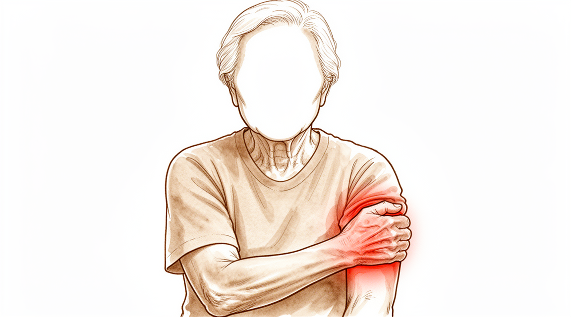
您可能会注意到肘部出现酸痛,尤其是对于从事重体力劳动的40岁以上男性而言。这种退行性关节炎在老年人和男性中更为常见。既往的肘部损伤也会增加患病风险。当您尝试完全伸直或弯曲手臂时,可能会感到疼痛。关节可能感觉僵硬,导致难以完成全范围活动。
简单的日常任务可能变得令人沮丧。您可能难以将手伸到背后扣上胸罩或塞好衬衫。提举物体可能引发剧烈疼痛。部分人还会在小指和无名指感到麻木或刺痛。这是因为靠近肘部的尺神经可能因关节结构的变化而受到刺激。
您的症状可能在手臂使用一段时间后加重。休息通常能带来缓解。然而,您也可能在夜间因不适而醒来。如果您侧卧于患侧,这种疼痛可能会干扰睡眠。您的外科医生将根据您的具体症状、年龄和日常需求来决定最佳的治疗方案。
在早期阶段,非手术治疗通常是首选。这些保守措施可以为许多人提供缓解。如果疼痛在活动的极限位置持续存在,您的外科医生可能会讨论关节镜手术。这种微创手术有助于清除碎屑并改善活动度。对于在所有动作中均出现疼痛的严重病例,可能会考虑全肘关节置换术。由于存在潜在并发症,这是一项仅保留用于特定情况的主要治疗选择。您的治疗方案将根据您的独特情况和功能需求量身定制。
实际发生了什么
在您肘部,骨端的光滑覆盖层磨损。这就是磨损性关节炎。在大多数情况下,这种情况首先发生在关节的主要铰链部分。骨头之间的间隙变窄,特别是在前臂骨与上臂骨相接的地方。随着关节发生变化,会形成小的骨性增生。这些被称为骨刺。它们可能会阻碍您的活动。例如,当您伸直手臂时,骨刺可能会撞击另一块骨头。
您的肘部依靠强韧的韧带来保持稳定。这些韧带就像将骨头捆绑在一起的绳索。如果韧带薄弱或撕裂,骨头就会以错误的方式相互摩擦。这种额外的压力会加速磨损。它还可能引起疼痛和不稳定。您的外科医生会同时检查骨性变化和韧带健康状况,以了解您的具体情况。
有时,受伤或手术后,关节周围的软组织中会形成额外的骨头。这被称为异位骨化。它可能会形成坚硬的阻挡物,使您无法完全活动肘部。这与正常的磨损过程不同。它是一个独立的问题,如果您存在活动机械性阻挡,您的外科医生会对此进行检查。
关节的这些变化解释了您为何感到疼痛和僵硬。光滑软骨的丧失意味着骨头相互研磨。骨刺在物理上阻止您的手臂自由活动。韧带问题使关节感觉松弛或不稳定。了解这些具体变化有助于您的外科医生为您选择正确的治疗方案。
我们能采取的措施
非手术治疗仍是肘骨关节炎早期管理的第一步。非手术管理可能在肘关节炎的早期阶段提供缓解。您的外科医生在考虑其他选项之前,可能会建议您进行自我护理和物理治疗。这些措施旨在减轻疼痛并维持您当前的活动范围。您可以使用冰敷或热敷来缓解僵硬。温和的锻炼有助于保持关节活动,同时不会造成进一步损伤。在转向更强效的治疗方案之前,请给予这种方法足够的时间以发挥作用。
如果简单措施效果不足,您的外科医生可能会讨论药物治疗方案。这通常包括止痛药和抗炎药,以帮助您在日常生活中管理不适。在某些情况下,可能会提供注射治疗。皮质类固醇注射可在有限时间内减轻炎症和疼痛。透明质酸注射旨在润滑关节,尽管其长期疗效的证据各不相同。富血小板血浆(PRP)注射利用您自身的血液成分来促进愈合,但结果并不保证。您的外科医生将根据您的具体病情和疼痛程度建议最合适的方案。
当保守治疗达到极限且症状仍然致残时,才会考虑手术。肘关节炎的适当治疗方案取决于病因、严重程度、患者年龄和功能需求。对于许多患者而言,对于伴有可接受并发症率的原发性肘骨关节炎的致残性症状,手术清创是一种有效的治疗方法。该手术涉及去除骨刺和发炎的组织,以改善活动并减轻疼痛。它在肘骨关节炎的治疗中是安全且有效的。关节镜清创可提供令人满意的疼痛缓解、肘关节活动度的改善以及良好的功能结果。在关节严重受损的更严重病例中,全肘关节置换术可能是一个选择。这涉及用人工组件替换受损的关节面。间置成形术是部分患者的另一种选择,使用组织移植物来缓冲关节。您的外科医生将根据您的个人需求和疾病阶段确定最佳的手术路径。
预期情况
您的肘部磨损性关节炎治疗之旅通常从非手术治疗开始。这是早期症状的标准第一步。您的外科医生将根据您的年龄、损伤的严重程度以及您对手臂功能的需求来制定个性化方案。在早期阶段,休息、康复训练及其他非手术方法通常能带来有意义的缓解。许多人发现其症状足以控制,从而无需手术即可维持日常生活。
如果保守措施无法缓解疼痛或僵硬,您的外科医生可能会讨论手术选项。正确的选择取决于关节炎是由旧伤引起还是自然发展所致,以及您目前保留的活动度。对于轻至中度病例,关节镜下骨膜成形术(一种通过钥匙孔切口去除骨刺并收紧关节囊的手术)是一条安全有效的途径。该手术通常能在并发症风险较低的情况下改善疼痛和功能。在经验丰富的医生操作下,开放手术也能取得类似结果,尽管部分患者可能会随时间推移出现僵硬复发。
对于更晚期的疾病,关节置换是部分患者的可选方案。然而,全肘关节置换术通常不推荐用于活跃人群。其在重度使用下并发症发生率较高,且使用寿命较短。如果您较年轻或非常活跃,且患有严重的创伤后关节炎,可能会考虑部分置换(半关节置换术)。请注意,这种方法后期需要翻修手术的比例较高。
总体而言,大多数接受清创术(清理关节腔)的患者在短期至中期随访中报告功能满意且疼痛得到缓解。尽管在某些关节镜手术后,短期与中期随访之间的活动度可能会有轻微下降,但为了减轻疼痛,这种权衡通常是值得的。如果不进行治疗,关节炎往往会进展,导致僵硬和疼痛加剧。通过适当的管理,您可以期待生活质量的显著改善,但必须接受根据病情的严重程度,可能会出现一定程度的活动度丧失或未来需要其他手术的情况。
何时就医
若肘部持续性疼痛经休息后无改善,请咨询全科医生。若出现关节无力、不稳或卡顿,请寻求专科医生评估。影响睡眠或工作的症状也需引起重视。疼痛突然加重是寻求医疗帮助的另一个原因。肘部骨关节炎较为常见,在40岁及以上人群中患病率为55.0%。它常导致疼痛和活动受限。早期非手术治疗有助于早期阶段的管理。您的外科医生将根据您的年龄、严重程度及日常需求制定个体化治疗方案。请勿忽视影响日常生活的症状。
Evidence & references
Overview
- Nonoperative treatment is the first step in the early management of elbow osteoarthritis [2].
- Elbow arthroscopic osteocapsular arthroplasty is a safe and efficacious treatment for patients with mild to moderate osteoarthritis [1].
- Arthroscopic osteocapsular arthroplasty can be recommended for its favorable overall treatment outcomes for elbow osteoarthritis [29].
- Arthroscopic debridement for primary degenerative osteoarthritis of the elbow results in statistically significant and clinically relevant improvement in elbow range of motion and clinical outcomes with low complication and reoperation rates [16].
- Computer simulation studies recommend arthroscopic debridement in the surgical management of patients with osteoarthritis of the elbow [5].
- Open and arthroscopic debridement procedures are safe and effective in the treatment of elbow osteoarthritis [27].
- Capsulectomy and debridement for primary osteoarthritis of the elbow through a medial trans-flexor approach is associated with a low rate of complications and is safe and effective [3].
- Shoulder arthroplasty is a reasonable option for select patients with osteoarthritis [7].
- Elbow arthroplasty remains a poor option for active patients due to high rates of complications and limited durability [7].
- Total elbow replacement is a viable option for severe arthropathy in patients with congenital afibrinogenemia, achieving satisfactory results with no complications [11].
- The indications for total elbow arthroplasty are broadening, with use for acute trauma and osteoarthritis becoming increasingly common [14].
- Elbow hemiarthroplasty is an option for young or active patients with end-stage posttraumatic arthritis who are unwilling to accept activity limitations, though high rates of revision surgery and revision to total elbow arthroplasty occur [22].
- Interposition elbow arthroplasty using fascia lata graft for post-traumatic elbow osteoarthritis without ligament reconstruction provides favorable surgical outcomes with high satisfaction rates among young patients [26].
Anatomy & Pathophysiology
- Osteophytic change occurs predominantly in the ulnohumeral compartment of the elbow [33].
- Joint space narrowing more frequently affects the radiocapitellar articulation [33].
- Normal kinematics was preserved in the osteoarthritic elbow with a normal radiocapitellar joint [13].
- Three-dimensional computational models identified unique regions of impingement, such as between the radial head and a posterior capitellar osteophyte in extension [34].
- An increase in carrying angle is associated with radial deviation of stress [23].
- Deficiency of the ulnar collateral ligament (UCL) increased contact pressure within the posteromedial compartment of the elbow with an associated decrease in the contact area [46].
- Elbow ligament reconstruction effectively restores stability and limits progression to osteoarthritis in the long term [32].
- Both posterolateral and posteromedial rotatory instability must be addressed surgically to restore elbow stability [40].
- Heterotopic ossification should be considered in patients with a mechanical block to function after injury to the elbow or surgery [39].
- Flexion contracture is not primarily related to bony changes of the elbow [44].
Classification
- The prevalence of elbow osteoarthritis is 55.0% in respondents aged 40 years or older [8].
- The symptomatic prevalence of elbow osteoarthritis is 22.6% in respondents aged 40 years or older [8].
- Older age is a significant risk factor for elbow osteoarthritis [8].
- Male sex is a significant risk factor for elbow osteoarthritis [8].
- A history of elbow trauma is a significant risk factor for elbow osteoarthritis [8].
- Primary osteoarthritis of the elbow is characterized by relative preservation of articular cartilage [10].
- Primary osteoarthritis of the elbow is characterized by maintenance of joint space [10].
- Primary osteoarthritis of the elbow is characterized by hypertrophic osteophyte formation [10].
- Normal kinematics is preserved in the osteoarthritic elbow with a normal radiocapitellar joint (OAN group) [13].
- Both the BM and HR classification systems demonstrated substantial intraobserver and interobserver reliability for evaluating radiographic severity of post-traumatic arthritis and primary osteoarthritis of the elbow [25].
- The bony landmarks classification system effectively delineated osteophyte distribution in elbow patients [36].
Clinical Presentation
- Nonoperative treatment remains the first step in the early management of elbow osteoarthritis [2].
- Nonsurgical management may provide relief in early stages of elbow arthritis [15].
- The appropriate treatment for elbow arthritis depends on the etiology, severity, patient age, and functional demands [4].
- Surgical treatment for elbow arthritis is based on disease etiology, severity of degeneration, and patient age [6].
- Treatment of elbow arthritis must be individualized based on etiology, severity, patient age, and functional demands [15].
- The prevalence of elbow osteoarthritis was 55.0% in respondents aged 40 years or older [8].
- The symptomatic prevalence of elbow osteoarthritis was 22.6% in respondents aged 40 years or older [8].
- Older age is a significant risk factor for elbow osteoarthritis [8].
- Male sex is a significant risk factor for elbow osteoarthritis [8].
- A history of elbow trauma is a significant risk factor for elbow osteoarthritis [8].
- Primary osteoarthritis of the elbow is characterized by relative preservation of articular cartilage and maintenance of joint space with hypertrophic osteophyte formation [10].
- Primary elbow osteoarthritis predominantly affects middle-aged men undertaking heavy manual work [28].
- Primary elbow osteoarthritis presents with pain, limited movement, and potential ulnar nerve symptoms [28].
- Arthroscopic treatment of elbow osteoarthritis addresses pathologic processes associated with arthritis of the elbow [20].
- Arthroscopic treatment of elbow osteoarthritis is indicated for pain at motion extremes [15].
- Total elbow arthroplasty is indicated for pain throughout the arc of motion in elbow arthritis [15].
Investigations
- Nonoperative treatment remains the first step in the early management of elbow osteoarthritis [2].
- Primary osteoarthritis of the elbow is unique due to relative preservation of articular cartilage and maintenance of joint space with hypertrophic osteophyte formation [10].
- Osteophytic change occurs predominantly in the ulnohumeral compartment of the elbow, whereas joint space narrowing more frequently affects the radiocapitellar articulation [33].
- An increase in carrying angle is associated with radial deviation of stress [23].
- Both the BM and HR classification systems demonstrated substantial intraobserver and interobserver reliability for evaluating radiographic severity of post-traumatic arthritis and primary osteoarthritis of the elbow [25].
- CT has greater sensitivity than radiographs for the detection of osteophytes and loose bodies in primary elbow osteoarthritis [51].
- Three-dimensional computational models identified the locations and volumes of bony impingement in patients with osteoarthritis of the elbow and highlighted unique regions of impingement, such as between the radial head and a posterior capitellar osteophyte in extension [34].
- Normal kinematics was preserved in the osteoarthritic elbow with a normal radiocapitellar joint (OAN group) [13].
- The authors recommend performing an MRI if healing does not occur by a reasonable time despite successful bony healing to assess potential cartilage damage [50].
Treatment
Non-Operative Management
- Nonoperative treatment remains the first step in the early management of elbow osteoarthritis [2].
- Nonsurgical management may provide relief in early stages of elbow arthritis [15].
Surgical Decision-Making and Indications
- The appropriate treatment for elbow arthritis depends on the etiology, severity, patient age, and functional demands [4].
- Surgical treatment for elbow arthritis is based on disease etiology, severity of degeneration, and patient age [6].
- Treatment of elbow arthritis must be individualized based on etiology, severity, patient age, and functional demands [15].
- Primary elbow osteoarthritis predominantly affects middle-aged men undertaking heavy manual work, presenting with pain, limited movement, and potential ulnar nerve symptoms [28].
Arthroscopic and Open Debridement
- Elbow arthroscopic osteocapsular arthroplasty (AOA) is a safe, efficacious treatment for patients with mild to moderate osteoarthritis [1].
- Capsulectomy and debridement for primary osteoarthritis of the elbow through a medial trans-flexor approach is associated with a low rate of complications and is safe and effective [3].
- Arthroscopic debridement is recommended in the surgical management of patients with osteoarthritis of the elbow based on computer simulation [5].
- Elbow arthroscopic debridement for primary degenerative osteoarthritis results in statistically significant and clinically relevant improvement in elbow range of motion and clinical outcomes with low complication and reoperation rates [16].
- Open and arthroscopic debridement procedures are safe and effective in the treatment of elbow OA [27].
- Arthroscopic osteocapsular arthroplasty can be recommended for its favorable overall treatment outcomes for elbow osteoarthritis [29].
- Osteocapsular debridement is an effective surgical treatment option for patients with symptomatic primary elbow osteoarthritis who have failed conservative management [30].
- Osteocapsular debridement is an effective surgical treatment option for patients with symptomatic primary elbow osteoarthritis who have failed conservative management [31].
- Surgical debridement is an effective treatment for the disabling symptoms of primary elbow OA with an acceptable complication rate [37].
- Arthroscopic debridement for elbow osteoarthritis provides satisfactory pain relief, improvement of elbow motion, and good functional outcome [38].
- Osteocapsular debridement, a non-arthroplasty option, proves to be safe and effective in treating patients with elbow arthritis [47].
Total Elbow Arthroplasty
- Shoulder arthroplasty is a reasonable option for select patients with osteoarthritis, whereas elbow arthroplasty remains a poor option for active patients due to high rates of complications and limited durability [7].
- Total elbow replacement is a viable option for severe arthropathy in patients with congenital afibrinogenemia, achieving satisfactory results with no complications [11].
- The range of indications for total elbow arthroplasty is broadening, with use for acute trauma and osteoarthritis becoming increasingly more common [14].
- Total elbow arthroplasty is a surgical option for pain throughout the arc of motion in elbow arthritis [15].
Interposition Arthroplasty
- Interposition elbow arthroplasty using fascia lata graft for post-traumatic elbow osteoarthritis without ligament reconstruction provides favorable surgical outcomes with high satisfaction rates among young patients [26].
Complications
- Elbow arthroplasty remains a poor option for active patients due to high rates of complications and limited durability [7].
- Total elbow arthroplasty has an acceptable complication rate [42].
- Total elbow replacement is a viable option for severe arthropathy in patients with congenital afibrinogenemia, with no complications reported in the case [11].
Recovery
- Nonoperative treatment is the first step in the early management of elbow osteoarthritis [2].
- Treatment of elbow arthritis must be individualized based on etiology, severity, patient age, and functional demands [15].
- Nonsurgical management may provide relief in early stages of elbow arthritis [15].
- Surgical options for elbow arthritis range from arthroscopic debridement for pain at motion extremes to total elbow arthroplasty for pain throughout the arc of motion [15].
- Elbow arthroplasty remains a poor option for active patients due to high rates of complications and limited durability [7].
- Elbow arthroscopic osteocapsular arthroplasty (AOA) is a safe and efficacious treatment for patients with mild to moderate osteoarthritis [1].
- Arthroscopic debridement provides satisfactory elbow function and improvement in pain with little chance of reoperation at midterm follow-up for both posttraumatic and primary degenerative osteoarthritis [9].
- Clinical outcomes for primary elbow osteoarthritis patients undergoing arthroscopic OCA improve from preoperative assessment to short- and medium-term follow-up [18].
- Range of motion (ROM) decreases between short- and medium-term follow-up after arthroscopic OCA for primary elbow osteoarthritis [18].
- Open and arthroscopic treatment yields similar results in experienced hands, though patients may experience some degree of recurrence or motion loss [56].
- Capsulectomy and debridement through a medial trans-flexor approach is associated with a low rate of complications and is safe and effective for primary osteoarthritis of the elbow [3].
- Most patients undergoing open debridement and radiocapitellar replacement have an uneventful postoperative course, painless elbow joint, and satisfactory functional recovery at short-term follow-up [19].
- Radiocapitellar prosthetic arthroplasty largely preserves elbow kinematics and stability [24].
- Revision radiocapitellar arthroplasty by mismatched implant components is a salvage option that improved Mayo elbow performance scores from poor to good/excellent with no signs of implant failure at a minimum three-year follow-up [12].
- Elbow hemiarthroplasty is an option for young or active patients with end-stage posttraumatic arthritis who are unwilling to accept activity limitations, though high rates of revision surgery and revision to total elbow arthroplasty occur [22].
- Elbow ligament reconstruction effectively restores stability and limits progression to osteoarthritis in the long term [32].
Key Evidence
- [L4] Elbow AOA is a safe, efficacious treatment for patients with mild to moderate osteoarthritis. [1] (10.1016/j.jhsa.2015.11.018)
- [L5] Nonoperative treatment remains the first step in the early management of elbow osteoarthritis. [2] (10.2106/jbjs.e.00568)
- [L4] This approach is associated with a low rate of complications and is safe and effective for the treatment of primary osteoarthritis of the elbow. [3] (10.1016/j.jhsa.2011.07.018)
- [L5] The appropriate treatment for elbow arthritis depends on the etiology, severity, patient age, and functional demands. [4] (10.1016/j.jhsa.2009.02.019)
- [L4] The study recommends this technique in the surgical management of patients with osteoarthritis of the elbow. [5] (10.1302/0301-620x.96b2.30714)
- [L5] Surgical treatment for elbow arthritis is based on disease etiology, severity of degeneration, and patient age. [6] (10.1016/j.jhsa.2007.12.022)
- [L5] Shoulder arthroplasty is a reasonable option for select patients with osteoarthritis, whereas elbow arthroplasty remains a poor option for active patients due to high rates of complications and limited durability. [7] (10.1016/j.jht.2022.02.002)
- [L3] The prevalence of elbow OA was 55.0% in respondents aged 40 years or older, with a symptomatic prevalence of 22.6%; older age, male sex, and a history of elbow trauma were identified as significant risk factors. [8] (10.1016/j.jse.2018.02.049)
- [L3] Patients with either pathology can expect satisfactory elbow function and an improvement in pain with little chance of reoperation at the midterm of the follow-up duration. [9] (10.1016/j.jseint.2021.07.018)
- [L4] Primary osteoarthritis of the elbow is unique due to relative preservation of articular cartilage and maintenance of joint space with hypertrophic osteophyte formation. [10] (10.5435/00124635-200802000-00005)
- [L4] The patient achieved satisfactory results with no complications, demonstrating that total elbow replacement is a viable option for severe arthropathy in this rare condition. [11] (10.2106/jbjs.i.00149)
- [L4] At a minimum of three-year follow-up, both cases improved from poor to good and excellent Mayo elbow performance scores with no signs of implant failure on standard radiographs. [12] (10.1177/17585732241297152)
- [L4] Normal kinematics was preserved in the osteoarthritic elbow with a normal radiocapitellar joint (OAN group). [13] (10.1016/j.jhsa.2013.02.006)
- [L2] The range of indications for total elbow arthroplasty is broadening; total elbow arthroplasty for acute trauma and osteoarthritis is becoming increasingly more common. [14] (10.1302/2058-5241.5.190036)
- [L5] Treatment of elbow arthritis must be individualized based on etiology, severity, patient age, and functional demands; nonsurgical management may provide relief in early stages, while surgical options range from arthroscopic debridement for pain at motion extremes to total elbow arthroplasty for pain throughout the arc of motion. [15] (10.1016/j.jhsa.2012.12.037)
- [L1] Elbow arthroscopic debridement for primary degenerative osteoarthritis results in statistically significant and clinically relevant improvement in elbow range of motion and clinical outcomes with low complication and reoperation rates. [16] (10.1016/j.arthro.2017.08.247)
- [L4] Serial assessment of patients with primary elbow OA who underwent arthroscopic OCA showed that the clinical outcomes improved from preoperative assessment to short- and medium-term follow-up, although ROM decreased between short- and medium-term follow-up. [18] (10.1177/23259671231162398)
- [L4] Most patients had an uneventful postoperative course and have shown a painless elbow joint, with satisfactory functional recovery at short-term follow-up. [19] (10.1016/j.jse.2011.08.071)
- [L4] This procedure addresses the pathologic processes associated with arthritis of the elbow and was safe and effective in this series. [20] (10.1016/j.jse.2007.04.005)
- [L4] Elbow hemiarthroplasty is an option for young or active patients with end-stage posttraumatic arthritis who are unwilling to accept activity limitations, though high rates of revision surgery and revision to total elbow arthroplasty occur. [22] (10.5435/jaaos-d-18-00055)
- [L3] An increase in carrying angle is associated with radial deviation of stress. [23] (10.1097/corr.0000000000002921)
- [L4] The procedure largely preserves elbow kinematics and stability. [24] (10.1016/j.jse.2014.01.042)
- [L3] Both the BM and HR classification systems demonstrated substantial intraobserver and interobserver reliability for evaluating radiographic severity of post-traumatic arthritis and primary osteoarthritis of the elbow. [25] (10.1016/j.jse.2014.10.015)
- [L4] It provides favorable surgical outcomes with high satisfaction rates among young patients with elbow osteoarthritis. [26] (10.1016/j.jhsg.2024.05.002)
- [L5] However, from the data we obtained the open and arthroscopic debridement procedures seem to be safe and effective in the treatment of elbow OA. [27] (10.1186/s12891-018-2318-x)
- [L4] Primary elbow osteoarthritis predominantly affects middle-aged men undertaking heavy manual work, presenting with pain, limited movement, and potential ulnar nerve symptoms. [28] (10.1111/j.1758-5740.2010.00089.x)
- [L4] Arthroscopic osteocapsular arthroplasty can be recommended for its favorable overall treatment outcomes for elbow osteoarthritis. [29] (10.1016/j.jse.2019.09.036)
- [L1] Osteocapsular debridement is an effective surgical treatment option for patients with symptomatic primary elbow osteoarthritis who have failed conservative management. [30] (10.1016/j.jse.2020.01.061)
- [L2] Osteocapsular debridement is an effective surgical treatment option for patients with symptomatic primary elbow osteoarthritis who have failed conservative management. [31] (10.1016/j.jse.2020.01.060)
- [L4] Elbow ligament reconstruction by the technique of O'Driscoll et al effectively restores stability and limits progression to osteoarthritis in the long term. [32] (10.1016/j.jseint.2022.12.009)
- [L3] Osteophytic change occurs predominantly in the ulnohumeral compartment of the elbow, whereas joint space narrowing more frequently affects the radiocapitellar articulation. [33] (10.1016/j.jse.2006.08.005)
- [L4] Three-dimensional computational models identified the locations and volumes of bony impingement in patients with osteoarthritis of the elbow and highlighted unique regions of impingement, such as between the radial head and a posterior capitellar osteophyte in extension. [34] (10.1016/j.jhsa.2013.03.035)
- [L3] The bony landmarks classification system effectively delineated osteophyte distribution in elbow patients. [36] (10.1186/s13018-025-06145-9)
- [L1] Surgical debridement is an effective treatment for the disabling symptoms of primary elbow OA with an acceptable complication rate. [37] (10.1302/2058-5241.5.190095)
- [L4] Arthroscopic debridement for elbow osteoarthritis provides satisfactory pain relief, improvement of elbow motion, and good functional outcome. [38] (10.1016/j.jse.2014.01.009)
- [L4] Its presence should be considered in patients in whom there is a mechanical block to function after injury to the elbow or surgery. [39] (10.1016/j.jse.2007.06.018)
- [L4] Both directions of instability must be addressed surgically to restore elbow stability. [40] (10.1016/j.injury.2007.01.039)
- [L5] Total elbow arthroplasty has become a successful reconstruction for painful destructive arthritis about the elbow with an acceptable complication rate. [42] (10.1016/j.jhsa.2009.02.021)
- [L4] This indicates that flexion contracture is not primarily related to bony changes of the elbow. [44] (10.5435/jaaos-d-17-00110)
- [L5] Under the conditions tested, deficiency of the UCL increased contact pressure within the posteromedial compartment of the elbow with associated decrease in the contact area. [46] (10.1016/j.arthro.2013.03.046)
- [Commentary] Osteocapsular debridement, a non-arthroplasty option, proves to be safe and effective in treating patients with elbow arthritis, making the elbow more forgiving than previously thought. [47] (10.1016/j.arthro.2020.10.029)
- [Case_report] The authors recommend performing an MRI if healing does not occur by a reasonable time despite successful bony healing to assess potential cartilage damage. [50] (10.1007/s00402-005-0018-0)
- [L1] CT has greater sensitivity than radiographs for the detection of osteophytes and loose bodies in primary elbow osteoarthritis. [51] (10.1016/j.jse.2021.04.001)
- [Commentary] Open and arthroscopic treatment yields similar results in experienced hands, but patients may have some degree of recurrence or motion loss. [56] (10.1016/j.arthro.2019.02.004)
References
[1] Outcomes of Elbow Arthroscopic Osteocapsular Arthroplasty. The Journal of Hand Surgery. 2016. DOI: 10.1016/j.jhsa.2015.11.018 [2] Management of Elbow Osteoarthritis. The Journal of Bone & Joint Surgery. 2006. DOI: 10.2106/jbjs.e.00568 [3] Capsulectomy and Debridement for Primary Osteoarthritis of the Elbow Through a Medial Trans-Flexor Approach. The Journal of Hand Surgery. 2011. DOI: 10.1016/j.jhsa.2011.07.018 [4] Elbow Arthritis: Current Concepts. The Journal of Hand Surgery. 2009. DOI: 10.1016/j.jhsa.2009.02.019 [5] Arthroscopic debridement in the treatment of patients with osteoarthritis of the elbow, based on computer simulation. The Bone & Joint Journal. 2014. DOI: 10.1302/0301-620x.96b2.30714 [6] Surgical Options for the Arthritic Elbow. The Journal of Hand Surgery. 2008. DOI: 10.1016/j.jhsa.2007.12.022 [7] Surgical management of osteoarthritis in the shoulder and elbow. Journal of Hand Therapy. 2022. DOI: 10.1016/j.jht.2022.02.002 [8] The prevalence of elbow osteoarthritis in Japanese middle-aged and elderly populations: the relationship between risk factors and function. Journal of Shoulder and Elbow Surgery. 2018. DOI: 10.1016/j.jse.2018.02.049 [9] Midterm outcomes and survivorship of arthroscopic elbow debridement: a comparison of posttraumatic versus primary degenerative osteoarthritis. JSES International. 2022. DOI: 10.1016/j.jseint.2021.07.018 [10] Primary Osteoarthritis of the Elbow: Current Treatment Options. Journal of the American Academy of Orthopaedic Surgeons. 2008. DOI: 10.5435/00124635-200802000-00005 [11] Severe Elbow Arthropathy in a Patient with Congenital Afibrinogenemia. The Journal of Bone & Joint Surgery. 2010. DOI: 10.2106/jbjs.i.00149 [12] Revision radiocapitellar arthroplasty by mismatched implant components – A salvage option: A report of two cases with a minimum three-year follow-up. Shoulder & Elbow. 2024. DOI: 10.1177/17585732241297152 [13] Kinematic Changes in Elbow Osteoarthritis: In Vivo and 3-Dimensional Analysis Using Computed Tomographic Data. The Journal of Hand Surgery. 2013. DOI: 10.1016/j.jhsa.2013.02.006 [14] Global trends in indications for total elbow arthroplasty: a systematic review of national registries. EFORT Open Reviews. 2020. DOI: 10.1302/2058-5241.5.190036 [15] Elbow Arthritis: Current Concepts. The Journal of Hand Surgery. 2013. DOI: 10.1016/j.jhsa.2012.12.037 [16] Arthroscopic Debridement for Primary Degenerative Osteoarthritis of the Elbow Leads to Significant Improvement in Range of Motion and Clinical Outcomes: A Systematic Review. Arthroscopy. 2017. DOI: 10.1016/j.arthro.2017.08.247 [18] Serial Changes in Clinical Outcomes After Arthroscopic Osteocapsular Arthroplasty for Primary Elbow Osteoarthritis: A Medium-term Follow-up Study. Orthopaedic Journal of Sports Medicine. 2023. DOI: 10.1177/23259671231162398 [19] Open debridement and radiocapitellar replacement in primary and post-traumatic arthritis of the elbow: a multicenter study. Journal of Shoulder and Elbow Surgery. 2012. DOI: 10.1016/j.jse.2011.08.071 [20] Osteoarthritis of the elbow: Results of arthroscopic osteophyte resection and capsulectomy. Journal of Shoulder and Elbow Surgery. 2008. DOI: 10.1016/j.jse.2007.04.005 [22] Outcomes After Hemiarthroplasty of the Elbow for the Management of Posttraumatic Arthritis: Minimum 2-Year Follow-up. Journal of the American Academy of Orthopaedic Surgeons. 2019. DOI: 10.5435/jaaos-d-18-00055 [23] How Does the Subchondral Bone Density Distribution of the Distal Humerus Change Between Early and Advanced Stages of Osteoarthritis?. Clinical Orthopaedics & Related Research. 2023. DOI: 10.1097/corr.0000000000002921 [24] Radiocapitellar prosthetic arthroplasty: a report of 6 cases and review of the literature. Journal of Shoulder and Elbow Surgery. 2014. DOI: 10.1016/j.jse.2014.01.042 [25] Reliability testing of two classification systems for osteoarthritis and post-traumatic arthritis of the elbow. Journal of Shoulder and Elbow Surgery. 2015. DOI: 10.1016/j.jse.2014.10.015 [26] Functional Outcomes Following Interposition Elbow Arthroplasty Using Fascia Lata Graft for Post-Traumatic Elbow Osteoarthritis Without Ligament Reconstruction: A Minimum 3-Year Follow-Up Study. Journal of Hand Surgery Global Online. 2024. DOI: 10.1016/j.jhsg.2024.05.002 [27] Treatment of osteoarthritis of the elbow with open or arthroscopic debridement: a narrative review. BMC Musculoskeletal Disorders. 2018. DOI: 10.1186/s12891-018-2318-x [28] Primary Elbow Osteoarthritis: An Updated Review. Shoulder & Elbow. 2011. DOI: 10.1111/j.1758-5740.2010.00089.x [29] Arthroscopic osteocapsular arthroplasty for advanced-stage primary osteoarthritis of the elbow using a computed tomography–based classification. Journal of Shoulder and Elbow Surgery. 2020. DOI: 10.1016/j.jse.2019.09.036 [30] Patient and Procedure Specific Variables Associated with Revision or Removal of Radial Head Arthroplasty. Journal of Shoulder and Elbow Surgery. 2020. DOI: 10.1016/j.jse.2020.01.061 [31] The Clinical Impact of Different Approaches to Osteocapsular Debridement for Primary Osteoarthritis of the Elbow: A Systematic Review. Journal of Shoulder and Elbow Surgery. 2020. DOI: 10.1016/j.jse.2020.01.060 [32] Lateral elbow ligament reconstruction for posterolateral rotatory instability: 10 years follow-up in 32 patients. JSES International. 2023. DOI: 10.1016/j.jseint.2022.12.009 [33] Radiographic changes at the elbow in primary osteoarthritis: A comparison with normal aging of the elbow joint. Journal of Shoulder and Elbow Surgery. 2007. DOI: 10.1016/j.jse.2006.08.005 [34] Identifying the Location and Volume of Bony Impingement in Elbow Osteoarthritis by 3-Dimensional Computational Modeling. The Journal of Hand Surgery. 2013. DOI: 10.1016/j.jhsa.2013.03.035 [36] Bony landmarks guided mapping of the osteophytes of the elbow osteoarthritis patients: a three dimensional computed tomograph based study. Journal of Orthopaedic Surgery and Research. 2025. DOI: 10.1186/s13018-025-06145-9 [37] Arthroscopic and open debridement in primary elbow osteoarthritis: a systematic review and meta-analysis. EFORT Open Reviews. 2020. DOI: 10.1302/2058-5241.5.190095 [38] Arthroscopic débridement for primary osteoarthritis of the elbow: analysis of preoperative factors affecting outcome. Journal of Shoulder and Elbow Surgery. 2014. DOI: 10.1016/j.jse.2014.01.009 [39] Heterotopic ossification after the Outerbridge-Kashiwagi procedure in the elbow. Journal of Shoulder and Elbow Surgery. 2008. DOI: 10.1016/j.jse.2007.06.018 [40] Combined posterolateral and posteromedial rotatory instability of the elbow. Injury Extra. 2007. DOI: 10.1016/j.injury.2007.01.039 [42] Total Elbow Arthroplasty: Surgical Technique. The Journal of Hand Surgery. 2009. DOI: 10.1016/j.jhsa.2009.02.021 [44] Natural History of the Elbow Bony Architecture in Patients With Obstetric Brachial Plexus Injury and the Association With Flexion Contractures. Journal of the American Academy of Orthopaedic Surgeons. 2018. DOI: 10.5435/jaaos-d-17-00110 [46] Impact of Ulnar Collateral Ligament Tear on Contact Pressure and Contact Area in the Posteromedial Compartment of the Elbow (SS‐39). Arthroscopy. 2013. DOI: 10.1016/j.arthro.2013.03.046 [47] Editorial Commentary: Arthroscopic Elbow Arthritis Treatment With Osteocapsular Debridement Yields Favorable Results: On Second Thought, the Elbow Isn’t That Unforgiving. Arthroscopy: The Journal of Arthroscopic & Related Surgery. 2021. DOI: 10.1016/j.arthro.2020.10.029 [50] Are bone bruises a possible cause of osteochondritis dissecans of the capitellum? a case report and review of the literature. Archives of Orthopaedic and Trauma Surgery. 2005. DOI: 10.1007/s00402-005-0018-0 [51] Effectiveness of radiographs and computed tomography in evaluating primary elbow osteoarthritis. Journal of Shoulder and Elbow Surgery. 2021. DOI: 10.1016/j.jse.2021.04.001 [56] Editorial Commentary: Open Versus Arthroscopic Elbow Osteocapsular Arthroplasty. Arthroscopy: The Journal of Arthroscopic & Related Surgery. 2019. DOI: 10.1016/j.arthro.2019.02.004


