Giải nén thần kinh trên gai
Patients › Rehabilitation
Rehabilitation after isolated arthroscopic suprascapular nerve decompression, with honest framing of nerve recovery.
Giao thức này bao gồm quá trình phục hồi chức năng sau phẫu thuật giải phóng chằng thần kinh trên gai bằng nội soi đơn độc với Dr Kieran Hirpara tại Bệnh viện tư nhân Mater Rockhampton: giải phóng thần kinh tại rãnh trên gai và/hoặc rãnh gai vai, mà không thực hiện bất kỳ sửa chữa nào khác. Hãy mang theo trang này hoặc bản PDF của nó đến buổi vật lý trị liệu đầu tiên để quá trình phục hồi chức năng của bạn được phối hợp nhịp nhàng. Quá trình phục hồi chức năng của bạn sẽ được điều chỉnh cá nhân hóa bởi bác sĩ vật lý trị liệu thông qua các giai đoạn dưới đây, tùy thuộc vào mức độ hồi phục của vai bạn.
Quan trọng: vui lòng đọc trước. Giải phóng thần kinh trên gai thường được thực hiện cùng với phẫu thuật sửa chữa gân quay. Giao thức này chỉ dành cho trường hợp giải phóng thần kinh đơn độc. Nếu ca phẫu thuật của bạn cũng bao gồm sửa chữa gân quay, vui lòng tuân theo giao thức sửa chữa gân quay: gân được sửa chữa sẽ đặt ra nhịp độ chậm hơn, và giao thức đó sẽ được ưu tiên áp dụng hơn giao thức này. Nếu bạn không chắc chắn về loại phẫu thuật mình đã thực hiện, hãy liên hệ với phòng khám trước khi bắt đầu.
Nếu bạn có bất kỳ lo ngại nào về vết mổ sau phẫu thuật, hãy liên hệ với phòng khám. Việc chụp ảnh vết mổ và gửi email để được xem xét thường rất hữu ích.
Những điều cần biết
Khi dây thần kinh được giải phóng, không cần bảo vệ bất kỳ quá trình sửa chữa gân nào, do đó quá trình phục hồi chức năng có thể diễn ra nhanh chóng. Băng cố định chỉ được đeo để giảm khó chịu và trong thời gian ngắn (thường là trong khoảng một tuần đầu tiên, và tối đa là hai tuần), và sẽ được tháo bỏ càng nhiều càng tốt khi vai ổn định. Các cử động nhẹ nhàng bắt đầu sớm, tùy theo mức độ thoải mái, và hầu hết mọi người có thể trở lại các hoạt động hàng ngày bình thường sau vài tuần.
Quá trình hồi phục của chính dây thần kinh tuân theo một tiến trình riêng, tách biệt với chuyển động của vai. Phẫu thuật giúp giảm áp lực lên dây thần kinh; cơn đau do áp lực này thường giảm đi tương đối nhanh. Việc hồi phục sức mạnh và khối lượng cơ ở các cơ do dây thần kinh chi phối (cơ trên gai và cơ dưới gai, nằm trên xương bả vai) diễn ra chậm hơn và tính theo thang đo là vài tháng. Mức độ hồi phục hoàn toàn của sức mạnh và khối lượng cơ khác nhau tùy từng người: ở một số người, quá trình hồi phục là hoàn toàn, ở những người khác chỉ là một phần, và các vấn đề dây thần kinh kéo dài có thể không hồi phục hoàn toàn. Vật lý trị liệu của bạn bao gồm các bài tập cụ thể để kích hoạt lại các cơ này khi dây thần kinh hồi phục. Chuyên gia vật lý trị liệu và phòng khám sẽ hướng dẫn bạn về những gì cần mong đợi trong trường hợp của bạn.
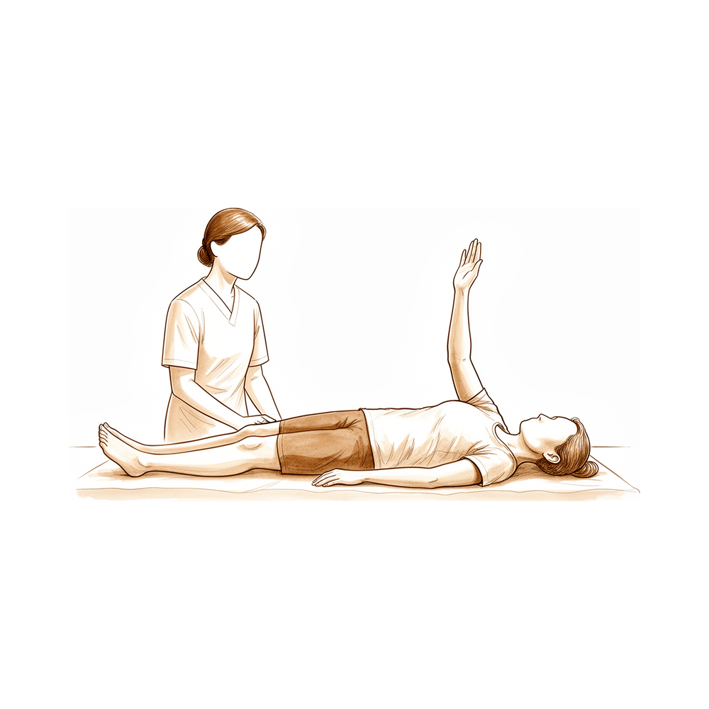
Giai đoạn I — Vận động sớm (Tuần 0–2)
Mục tiêu đầu tiên là sự thoải mái và vận động nhẹ nhàng, sớm. Nẹp cố định vai chỉ dùng để giảm khó chịu và nên được tháo bỏ càng nhiều càng tốt khi vai đã ổn định; bạn không cần phải đeo khi ngủ. Không lái xe khi đang đeo nẹp. Uống thuốc giảm đau đều đặn trong những ngày đầu để bạn có thể bắt đầu vận động cánh tay. Giữ cho bàn tay, cổ tay và khuỷu tay vận động ngay từ đầu, và bắt đầu vận động vai nhẹ nhàng trong phạm vi thoải mái theo hướng dẫn.
Dành cho nhà vật lý trị liệu của bạn:
Mục tiêu
- Thoải mái và bảo vệ vết mổ
- Vận động phạm vi chuyển động nhẹ nhàng sớm trong giới hạn không đau
- Duy trì vận động bàn tay, cổ tay và khuỷu tay
Quản lý điều trị
- Nẹp cố định chỉ dùng để giảm khó chịu, thường trong khoảng 7 ngày sau phẫu thuật (tối đa hai tuần nếu cần thiết cho sự thoải mái), giảm dần theo mức độ triệu chứng cho phép
- Vận động phạm vi chuyển động nhẹ nhàng sớm theo mức độ thoải mái: bài tập con lắc, nâng đỡ thụ động và chủ động hỗ trợ, xoay ngoài và xoay trong, và gập/mở rộng khuỷu tay
- Co cơ đẳng trương cơ delta và cơ bả vai khi cảm thấy thoải mái
- Dùng thuốc giảm đau trước khi tập; chườm lạnh để giảm đau khi cần thiết
Cẩn trọng
- Giữ vận động sớm trong phạm vi thoải mái, không đau
- Không nâng vật nặng, đẩy hoặc kéo mạnh
- Không lái xe khi đang đeo nẹp
Tiêu chí để chuyển sang giai đoạn tiếp theo
- Đau đỡ và ổn định hơn
- Vết mổ lành tốt
- Dung nạp được vận động phạm vi chuyển động sớm
Giai đoạn II — Khôi phục tầm vận động và kích hoạt lại cơ (Tuần 2–6)
Sau khi bỏ nạng, giai đoạn này khôi phục tầm vận động hoàn toàn và bắt đầu tăng cường sức mạnh nhẹ, bao gồm các bài tập cụ thể để kích hoạt lại cơsupraspinatus và infraspinatus khi dây thần kinh hồi phục. Hầu hết mọi người trở lại các hoạt động hàng ngày bình thường trong giai đoạn này. Tiến độ được hướng dẫn bởi sự thoải mái, không phải theo lịch trình.
Đối với nhà vật lý trị liệu của bạn:
Mục tiêu
- Tầm vận động chủ động hoàn toàn trong tất cả các mặt phẳng
- Bắt đầu tăng cường sức mạnh nhẹ và kích hoạt lại cơ vòng xoay (supraspinatus/infraspinatus)
- Tự lập trong các hoạt động sinh hoạt hàng ngày
Quản lý
- Tiến tới tầm vận động chủ động hoàn toàn trong tất cả các hướng
- Bắt đầu tăng cường sức mạnh nhẹ từ khoảng tuần thứ 2: đẳng trương tiến tới bài tập với dây đàn hồi cho cơ vòng xoay, cơ delta và các cơ ổn định xương bả vai, tải trọng thấp và số lần lặp lại cao hơn
- Đặc biệt chú ý đến xoay ngoài không đau và kích hoạt lại cơ supraspinatus và infraspinatus khi dây thần kinh hồi phục
- Trở lại dần dần các hoạt động hàng ngày bình thường, thường là vào khoảng bốn tuần
Cẩn trọng
- Tăng cường sức mạnh chỉ trong phạm vi thoải mái và không nên gây đau kéo dài
- Tránh đẩy, kéo mạnh và nâng vật nặng trong khi sức mạnh đang hồi phục
- Dự kiến sức mạnh sẽ trở lại dần dần: công việc kích hoạt lại được điều chỉnh theo tốc độ hồi phục của dây thần kinh, không ép buộc
Tiêu chí để tiến độ
- Tầm vận động chủ động hoàn toàn hoặc gần hoàn toàn, không đau
- Tăng cường sức mạnh nhẹ được dung nạp mà không gây bùng phát
Giai đoạn III — Tăng cường sức mạnh và trở lại hoạt động (Tuần 6–12 và xa hơn)
Từ khoảng sáu tuần, việc tăng cường sức mạnh được tiến triển mà không có hạn chế cụ thể, hướng tới việc trở lại các hoạt động trên đầu, công việc nặng hơn và thể thao. Việc tăng cường sức mạnh cô lập cơ supraspinatus và infraspinatus được nâng cao khi dây thần kinh tiếp tục hồi phục, quá trình này thường kéo dài trong vài tháng.
Đối với nhà vật lý trị liệu của bạn:
Mục tiêu
- Tăng cường sức mạnh toàn diện mà không có hạn chế
- Trở lại dần dần các hoạt động trên đầu, công việc nặng hơn và thể thao
- Tiếp tục phục hồi sức mạnh của nhóm cơ xoay cuff khi dây thần kinh hồi phục
Quản lý
- Từ khoảng tuần 6, tiến triển đến tăng cường sức mạnh toàn diện, bao gồm các bài tập chuỗi kín và kháng lực tiến triển
- Từ khoảng tuần 12, nâng cao việc tăng cường sức mạnh cô lập cơ supraspinatus và infraspinatus
- Chia giai đoạn việc trở lại công việc trên đầu và thể thao; việc trở lại hoàn toàn các hoạt động trên đầu thường đạt được vào khoảng bốn đến sáu tuần đối với các nhiệm vụ nhẹ hơn, với việc trở lại thể thao dần dần trong các tuần đến tháng tiếp theo khi sức mạnh cho phép và khi không còn đau
- Tiếp tục một chương trình duy trì khi dây thần kinh và cơ bắp tiếp tục hồi phục
Cẩn trọng
- Sự tiến triển vẫn dựa trên triệu chứng
- Sức mạnh và khối lượng cơ của các cơ bị ảnh hưởng có thể tiếp tục hồi phục trong vài tháng, và sự hồi phục có thể chỉ một phần: điều chỉnh kỳ vọng cho phù hợp và tránh gây căng thẳng quá mức trong khi sức mạnh chưa hoàn toàn
Sau giao thức của bạn
Các giai đoạn trên được điều chỉnh từ các bài báo kỹ thuật và nghiên cứu lâm sàng đã công bố về giải phóng chằng chằng dưới đòn nội soi. Các khoảng thời gian theo tuần là điển hình chứ không cố định, và quá trình phục hồi chức năng liên tục của bạn được hướng dẫn cá nhân bởi bác sĩ vật lý trị liệu của bạn, phối hợp với phòng khám, dựa trên mức độ hồi phục của vai và dây thần kinh. Trang này hoạt động cùng với lời khuyên chung về phục hồi của phòng khám; xem quản lý đau sau phẫu thuật và chăm sóc vết thương. Đối với chính cuộc phẫu thuật và tình trạng mà nó điều trị, xem giải phóng dây thần kinh dưới đòn. Bằng chứng đằng sau giao thức này (văn học về giảm đau và phục hồi sức mạnh trong giải phóng dây thần kinh) được tóm tắt trong phần bằng chứng, có sẵn dưới dạng PDF từ đầu trang này.
Evidence & references
Suprascapular Nerve Decompression — Post-operative Rehabilitation (Arthroscopic Release)
Topic scope: Post-operative rehabilitation after an isolated arthroscopic suprascapular nerve decompression / release — release of the nerve at the suprascapular notch (division of the transverse/superior scapular ligament) and/or the spinoglenoid notch, performed for nerve entrapment, often with excision of an associated paralabral / spinoglenoid ganglion cyst. This page covers the isolated decompression only; when the procedure is combined with a rotator cuff repair the slower, protected rotator-cuff-repair pathway takes precedence.
Defining principle of the rehab here: decompression relieves pressure on a nerve and creates no construct that needs months of protection — there is no tendon repair or capsular reconstruction to safeguard. So (like a debridement/decompression, and unlike a cuff repair or labral repair) this is an early-movement pathway: a short sling for comfort only (about the first week, two at most), early range of motion as comfort allows, and return to daily activities within a few weeks. The crucial separate timeline is the nerve itself: the compression pain often settles relatively quickly, but recovery of strength and bulk in the muscles the nerve supplies (supraspinatus and infraspinatus) is paced over weeks to months and is frequently only partial — functional recovery follows the nerve, not the calendar. The single branch point is whether a rotator cuff repair was also performed — if so, the recovery converts to the protected rotator-cuff-repair pathway.
The procedure
The suprascapular nerve can be entrapped at two fibro-osseous tunnels as it crosses the scapula: the suprascapular notch (under the superior transverse scapular ligament — entrapment here affects both supraspinatus and infraspinatus) and the spinoglenoid notch (under the spinoglenoid ligament — entrapment here is more selective for the infraspinatus). A paralabral ganglion cyst, often arising from a posterosuperior labral tear, is a common space-occupying cause at the spinoglenoid notch.
Arthroscopic decompression releases the offending ligament (and decompresses/excises any cyst); where the cyst arises from a labral tear, the labral source may be addressed at the same sitting. Because the operation removes a compressive lesion rather than creating a repair, there is no healing construct that dictates a protected immobilisation period — the rehab is governed by comfort and by the nerve's own recovery.
Evidence by theme
Pain relief is the most reliable benefit
Across cohorts and a systematic review, decompression gives good pain relief and functional improvement in the majority. In a retrospective series of 112 arthroscopic decompressions, VAS pain fell from a mean of 6.5 to 2.9 (p < 0.0001) at a mean follow-up of ~9 months, with no neurovascular injuries, infections or fractures [112-patient series, PMC6994808]. A 2018 systematic review of decompression outcomes reported broad improvements in patient-reported scores and high rates of return to sport/duty [systematic review, JSES 2018, DOI 10.1016/j.jse.2017.09.025]. A volleyball-player cohort and a spinoglenoid-notch technique series likewise report reliable return of arm function [Brzoska 2023; Plancher 2021]. Moderate (cohorts + SR of level III–IV studies).
Strength recovery follows the nerve — slower, and often incomplete
This is the key counselling point. The same 112-patient series showed measurable strength gains (supraspinatus 3.3 → 4.9; infraspinatus 3.3 → 4.8 on the 0–5 scale) but over months, not weeks [PMC6994808]. A systematic review of motor recovery after notch decompression found that full strength was NOT regained in the majority (~60%) of reported cases, and that established fatty (structural) muscle degeneration generally did not reverse — "patients should be informed about this" [motor-recovery SR, PubMed 32392599]. Open spinoglenoid-notch series report better external- rotation strength figures (e.g. ~66% regaining full ER strength) for cyst-related entrapment, where the lesion is discrete and recovery potential higher [open decompression, PubMed 23664748]. Earlier diagnosis and decompression, and a discrete compressive cause (cyst) rather than chronic idiopathic entrapment, predict more complete muscular recovery. Moderate–weak; consistent direction across studies.
Ganglion-cyst vs idiopathic entrapment
Cyst-related entrapment (a removable, space-occupying cause) tends to do well — decompression removes the cause and electrodiagnostic recovery of the nerve has been documented post- decompression [Feinberg 2019, Muscle Nerve]. Chronic idiopathic entrapment, longstanding denervation, and established fatty infiltration carry a more guarded prognosis for strength return. This distinction underlies the variable, partly-incomplete recovery seen in the pooled literature. Weak (case series / mechanistic).
The rehabilitation protocol itself is consensus/expert
The phased post-op programme below is drawn from published technique papers and patient-guidance protocols, not from a rehabilitation RCT — there is no trial defining the optimal post- decompression regimen. Phase timings are typical, not trial-derived. Weak/consensus.
Phased post-op timeline (isolated decompression — no cuff repair)
| Phase | Window | Sling | ROM / use | Strengthening | Notes |
|---|---|---|---|---|---|
| I — Early movement | Week 0–2 | Comfort only, ~first week (up to 2 wk), off ASAP; not worn to sleep | Early gentle ROM as comfort allows — pendulums, passive/active-assisted elevation, ER/IR, elbow flexion/extension; keep hand/wrist/elbow moving from day 1 | Isometric deltoid + scapular setting as comfortable | Settle post-op flare; no driving while in sling; no heavy lifting/forceful push-pull |
| II — Range & muscle reactivation | Week 2–6 | Off | Progress to full active ROM in all planes | Light strengthening from ~wk 2 (isometric → band), low-load/high-rep cuff, deltoid, scapular stabilisers; particular attention to pain-free ER and to reactivating supraspinatus/infraspinatus as the nerve recovers | Most return to normal daily activities (~wk 4); progress guided by comfort, not calendar |
| III — Strengthening / return | Week 6–12 and beyond | Off | Full ROM maintained | Full strengthening without restriction from ~wk 6; isolated supraspinatus/infraspinatus strengthening advanced from ~wk 12; staged return to overhead work and sport | Strength + muscle bulk may keep recovering over several months and may be only partial — pace expectations to the nerve |
Branch point — if a rotator cuff repair was also performed: recovery converts to the protected rotator-cuff-repair pathway (sling ~6 weeks, ROM restrictions, strengthening deferred). The surgeon confirms which pathway applies.
Key controversies / evidence quality
- Strength recovery is the honest weak point. Decompression reliably relieves pain but does not reliably restore full strength — a systematic review found ~60% of cases fell short of full strength recovery, and fatty muscle degeneration generally did not reverse [PubMed 32392599]. This is the single most important thing to counsel before surgery. Moderate (SR of level III–IV).
- Evidence is small cohorts and case series. The largest single series is ~112 patients; most are < 30; the systematic reviews pool level III–IV studies. There is no RCT for isolated decompression rehab, and no rehab trial at all. Weak overall evidence base — stated honestly.
- Indication / patient selection. When to decompress (especially for asymptomatic or mildly symptomatic cysts, or chronic idiopathic entrapment with established atrophy) remains debated — reflected in editorial commentary in the corpus ("should you have the nerve to do it?", Arthroscopy 2021, DOI 10.1016/j.arthro.2020.12.192). Consensus/expert.
- The rehab protocol is consensus, drawn from technique papers and surgeon patient-guidance documents rather than a rehab trial — phase timings are typical, not trial-derived.
Evidence-strength flags (summary)
- MODERATE (cohorts + SR): decompression relieves pain and improves function in the majority (112-patient series VAS 6.5→2.9; 2018 JSES SR; volleyball-player cohort).
- MODERATE–WEAK (SR of level III–IV): strength recovery is slower and often incomplete (~60% short of full strength; fatty degeneration usually does not reverse — motor-recovery SR).
- WEAK (case series / mechanistic): cyst-related entrapment outperforms chronic idiopathic entrapment; earlier decompression predicts fuller recovery; documented electrodiagnostic nerve recovery post-release.
- WEAK / CONSENSUS: the post-operative rehabilitation protocol itself (technique papers + surgeon patient-guidance; no defining rehab RCT).
Citations
RAG corpus (180,000+ Orthopaedic articles)
- Clinical outcomes of suprascapular nerve decompression. J Shoulder Elbow Surg. 2011. DOI: 10.1016/j.jse.2010.10.032
- Clinical outcomes of suprascapular nerve decompression: a systematic review. J Shoulder Elbow Surg. 2018. DOI: 10.1016/j.jse.2017.09.025
- Arthroscopic Decompression of the Suprascapular Nerve at the Spinoglenoid Notch and Suprascapular Notch Through the Subacromial Space. Arthroscopy. 2009. DOI: 10.1016/j.arthro.2008.10.024
- Arthroscopic suprascapular nerve decompression: indications and surgical technique. J Shoulder Elbow Surg. 2010. DOI: 10.1016/j.jse.2010.01.006
- Suprascapular neuropathy: what does the literature show? J Shoulder Elbow Surg. 2012. DOI: 10.1016/j.jse.2011.11.033
- The Evaluation and Management of Suprascapular Neuropathy. J Am Acad Orthop Surg. 2020. DOI: 10.5435/jaaos-d-19-00526
- Suprascapular Neuropathy. J Bone Joint Surg Am. 2010. DOI: 10.2106/jbjs.i.01743
- Outcomes of Arthroscopic Nerve Release in Patients Treated for Large or Massive Rotator Cuff Tears and Associated Suprascapular Neuropathy: A Prospective, Randomized, Double-Blinded Clinical Trial. Am J Sports Med. 2021. DOI: 10.1177/03635465211021834
- Editorial Commentary: Suprascapular Nerve Decompression Can Be Effective, But Should You Have the Nerve to Do It? Arthroscopy. 2021. DOI: 10.1016/j.arthro.2020.12.192
- Complete Fatty Infiltration of Intact Rotator Cuffs Caused by Suprascapular Neuropathy. Arthroscopy. 2014. DOI: 10.1016/j.arthro.2014.01.010
Literature (URLs)
- A retrospective review of 112 patients undergoing arthroscopic suprascapular nerve decompression (VAS 6.5→2.9; supraspinatus/infraspinatus strength gains; no neurovascular/infective/fracture complications). PMC. https://pmc.ncbi.nlm.nih.gov/articles/PMC6994808/
- Motor Recovery of the Suprascapular Nerve after Arthroscopic Decompression in the Scapular Notch — a Systematic Review (~60% do not regain full strength; fatty degeneration generally not reversed). PubMed. https://pubmed.ncbi.nlm.nih.gov/32392599/
- Suprascapular nerve entrapment isolated to the spinoglenoid notch: surgical technique and results of open decompression (~66% regained full ER strength). PubMed. https://pubmed.ncbi.nlm.nih.gov/23664748/
- Arthroscopic release of suprascapular nerve entrapment at the suprascapular notch: technique and preliminary results. PubMed. https://pubmed.ncbi.nlm.nih.gov/17210425/
- Compression of the suprascapular nerve by a ganglion cyst of the spinoglenoid notch: the arthroscopic solution. PubMed. https://pubmed.ncbi.nlm.nih.gov/14595536/
Published protocols / technique papers (basis for the phase structure)
- Plancher KD, Evely TB, Brite JE, Briggs KK, Petterson SC. Endoscopic/arthroscopic decompression of the suprascapular nerve at the spinoglenoid notch: indications and surgical technique. JSES Rev Rep Tech. 2021;1(3):198-206. https://www.sciencedirect.com/science/article/pii/S2666639121000250
- Harkin WE, Kerzner B, Scanaliato J, et al. Open Suprascapular Nerve Decompression at the Spinoglenoid Notch. Arthrosc Tech. 2024;13(9):103051. https://pmc.ncbi.nlm.nih.gov/articles/PMC11411363/
- Brzoska R, Laprus H, Klaptocz P, et al. Arm Function After Arthroscopic Decompression of the Suprascapular Nerve at the Spinoglenoid Notch and Suprascapular Notch in Volleyball Players. Orthop J Sports Med. 2023;11(2):23259671221147892. https://pmc.ncbi.nlm.nih.gov/articles/PMC9974621/
- Feinberg JH, Mehta P, Gulotta LV, et al. Electrodiagnostic evidence of suprascapular nerve recovery after decompression. Muscle Nerve. 2019;59(2):247-249. https://pubmed.ncbi.nlm.nih.gov/30291636/


