Rối loạn Màng Khớp Xoay
Patients › Shoulder
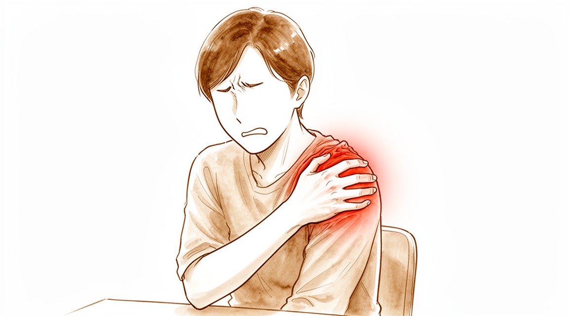
Rotator cuff disorders: common causes of shoulder pain, ranging from mild ache to debilitating injury.
Những gì bạn đang cảm thấy
Bệnh lý vòng xoay chóp (rotator cuff) là phổ biến và có xu hướng gia tăng theo tuổi tác. Bạn có thể cảm thấy đau ở phía trước vai hoặc mặt ngoài của cánh tay trên. Cơn đau này thường bắt nguồn từ các gân giúp nâng cánh tay của bạn. Bạn cũng có thể nhận thấy cảm giác âm ỉ ở phía trước vai nếu gân cơ nhị đầu (biceps tendon) liên quan. Gân này chạy dọc theo mặt trước của cánh tay và hoạt động cùng với vòng xoay chóp.
Các triệu chứng của bạn có thể mang tính cơ học, như cảm giác kẹt hoặc ma sát trong khớp. Đây là một phàn nàn phổ biến. Cơn đau thường bùng phát vào ban đêm, gây khó khăn cho việc ngủ nghiêng về bên đó. Bạn cũng có thể cảm thấy cứng khớp khi vừa thức dậy. Sau khi sử dụng cánh tay, cảm giác âm ỉ có thể trở nên trầm trọng hơn. Tuy nhiên, thời gian bạn trải qua các triệu chứng này không cho chúng ta biết mức độ nghiêm trọng của vết rách. Một số người có vết rách lớn nhưng ít đau, trong khi những người khác có vết rách nhỏ nhưng chịu nhiều khó chịu đáng kể.
Các hoạt động hàng ngày có thể trở nên khó khăn. Việc với tay ra sau lưng để cài áo ngực hoặc nhét áo vào quần có thể cảm thấy bất tiện hoặc gây đau. Nâng vật dụng lên cao có thể kích hoạt cơn đau nhói. Bạn có thể nhận thấy mình tránh một số cử động nhất định để bảo vệ vai. Cảm giác chủ quan về sự mất ổn định hoặc yếu sức cũng rất phổ biến. Những cảm giác này có thể khiến bạn cảm thấy như vai của mình không hoạt động đúng cách.
Điều quan trọng cần biết là sức khỏe tinh thần của bạn đóng một vai trò lớn trong mức độ đau đớn mà bạn cảm nhận và khả năng hoạt động của vai. Mối liên hệ này có thể mạnh hơn cả kích thước thực tế của vết rách. Bác sĩ phẫu thuật của bạn sẽ xem xét toàn bộ tình trạng của bạn, bao gồm tuổi tác và mức độ hoạt động, để quyết định hướng điều trị tốt nhất. Dù bạn chọn phẫu thuật hay điều trị không phẫu thuật, cả hai lựa chọn đều có thể hiệu quả. Mục tiêu là giảm đau và giúp bạn quay trở lại các hoạt động mà bạn yêu thích. Nếu chẩn đoán chưa rõ ràng, bác sĩ phẫu thuật có thể tập trung vào việc điều trị trực tiếp cơn đau vai để tránh các thủ thuật không cần thiết.
Những gì thực sự đang xảy ra
Vai của bạn là một khớp cầu và hốc. Đầu cầu nằm trong một hốc nông. Một nhóm bốn gân, được gọi là vòng xoay vai (rotator cuff), bao quanh đầu cầu như một chiếc áo. Những gân này giữ đầu cầu tại chỗ và giúp bạn nâng cánh tay. Tình trạng này phổ biến và trở nên phổ biến hơn khi bạn già đi.
Khi bạn bị rách một gân, sự cân bằng của vai bạn thay đổi. Đầu cầu có thể trượt lên trên thay vì vẫn nằm ở trung tâm của hốc. Điều này được gọi là di chuyển lên trên (superior migration). Nó xảy ra vì gân không còn có thể kéo đầu cầu xuống. Theo thời gian, sự sai lệch này gây ra ma sát và hao mòn. Các vết rách mãn tính không được điều trị có thể dẫn đến thoái hóa khớp (arthrosis), là viêm khớp do hao mòn của khớp.
Cơn đau và sự suy yếu mà bạn cảm thấy đến từ sự mất ổn định này. Bao khớp, là lớp áo bao quanh vai, có thể bị giãn hoặc rách. Điều này cho phép các xương cọ xát vào nhau. Trong một số trường hợp, các cơ xung quanh vai thay đổi vị trí để bù đắp. Điều này làm thay đổi cách vai của bạn di chuyển trong các nhiệm vụ hàng ngày.
Bác sĩ phẫu thuật xem xét những thay đổi cơ học này để quyết định phương pháp điều trị. Cả các lựa chọn phẫu thuật và không phẫu thuật đều có thể hiệu quả. Mục tiêu là khôi phục chuyển động bình thường và giảm đau. Trong một số trường hợp, các thủ thuật nhằm khôi phục vị trí tự nhiên của đầu cầu trong hốc. Điều này giúp giảm áp lực lên khớp.
Sức khỏe tâm thần đóng một vai trò trong cách bạn trải nghiệm cơn đau này. Nó có thể quan trọng không kém kích thước của vết rách. Kỳ vọng của bạn cũng ảnh hưởng trực tiếp đến kết quả điều trị. Hiểu những gì đang xảy ra bên trong vai của bạn giúp bạn chuẩn bị cho quá trình hồi phục.
Những gì chúng tôi có thể làm về vấn đề này
Bạn có các lựa chọn để quản lý bệnh lý chóp xoay, và cả hai phương pháp điều trị không phẫu thuật và phẫu thuật đều có hiệu quả. Lựa chọn phù hợp phụ thuộc vào vết rách cụ thể của bạn, tuổi tác và tình trạng sức khỏe tổng thể. Đối với nhiều bệnh nhân, bắt đầu với tự chăm sóc và vật lý trị liệu là một hướng đi khả thi. Sau 13 năm kể từ khi chẩn đoán, khoảng 90% bệnh nhân được điều trị bảo tồn cho vết rách chóp xoay không có hoặc chỉ có đau nhẹ. Sau 13 năm kể từ khi chẩn đoán, khoảng 70% bệnh nhân được điều trị bảo tồn cho vết rách chóp xoay không có sự gián đoạn trong các hoạt động sống hàng ngày.
Vật lý trị liệu nhằm mục đích tăng cường các cơ xung quanh vai của bạn để cải thiện chức năng và giảm đau. Một phác đồ vật lý trị liệu cụ thể có hiệu quả trong việc điều trị vết rách chóp xoay toàn phần không do chấn thương ở khoảng 75% bệnh nhân được theo dõi trong 2 năm. Nếu bạn có vết rách cơ trên gai không do chấn thương, điều trị bảo tồn nên được xem xét là phương pháp điều trị chính. Bác sĩ phẫu thuật của bạn có thể khuyên bạn tránh các cử động vai gây chèn ép, đó là khi các mô bị kẹp trong khớp vai. Sức khỏe tâm thần cũng đóng một vai trò đáng kể; nó có mối liên hệ mạnh mẽ hơn với đau vai và chức năng do bệnh nhân báo cáo so với kích thước vết rách ở những bệnh nhân có vết rách chóp xoay toàn phần.
Quản lý y khoa tập trung vào giảm đau và giảm viêm. Trong khi tiêm corticosteroid đôi khi được sử dụng, có rất ít bằng chứng tái lập để hỗ trợ hiệu quả của chúng trong việc quản lý bệnh lý chóp xoay. Bạn nên thận trọng nếu xem xét các mũi tiêm này, vì chúng nên được trì hoãn nếu việc sửa chữa chóp xoay sẽ được thực hiện trong vòng 6 tháng tiếp theo. Bằng chứng hạn chế hiện tại cho thấy rằng tiêm huyết tương giàu tiểu cầu (PRP) có thể không mang lại lợi ích cho điều trị bảo tồn bệnh lý chóp xoay mạn tính trong ngắn hạn. Thuốc giảm đau và thuốc chống viêm có thể giúp quản lý các triệu chứng trong khi bạn tham gia liệu pháp.
Phẫu thuật được xem xét khi chăm sóc bảo tồn đã đạt đến giới hạn hoặc nếu bạn có vết rách do chấn thương không lành. Sửa chữa chóp xoay nội soi là một lựa chọn hiệu quả và an toàn để điều trị các triệu chứng của vết rách chóp xoay, với kết quả lâm sàng bền vững theo thời gian. Thủ thuật này được ưa chuộng để cải thiện chức năng vai, và các thủ thuật khác hoặc điều trị trong khớp không mang lại lợi ích đáng kể so với sửa chữa chóp xoay nội soi cho mục tiêu này. Đối với các vết rách lớn hoặc không thể sửa chữa, sửa chữa một phần có thể hiệu quả trong việc giảm đau và cải thiện chức năng bằng cách khôi phục sự cân bằng cho cặp lực. Trong các trường hợp khớp bị tổn thương, chẳng hạn như với bệnh khớp, khớp vai đảo chiều có thể được dự trữ để điều trị, mặc dù nó chống chỉ định ở những bệnh nhân bị viêm khớp dạng thấp. Bác sĩ phẫu thuật của bạn sẽ xem xét cẩn thận các yếu tố nguy cơ của bạn đối với thất bại sửa chữa khi lập kế hoạch điều trị cho bạn.
Những điều cần mong đợi
Tiên lượng của bạn phụ thuộc phần lớn vào việc bạn lựa chọn phẫu thuật hay điều trị bảo tồn. Đối với các rách nhỏ đến trung bình, phẫu thuật mang lại giảm đau và cải thiện chức năng dài hạn tốt hơn so với chỉ vật lý trị liệu, với những lợi ích kéo dài lên đến 15 năm. Nếu bạn có một vết rách lớn hoặc rách khổng lồ, phẫu thuật vẫn có thể mang lại kết quả dài hạn thỏa đáng. Ngay cả khi bạn cần phẫu thuật sửa chữa lại, kết quả ngắn hạn của bạn tương tự như kết quả của lần sửa chữa đầu tiên.
Nếu bạn quản lý vết rách mà không phẫu thuật, diễn biến thường diễn ra nhẹ nhàng. Khoảng 90% bệnh nhân không có hoặc chỉ có đau nhẹ sau 13 năm kể từ khi chẩn đoán. Khoảng 70% không có sự gián đoạn trong các hoạt động hàng ngày tại mốc thời gian 13 năm này. Tuy nhiên, các vết rách mạn tính không được điều trị cuối cùng có thể dẫn đến thoái hóa khớp, là dạng viêm xương khớp do hao mòn. Phẫu thuật có thể thay đổi diễn biến tự nhiên sớm này, mang lại những khác biệt có ý nghĩa lâm sàng về đau và chức năng so với điều trị không phẫu thuật.
Nếu bạn trải qua phẫu thuật sửa chữa, quá trình hồi phục của bạn là một quá trình diễn ra dần dần. Bạn sẽ đạt được khoảng 60% mức độ hồi phục chức năng cuối cùng sau 3 tháng phẫu thuật. Đến 6 tháng, bạn sẽ đạt được khoảng 75% mức độ hồi phục chức năng. Một năm sau phẫu thuật không xác định kết quả dài hạn của bạn, vì vậy sự kiên nhẫn là chìa khóa.
Tuổi tác và kỳ vọng của bạn định hình kết quả điều trị. Nếu bạn từ 50 tuổi trở xuống, bạn có thể mong đợi giảm đau dài hạn. Tuy nhiên, bạn có thể không thấy sự cải thiện đáng kể về tầm vận động trong dài hạn. Một tỷ lệ lớn bệnh nhân trong nhóm tuổi này có kết quả dài hạn không thỏa đáng. Sức khỏe tinh thần của bạn cũng đóng một vai trò mạnh mẽ trong cách bạn cảm nhận đau đớn và chức năng, đôi khi còn nhiều hơn cả kích thước của vết rách. Việc trực tiếp điều chỉnh kỳ vọng của bạn phù hợp với bác sĩ phẫu thuật giúp đảm bảo kết quả điều trị tốt hơn.
Khi nào cần gặp bác sĩ
Hãy gặp bác sĩ đa khoa nếu bạn bị đau vai không cải thiện sau khi nghỉ ngơi. Yêu cầu đánh giá bởi bác sĩ chuyên khoa nếu bạn nhận thấy yếu hoặc mất vững khớp. Hãy tìm kiếm chăm sóc y tế nếu vai của bạn bị kẹt hoặc đột ngột mất sức. Liên hệ với bác sĩ nếu các triệu chứng ảnh hưởng đến giấc ngủ hoặc công việc của bạn. Sự gia tăng đột ngột về mức độ đau cũng là lý do để tìm kiếm sự giúp đỡ. Lưu ý rằng, thời gian bạn đã có các triệu chứng không cho chúng tôi biết mức độ nghiêm trọng của vết rách. Các vết rách mạn tính không được điều trị có thể dẫn đến viêm xương khớp do hao mòn theo thời gian. Đánh giá sớm giúp ngăn ngừa tổn thương thêm và đảm bảo bạn nhận được kế hoạch điều trị phù hợp với nhu cầu cụ thể của mình.
Evidence & references
Overview
- The majority of rotator cuff disorders are amenable to conservative treatment [1].
- Rotator cuff dysfunction may necessitate surgical treatment [1].
- The major indication for revision rotator cuff repair is the persistence of clinical symptoms despite nonsurgical management in the absence of substantial risk factors for failure [2].
- Rotator cuff injuries are most accurately diagnosed with a combination of cuff- and impingement-specific clinical tests [3].
- Deltoid complications combined with rotator cuff pathology represent a rare but devastating complication with no well-described surgical option [4].
- Treatment of chronic massive rotator cuff tears is challenging, and results are comparatively inferior to those of treating patients with smaller rotator cuff tears [17].
- Shoulder arthroscopy literature remains controversial, with conclusions often unsupported due to bias and limitations [19].
- No clinical guidelines for shoulder arthroscopy are definitive pending higher levels of evidence [19].
- No recommendations regarding suprascapular nerve release in conjunction with rotator cuff repair can be made at this time [22].
- Further research is necessary to better delineate the indications for suprascapular nerve release in conjunction with rotator cuff repair [22].
- Open approaches for rotator cuff repairs continue to have indications in certain circumstances, such as complete rotator cuff tendon avulsion and glenohumeral joint incarceration after high-velocity trauma [23].
- There is no statistically significant difference in subjective outcome after arthroscopic rotator cuff repair with or without acromioplasty at intermediate follow-up [26].
- Critical shoulder angle and acromial index do not appear to influence 24-month functional outcomes postoperatively [47].
- Critical shoulder angle and acromial index are not contraindications to arthroscopic rotator cuff repair [47].
- Predictors of pain and functional outcomes after operative treatment for rotator cuff tears can be used to select optimal candidates for operative treatment [54].
- Predictors of pain and functional outcomes after operative treatment for rotator cuff tears can assist with patient education and expectations before treatment [54].
- There were no differences of clinically relevant size between arthroscopic and open rotator cuff surgery in postoperative pain in a comparative series [71].
Anatomy & Pathophysiology
- Understanding the function and pathology surrounding the teres minor is paramount in comprehensive management of patients with shoulder pathology [6].
- A systematic approach to magnetic resonance imaging interpretation of shoulder injuries describes the normal imaging appearance of each anatomical structure, the most useful pulse sequences and imaging planes, and signs of injury [28].
- Imaging is an essential tool for evaluation of patients with shoulder pain, and understanding the extent of an injury with imaging is key to successful management [29].
- Tears of the subscapularis have greater biomechanical consequences than do tears of the infraspinatus [31].
- Dynamic superior migration of the humeral head during abduction occurs in patients with rotator cuff tears, as confirmed by in vivo 3D kinematic analysis [37].
- In massive rotator cuff tears, the pectoralis major and latissimus dorsi muscles are effective in improving glenohumeral kinematics and reducing acromiohumeral pressures [40].
- Biomechanical studies indicate that the long head of the biceps contributes to stability of the glenohumeral joint in all directions, although in vivo studies have not yet established this stabilizing effect or the physiologic load required [46].
- Increasing supraspinatus tendon loading causes a mechanical interaction between the supraspinatus and infraspinatus tendons, paralleling the increase in supraspinatus tendon strain [51].
- The physiopathology of symptomatic anterior instabilities is related to dysfunction of the anterosuperior glenohumeral capsular ligament rather than the inferior glenohumeral ligament [52].
- Additional repair of a partial subscapularis tear combined with a supraspinatus tear did not affect external rotation or glenohumeral kinematics [53].
- Shoulders with rotator cuff tears require considerable compensatory deltoid function to prevent abduction motion loss [57].
- Pain reduction from subacromial injection causes shifts in scapulohumeral rhythm, resulting in increased glenohumeral motion and reduced reliance on scapular rotation [59].
- The clinical evidence to support correction of the critical shoulder angle with lateral acromioplasty is insufficient, and further research is required to demonstrate an association between critical shoulder angle and clinical outcomes before treatment algorithms should be altered [61].
- The critical shoulder angle, posterior acromial height, and posterior acromial tilt do not change significantly over a long-term follow-up of at least 10 years, supporting the hypothesis that these scapular morphologic parameters are stable anthropometric characteristics [62].
- Simulated isolated supraspinatus cord and strap tears significantly reduced shoulder abduction force, with cord tears causing a larger decline than strap tears [63].
- The critical shoulder angle may not be responsible for rotator cuff tears; rather, patient activities throughout several decades could induce both cuff lesions and bone remodeling at the acromial level [66].
- The human scapula has two distinctive characteristics: a lateral orientation of the glenoid cavity and a narrow coraco-acromial arch [73].
- Cervical spine position may cause decreased shoulder rotation strength, meaning clinicians should assess shoulder strength in the position the patient requires to use their shoulder because weakness may be missed in standard testing positions [78].
Classification
- Rotator cuff disorders are recognized and managed conditions among patients with shoulder pain [1].
- Rotator cuff dysfunction may necessitate surgical treatment [1].
- The majority of rotator cuff conditions are amenable to conservative treatment [1].
- The major indication for revision rotator cuff repair is the persistence of clinical symptoms despite nonsurgical management in the absence of substantial risk factors for failure [2].
- Rotator cuff injuries are most accurately diagnosed with a combination of cuff- and impingement-specific clinical tests [3].
- Deltoid complications combined with rotator cuff pathology represent a rare but devastating complication with no well-described surgical option [4].
- Increasing knowledge about rotator cuff syndrome, including better imaging, has facilitated patient treatment for a stable spectrum of rotator cuff pathology [5].
- The application of endoscopic surgery has facilitated patient treatment for a stable spectrum of rotator cuff pathology [5].
- Rotator cuff disease, shoulder instability, and associated lesions are common pathologic conditions of the shoulder involving soft tissues [7].
- In the context of rotator cuff disease, the etiology of anterior shoulder pain with macroscopic changes in the biceps tendon is related to the complex interaction of the tendon and surrounding soft tissues, rather than a single entity [8].
- The Korean Shoulder Scoring System (KSS) is a useful measurement tool that combines subjective and objective evaluations for shoulder function related to rotator cuff disorders [9].
- The acromial morphology classification system is an unreliable method to assess the acromion [14].
- The acromial index shows no association with the presence of rotator cuff disease [14].
- A classification system exists to divide coracoids according to their morphology and relative risk of associated subscapularis tears [30].
- Comparing histopathological data with demographical information allows for the identification of rotator cuff tears at risk of repair failure [50].
- Gene expression in human rotator cuff muscles varied according to tendon injury severity [67].
Clinical Presentation
- Rotator cuff disorders are amenable to conservative treatment in the majority of cases [1].
- Rotator cuff dysfunction may necessitate surgical treatment [1].
- The major indication for revision rotator cuff repair is the persistence of clinical symptoms despite nonsurgical management in the absence of substantial risk factors for failure [2].
- Rotator cuff injuries are most accurately diagnosed with a combination of cuff- and impingement-specific clinical tests [3].
- Deltoid complications combined with rotator cuff pathology represent a rare but devastating complication with no well-described surgical option [4].
- Increasing knowledge about rotator cuff syndrome, including better imaging, has facilitated patient treatment for a stable spectrum of rotator cuff pathology [5].
- The application of endoscopic surgery has facilitated patient treatment for a stable spectrum of rotator cuff pathology [5].
- Understanding the function and pathology surrounding the teres minor is paramount in comprehensive management of the patient with shoulder pathology [6].
- Rotator cuff disease, shoulder instability, and associated lesions are common pathologic conditions of the shoulder involving soft tissues [7].
- In the context of rotator cuff disease, the etiology of anterior shoulder pain with macroscopic changes in the biceps tendon is related to the complex interaction of the tendon and surrounding soft tissues, rather than a single entity [8].
- The Korean Shoulder Scoring System (KSS) is a useful measurement tool that combines subjective and objective evaluations for shoulder function related to rotator cuff disorders [9].
- In one-quarter of patients with painful cuff tears, pain developed in a contralateral asymptomatic cuff tear that resulted in a measurable decline in function within 3 years [10].
- The acromial morphology classification system is an unreliable method to assess the acromion [14].
- The acromial index shows no association with the presence of rotator cuff disease [14].
- Ultrasound is an useful tool for discovering in pre-symptomatic stages subjects that may undergo shoulder symptomatic pathologies [15].
- Rotator cuff injuries in adolescents may be overlooked as a cause of disability, leading to significant delays in diagnosis [32].
- Intratendinous rotator cuff tears are difficult to diagnose preoperatively [34].
- Current physiotherapy practice in relation to rotator cuff disorders is variable [35].
- Current physiotherapy practice in relation to rotator cuff disorders is variable [36].
- The Functional Shoulder Score (FSS) is a patient-reported outcome measure that can easily be incorporated into clinical practice [38].
- The FSS provides a quick, reliable, valid and practical measure for rotator cuff problems [38].
Investigations
- Rotator cuff dysfunction may necessitate surgical treatment [1].
- Rotator cuff injuries are most accurately diagnosed with a combination of cuff- and impingement-specific clinical tests [3].
- Better imaging has facilitated patient treatment for a stable spectrum of rotator cuff pathology [5].
- Ultrasound is an useful tool for discovering subjects in pre-symptomatic stages that may undergo shoulder symptomatic pathologies [15].
- The use of MRI before a trial of conservative management in patients with atraumatic shoulder pain, minimal to no strength deficits on physical examination, and suspected cuff tendinopathy other than full-thickness tears provides negative value in the management of these patients, at both the individual and population level [24].
- A systematic approach to the interpretation of a magnetic resonance examination of the shoulder describes the normal imaging appearance of each anatomical structure, the most useful pulse sequences and imaging planes, and the signs of injury [28].
- Imaging is an essential tool for evaluation of patients with shoulder pain [29].
- Understanding the extent of an injury with imaging is key to successful management [29].
- Most abnormal MRI findings were not different in frequency between symptomatic and asymptomatic shoulders [43].
- MRI and US provide similar assessments of postoperative rotator cuff healing, although US is less sensitive [58].
- Shoulder MRI may be warranted for preoperative planning in the select population of patients with chronic calcific tendinopathy and prolonged refractory pain, although the probability of identifying additional cuff pathology requiring surgical intervention is very low [64].
- Preoperative MRI scans of the shoulder interpreted by orthopaedic surgeons with a systematic approach resulted in improved accuracy in diagnosing subscapularis tendon tears compared with previous studies [65].
- The diagnostic accuracy of US, MRI and MRA in the characterisation of full-thickness rotator cuff tears is high with overall estimates of sensitivity and specificity over 0.90 [68].
- A non-contrast shoulder MRI obtained in the community setting after non-dislocating shoulder trauma has a moderate sensitivity for most intraarticular pathologies when interpreted by musculoskeletal radiologists [70].
- The MRI tendinosis grade is associated with stiffness assessed using sonoelastography in patients with rotator cuff tendinopathy [72].
- Tendinosis severity assessed by preoperative MRI was the only factor associated with failure to heal in patients with partial-thickness and small full-thickness rotator cuff tears [75].
- A 3 T MRI protocol can be applied to evaluate morphological tendon outcomes after different treatment modalities [77].
- Shoulders with a symptomatic rotator cuff tear showed higher radioisotope uptake on bone scintigraphy than those with an asymptomatic tear [79].
- Unenhanced magnetic resonance imaging of the shoulder in asymptomatic high performance throwing athletes reveals abnormalities that may encompass a spectrum of nonclinical findings [80].
Treatment
Non-Operative Management
- The majority of rotator cuff disorders are amenable to conservative treatment [1].
- MRI use before a trial of conservative management in patients with atraumatic shoulder pain, minimal to no strength deficits, and suspected cuff tendinopathy (other than full-thickness tears) provides negative value at both individual and population levels [24].
- Nonoperative treatment is appropriate as initial therapy for partial-thickness rotator cuff tears [42].
- There is little reproducible evidence to support the efficacy of subacromial corticosteroid injection in managing rotator cuff disease [33].
- Orthobiologics offer a relatively safe management option for rotator cuff pathology, but evidence is inconclusive for or against its use [45].
- Limited evidence suggests that platelet-rich plasma (PRP) injections may not be beneficial in the short term for the nonoperative treatment of chronic rotator cuff disease [56].
- Subacromial PRP injections produce significantly worse improvement in functional outcomes in patients with partial supraspinatus tears compared to patients with isolated tendinopathy [44].
Operative Management
- Rotator cuff dysfunction may necessitate surgical treatment when conservative options are insufficient [1].
- Early operative treatment appears to be better for rotator cuff tears with a sudden onset of symptoms and poor function to achieve maximal return of shoulder function [18].
- Patients undergoing operative treatment for rotator cuff tears had significantly better pain and functional outcomes compared with patients undergoing nonoperative treatment in a prospective cohort study [76].
- Treatment results for chronic massive rotator cuff tears are comparatively inferior to those for smaller rotator cuff tears [17].
- Open approaches for rotator cuff repairs continue to have indications in certain circumstances, such as complete tendon avulsion and glenohumeral joint incarceration following high-velocity trauma [23].
- Arthroscopic revision rotator cuff repair is indicated for the persistence of clinical symptoms despite nonsurgical management in the absence of substantial risk factors for failure [2].
- Repair of partial- and full-thickness rotator cuff tears using a bioinductive implant shows safety and efficacy at 1-year follow-up [11].
- Arthroscopy is a safe and effective treatment for symptomatic calcific tendonitis of the shoulder, including or excluding patients with rotator cuff tears [48].
- For patients with intact rotator cuffs and calcific tendonitis, needling or extracorporeal shockwave therapy (ESWT) may be beneficial alternatives to arthroscopy in some cases [48].
Arthroscopic Subacromial Decompression (SAD)
- Following nonoperative treatment for at least 6 weeks, SAD is a viable and good surgical option for shoulder impingement with an intact rotator cuff [39].
- SAD without cuff repair appears to be a safe, efficacious, and sustainable procedure for patients with partial rotator cuff tears [41].
- Operative management for partial-thickness tears, including arthroscopic subacromial decompression, is considered when nonoperative treatment fails [42].
Adjunctive Procedures and Implants
- PRP does not have an effect on overall retear rates or shoulder-specific outcomes after arthroscopic rotator cuff repair [21].
- Routine arthroscopic suprascapular nerve release (SSNR) is not recommended when treating patients with rotator cuff tear [49].
- No recommendations regarding suprascapular nerve release in conjunction with rotator cuff repair can be made at this time, and further research is necessary [22].
Reverse Total Shoulder Arthroplasty (RTSA)
- Severely impaired deltoid function is a contraindication to RTSA [13].
- An isolated supraspinatus tear is a contraindication to RTSA [13].
- The presence of full active shoulder elevation with a massive rotator cuff tear and arthritis is a contraindication to RTSA [13].
Evidence Quality
- Conclusions in shoulder arthroscopy literature are often unsupported due to bias and limitations, and no clinical guidelines are definitive pending higher levels of evidence [19].
Complications
- Rotator cuff dysfunction may necessitate surgical treatment [1].
- Deltoid complications combined with rotator cuff pathology represent a rare but devastating complication with no well-described surgical option [4].
- In one-quarter of patients with painful cuff tears, pain developed in a contralateral asymptomatic cuff tear that resulted in a measurable decline in function within 3 years [10].
- Arthroscopic rotator cuff repair leads to a structural failure rate of 33% but satisfactory functional results with high patient satisfaction at midterm follow-up [27].
Recovery
- The majority of rotator cuff disorders are amenable to conservative treatment, although rotator cuff dysfunction may necessitate surgical treatment [1].
- Conservative versus surgical management for rotator cuff tears does not result in significantly improved shoulder function (evaluated by CMS) at a 2-year follow-up [12].
- Arthroscopic decompression is not recommended in the treatment of rotator cuff tendinopathy [16].
- The natural history of rotator cuff tendinopathy probably plays a significant role in long-term results [16].
- Arthroscopic acromioplasty significantly improves long-term clinical outcomes up to 2 years for chronic rotator cuff tendinopathy when used with platelet-rich plasma injection [69].
- Patients with rotator cuff disease treated without surgery experience a clinically important change in self-assessed outcome with a 2-point change in the Simple Shoulder Test (SST) score or a 12 to 17-point change in the American Shoulder and Elbow Surgeons (ASES) score [83].
- Younger age, lower BMI, more functional capacity, a shorter symptomatic period, reversible changes on MRI, and higher Constant and ASES scores at the first evaluation are good prognostic factors for the natural course of subacromial impingement syndrome [84].
- In one-quarter of patients with painful cuff tears, pain developed in a contralateral asymptomatic cuff tear that resulted in a measurable decline in function within 3 years [10].
- Early operative treatment appears to be better for rotator cuff tears with a sudden onset of symptoms and poor function to achieve maximal return of shoulder function [18].
- The major indication for revision rotator cuff repair is the persistence of clinical symptoms despite nonsurgical management in the absence of substantial risk factors for failure [2].
- Outcomes after repair of partial- and full-thickness rotator cuff tears using a bioinductive implant show safety and efficacy at 1-year follow-up [11].
- Arthroscopic rotator cuff repair leads to a structural failure rate of 33% but yields satisfactory functional results with high patient satisfaction at midterm follow-up [27].
- Increased age and longer duration of follow-up were associated with lower healing rates after double-row rotator cuff repair [20].
- The 'critical period' for healing following rotator cuff repair, during which risks of retears are high, extends to the first 6 months [85].
- Although functional status improved with time after 6 months, the structural status of repaired cuffs remained unchanged between 6 and 19 months postoperatively [55].
- Improvement in functional outcome after arthroscopic repair of a subscapularis tendon tear is maintained long-term [60].
Key Evidence
- [L4] The majority of conditions are amenable to conservative treatment, although rotator cuff dysfunction may necessitate surgical treatment. [1] (10.1002/art.20668)
- [L5] The major indication for revision rotator cuff repair is the persistence of clinical symptoms despite nonsurgical management in the absence of substantial risk factors for failure. [2] (10.5435/00124635-201111000-00002)
- [L5] Current consensus suggests rotator cuff injuries are most accurately diagnosed with a combination of cuff- and impingement-specific clinical tests. [3] (10.1016/j.arthro.2013.07.265)
- [Case_report] Deltoid complications combined with rotator cuff pathology represent a rare but devastating complication with no well-described surgical option. [4] (10.1016/j.jse.2011.09.023)
- [L3] Increasing knowledge about this syndrome, including better imaging, has facilitated patient treatment for a stable spectrum of rotator cuff pathology, as has the application of endoscopic surgery. [5] (10.1016/j.arthro.2010.02.029)
- [L5] Understanding the function and pathology surrounding the teres minor is paramount in comprehensive management of the patient with shoulder pathology. [6] (10.5435/jaaos-d-15-00258)
- [L4] In the context of rotator cuff disease, the etiology of anterior shoulder pain with macroscopic changes in the biceps tendon is related to the complex interaction of the tendon and surrounding soft tissues, rather than a single entity. [8] (10.1016/j.jse.2008.05.044)
- [L4] The KSS is a useful measurement tool that combines subjective and objective evaluations for shoulder function related to rotator cuff disorders. [9] (10.1016/j.jse.2008.11.019)
- [L2] In one-quarter of patients with painful cuff tears, pain developed in a contralateral asymptomatic cuff tear that resulted in a measurable decline in function within 3 years. [10] (10.1016/j.jse.2023.09.008)
- [L4] Outcomes after repair of partial- and full-thickness rotator cuff tears using a bioinductive implant show safety and efficacy at 1-year follow-up. [11] (10.1016/j.arthro.2019.02.019)
- [L1] At a 2-year follow-up, shoulder function evaluated in terms of CMS was not significantly improved. [12] (10.1186/s12891-020-03872-4)
- [L5] Severely impaired deltoid function, an isolated supraspinatus tear, and the presence of full active shoulder elevation with a massive rotator cuff tear and arthritis are contraindications to RTSA. [13] (10.1007/s11999-009-1188-9)
- [L3] The acromial morphology classification system is an unreliable method to assess the acromion, and the acromial index shows no association with the presence of rotator cuff disease. [14] (10.1016/j.jse.2011.09.028)
- [L3] Ultrasound is an useful tool for discovering in pre-symptomatic stages the subjects that may undergo shoulder symptomatic pathologies. [15] (10.1186/1471-2474-11-278)
- [L1] The natural history of rotator cuff tendinopathy probably plays a significant role in the results in the long-term. [16] (10.1302/0301-620x.99b6.bjj-2016-0569.r1)
- [L5] However, treatment of these patients is challenging, and results are comparatively inferior to those of treating patients with smaller rotator cuff tears. [17] (10.5435/00124635-200309000-00005)
- [L3] Early operative treatment appears to be better for rotator cuff tears with a sudden onset of symptoms and poor function to achieve maximal return of shoulder function. [18] (10.1016/j.jse.2005.07.006)
- [L5] The editorial states that shoulder arthroscopy literature remains controversial, conclusions are often unsupported due to bias and limitations, and no clinical guidelines are definitive pending higher levels of evidence. [19] (10.1016/j.arthro.2012.07.001)
- [L4] Increased age and longer duration of follow-up were associated with lower healing rates after double-row rotator cuff repair. [20] (10.1177/0363546510382835)
- [L1] PRP does not have an effect on overall retear rates or shoulder-specific outcomes after arthroscopic rotator cuff repair. [21] (10.1016/j.arthro.2012.03.007)
- [L4] No recommendations regarding suprascapular nerve release in conjunction with rotator cuff repair can be made at this time, and further research is necessary to better delineate the indications in the future. [22] (10.1016/j.jse.2011.11.033)
- [Case_report] This case highlights the importance of the initial workup after high-velocity trauma and that open approaches for rotator cuff repairs continue to have indications in certain circumstances. [23] (10.1016/j.jse.2009.07.014)
- [L4] The use of MRI before a trial of conservative management in patients with atraumatic shoulder pain, minimal to no strength deficits on physical examination, and suspected cuff tendinopathy other than full-thickness tears provides negative value in the management of these patients, at both the individual and population level. [24] (10.1016/j.jse.2019.04.003)
- [L1] On the basis of the currently available literature, there is no statistically significant difference in subjective outcome after arthroscopic rotator cuff repair with or without acromioplasty at intermediate follow-up. [26] (10.1016/j.arthro.2011.11.022)
- [L4] Arthroscopic rotator cuff repair leads to a structural failure rate of 33% but satisfactory functional results with high patient satisfaction at midterm follow-up. [27] (10.1016/j.jse.2015.05.051)
- [L5] This article provides a systematic approach to the interpretation of a magnetic resonance examination of the shoulder, describing the normal imaging appearance of each anatomical structure, the most useful pulse sequences and imaging planes, and the signs of injury. [28] (10.1177/0363546505278255)
- [L4] Imaging is an essential tool for evaluation of patients with shoulder pain; understanding the extent of an injury with imaging is key to successful management. [29] (10.1016/j.csm.2013.03.009)
- [L3] This study was the first to create a classification system to divide coracoids according to their morphology and relative risk of associated subscapularis tears. [30] (10.1016/j.jse.2020.01.074)
- [L5] Tears of the subscapularis have greater biomechanical consequences than do tears of the infraspinatus. [31] (10.1016/j.arthro.2009.09.007)
- [L4] Rotator cuff injuries in adolescents may be overlooked as a cause of disability, leading to significant delays in diagnosis. [32] (10.1177/0363546504269033)
- [L1] This systematic review of the available literature indicates that there is little reproducible evidence to support the efficacy of subacromial corticosteroid injection in managing rotator cuff disease. [33] (10.5435/00124635-200701000-00002)
- [L4] Intratendinous rotator cuff tears are difficult to diagnose preoperatively. [34] (10.1016/j.jse.2010.01.013)
- [L4] Current physiotherapy practice in relation to rotator cuff disorders is variable, which might reflect the lack of high-quality evidence available. [35] (10.1177/1758573217717103)
- [L4] Current physiotherapy practice in relation to rotator cuff disorders is variable, which might reflect the lack of high-quality evidence available. [36] (10.1111/j.1758-5740.2011.00164.x)
- [L3] This study confirms dynamic superior migration of the humeral head during abduction in patients with rotator cuff tears using in vivo 3D kinematic analysis. [37] (10.1016/j.arthro.2015.08.031)
- [L2] The FSS is a patient-reported outcome measure that can easily be incorporated into clinical practice, providing a quick, reliable, valid and practical measure for rotator cuff problems. [38] (10.1177/1758573215578589)
- [L5] Following nonoperative treatment for at least 6 weeks, SAD is a viable and good surgical option for the treatment of shoulder impingement with an intact rotator cuff. [39] (10.1016/j.arthro.2019.06.012)
- [L5] In massive rotator cuff tear, the pectoralis major and latissimus dorsi muscles are effective in improving glenohumeral kinematics and reducing acromiohumeral pressures. [40] (10.1016/j.jse.2013.11.030)
- [L4] ASD without cuff repair appears to be a safe, efficacious, and sustainable procedure for patients with partial rotator cuff tears. [41] (10.1016/j.arthro.2015.08.026)
- [L5] Nonoperative treatment is appropriate as initial therapy, while operative management including arthroscopic subacromial decompression, debridement, or repair is considered when nonoperative treatment fails. [42] (10.5435/00124635-199901000-00004)
- [L3] Most abnormal MRI findings were not different in frequency between symptomatic and asymptomatic shoulders. [43] (10.1016/j.jse.2019.04.001)
- [L2] However, improvement in symptoms and functional outcomes was significantly worse in patients who had a partial-thickness rotator cuff tear compared with patients who had an isolated tendinopathy. [44] (10.1016/j.arthro.2023.03.019)
- [L2] Orthobiologics offer a relatively safe management option with inconclusive evidence for or against its use for rotator cuff pathology. [45] (10.3233/bmr-201844)
- [L5] Biomechanical studies indicate that the long head of the biceps contributes to stability of the glenohumeral joint in all directions, though in vivo studies have yet to establish this stabilizing effect and the physiologic load required remains unknown. [46] (10.1016/j.arthro.2010.10.014)
- [L3] CSA and AI do not appear to influence 24-month functional outcomes postoperatively and hence are not contraindications to arthroscopic rotator cuff repair. [47] (10.1177/0363546517717947)
- [L5] Arthroscopy is a safe and effective treatment for symptomatic calcific tendonitis of the shoulder, excluding or including patients with rotator cuff tears, but patients with intact cuffs could benefit from needling or ESWT in some cases. [48] (10.1016/j.arthro.2015.11.003)
- [L1] Routine arthroscopic SSNR is not recommended when treating patients with rotator cuff tear. [49] (10.1007/s00167-022-07066-4)
- [L2] Comparing histopathological data with demographical information allows for the identification of rotator cuff tears at risk of repair failure. [50] (10.1007/s00167-011-1521-1)
- [L5] Increasing supraspinatus tendon loading causes a mechanical interaction between the two tendons, paralleling the increase in supraspinatus tendon strain. [51] (10.1016/j.jse.2009.10.003)
- [L3] The physiopathology is related to dysfunction of the anterosuperior glenohumeral capsular ligament rather than the inferior glenohumeral ligament. [52] (10.1016/j.jse.2022.10.005)
- [L5] Additional repair of the partial subscapularis tear with supraspinatus tear did not affect external rotation or glenohumeral kinematics. [53] (10.1016/j.jse.2013.09.015)
- [L2] These data can be used to select optimal candidates for operative treatment of rotator cuff tears and assist with patient education and expectations before treatment. [54] (10.1016/j.jse.2018.04.016)
- [L4] Although functional status improved with time after 6 months, the structural status of repaired cuffs remained unchanged between 6 and 19 months. [55] (10.1016/j.jse.2011.05.027)
- [L2] The currently limited available evidence on PRP for nonoperative treatment of chronic rotator cuff disease suggests that in the short term, PRP injections may not be beneficial. [56] (10.1016/j.arthro.2018.10.115)
- [L5] Shoulders with rotator cuff tears require considerable compensatory deltoid function to prevent abduction motion loss. [57] (10.1177/0363546518768276)
- [L3] MRI and US provide similar assessments of postoperative rotator cuff healing, although US is less sensitive. [58] (10.1016/j.otsr.2015.06.006)
- [L3] Pain reduction caused shifts in scapulohumeral rhythm resulting in an increase in glenohumeral motion and a reduced reliance on scapular rotation. [59] (10.1016/j.jse.2007.05.010)
- [L4] This study shows that improvement in functional outcome after arthroscopic repair of a subscapularis tendon tear is maintained long-term. [60] (10.1016/j.arthro.2012.02.031)
- [L5] The clinical evidence to support correction of the critical shoulder angle with lateral acromioplasty is insufficient at this time, and further research is required to demonstrate an association between critical shoulder angle and clinical outcomes before treatment algorithms should be altered. [61] (10.1016/j.arthro.2018.06.020)
- [L3] The critical shoulder angle, posterior acromial height, and posterior acromial tilt do not change significantly over a long-term follow-up of at least 10 years, supporting the hypothesis that these scapular morphologic parameters are stable anthropometric characteristics. [62] (10.1016/j.jse.2020.09.042)
- [L5] Simulated isolated supraspinatus cord and strap tears significantly reduced shoulder abduction force, with cord tears causing a larger decline than strap tears. [63] (10.1016/j.jse.2023.07.003)
- [Commentary] Shoulder MRI may be warranted for preoperative planning in the select population of patients with chronic calcific tendinopathy and prolonged refractory pain, although the probability of identifying additional cuff pathology requiring surgical intervention is very low. [64] (10.1016/j.arthro.2020.01.014)
- [L3] Preoperative MRI scans of the shoulder interpreted by orthopaedic surgeons with the described systematic approach resulted in improved accuracy in diagnosing subscapularis tendon tears compared with previous studies. [65] (10.1016/j.arthro.2012.04.142)
- [L5] The critical shoulder angle may not be responsible for rotator cuff tears; rather, patient activities throughout several decades could induce both cuff lesions and bone remodeling at the acromial level. [66] (10.1016/j.arthro.2020.04.030)
- [L4] Gene expression in human rotator cuff muscles varied according to tendon injury severity. [67] (10.2106/jbjs.m.01585)
- [L1] The diagnostic accuracy of US, MRI and MRA in the characterisation of full-thickness rotator cuff tears is high with overall estimates of sensitivity and specificity over 0.90. [68] (10.1136/bjsports-2014-094148)
- [L1] Arthroscopic acromioplasty significantly improves long-term clinical outcomes up to 2 years. [69] (10.1177/0363546515608485)
- [L4] A non-contrast shoulder MRI obtained in the community setting after non-dislocating shoulder trauma has a moderate sensitivity for most intraarticular pathologies when interpreted by musculoskeletal radiologists. [70] (10.1007/s00167-014-3102-6)
- [L2] There were no differences of clinically relevant size between arthroscopic and open rotator cuff surgery in this comparative series. [71] (10.1007/s11999-014-3715-6)
- [L3] The MRI tendinosis grade is associated with stiffness assessed using sonoelastography in patients with rotator cuff tendinopathy. [72] (10.1016/j.jse.2015.10.019)
- [L5] The study identified two distinctive characteristics of the human scapula: a lateral orientation of the glenoid cavity and a narrow coraco-acromial arch. [73] (10.1016/j.otsr.2014.09.011)
- [L3] Tendinosis severity assessed by preoperative MRI was the only factor associated with failure to heal in patients with partial-thickness and small full-thickness rotator cuff tears. [75] (10.1177/0363546514561004)
- [L3] In this prospective cohort study, patients undergoing operative treatment had significantly better pain and functional outcomes as compared with patients undergoing nonoperative treatment for rotator cuff tears. [76] (10.1177/0363546519873840)
- [L4] This rotator cuff MRI protocol can be applied to evaluate morphological tendon outcomes after different treatment modalities. [77] (10.1186/s13018-014-0128-x)
- [L3] Clinicians should assess shoulder strength in the position the patient requires to use their shoulder because cervical spine position may cause weakness that would be missed in standard testing positions. [78] (10.1097/corr.0000000000002212)
- [L3] Shoulders with a symptomatic rotator cuff tear showed higher radioisotope uptake on bone scintigraphy than those with an asymptomatic tear. [79] (10.1177/0363546513494741)
- [L4] Unenhanced magnetic resonance imaging of the shoulder in asymptomatic high performance throwing athletes reveals abnormalities that may encompass a spectrum of nonclinical findings. [80] (10.1177/03635465020300012501)
- [L2] Patients with rotator cuff disease who are treated without surgery and have a 2-point change in the SST score or a 12 to 17-point change in the ASES score experience a clinically important change in self-assessed outcome. [83] (10.2106/jbjs.h.01296)
- [L2] Younger age, lower BMI, more functional capacity, a shorter symptomatic period, reversible changes on MRI, and higher Constant and ASES scores at the first evaluation were good prognostic factors for the natural course of subacromial impingement syndrome. [84] (10.1016/j.jse.2015.06.007)
- [L3] The 'critical period' for healing following rotator cuff repair, during which risks of retears are high, extends to the first 6 months. [85] (10.1007/s00167-016-4276-x)
References
[1] Rotator cuff disorders: Recognition and management among patients with shoulder pain. Arthritis & Rheumatism. 2004. DOI: 10.1002/art.20668 [2] Arthroscopic Revision Rotator Cuff Repair. American Academy of Orthopaedic Surgeon. 2011. DOI: 10.5435/00124635-201111000-00002 [3] Management of Disorders of the Rotator Cuff: Proceedings of the ISAKOS Upper Extremity Committee Consensus Meeting. Arthroscopy. 2013. DOI: 10.1016/j.arthro.2013.07.265 [4] Rotator cuff tear arthropathy and deltoid avulsion treated with reverse total shoulder arthroplasty and latissimus dorsi transfer: case report and review of the literature. Journal of Shoulder and Elbow Surgery. 2012. DOI: 10.1016/j.jse.2011.09.023 [5] Arthroscopy and the Dramatic Increase in Frequency of Anterior Acromioplasty From 1980 to 2005: An Epidemiologic Study. Arthroscopy. 2010. DOI: 10.1016/j.arthro.2010.02.029 [6] Understanding the Importance of the Teres Minor for Shoulder Function: Functional Anatomy and Pathology. Journal of the American Academy of Orthopaedic Surgeons. 2018. DOI: 10.5435/jaaos-d-15-00258 [7] Chapter 24 Shoulder Instability, Rotator Cuff Disorders, Muscular Ruptures, Adhesive Capsulitis, Calcific Tendinitis. 2020. [8] Biceps tendinitis in chronic rotator cuff tears: A histologic perspective. Journal of Shoulder and Elbow Surgery. 2008. DOI: 10.1016/j.jse.2008.05.044 [9] The development and validation of an appraisal method for rotator cuff disorders: The Korean Shoulder Scoring System. Journal of Shoulder and Elbow Surgery. 2009. DOI: 10.1016/j.jse.2008.11.019 [10] Predictors of pain development for contralateral asymptomatic degenerative rotator cuff tears based on features of an ipsilateral painful cuff tear: a prospective longitudinal cohort study. Journal of Shoulder and Elbow Surgery. 2024. DOI: 10.1016/j.jse.2023.09.008 [11] Patient‐Reported Outcomes After Use of a Bioabsorbable Collagen Implant to Treat Partial and Full‐Thickness Rotator Cuff Tears. Arthroscopy. 2019. DOI: 10.1016/j.arthro.2019.02.019 [12] Conservative versus surgical management for patients with rotator cuff tears: a systematic review and META-analysis. BMC Musculoskeletal Disorders. 2021. DOI: 10.1186/s12891-020-03872-4 [13] Indications for Reverse Total Shoulder Arthroplasty in Rotator Cuff Disease. Clinical Orthopaedics & Related Research. 2010. DOI: 10.1007/s11999-009-1188-9 [14] Relationship of radiographic acromial characteristics and rotator cuff disease: a prospective investigation of clinical, radiographic, and sonographic findings. Journal of Shoulder and Elbow Surgery. 2012. DOI: 10.1016/j.jse.2011.09.028 [15] Sonographic evaluation of the shoulder in asymptomatic elderly subjects with diabetes. BMC Musculoskeletal Disorders. 2010. DOI: 10.1186/1471-2474-11-278 [16] Arthroscopic decompression not recommended in the treatment of rotator cuff tendinopathy. The Bone & Joint Journal. 2017. DOI: 10.1302/0301-620x.99b6.bjj-2016-0569.r1 [17] Chronic Massive Rotator Cuff Tears: Evaluation and Management. Journal of the American Academy of Orthopaedic Surgeons. 2003. DOI: 10.5435/00124635-200309000-00005 [18] Results of early operative treatment of rotator cuff tears with acute symptoms. Journal of Shoulder and Elbow Surgery. 2006. DOI: 10.1016/j.jse.2005.07.006 [19] Shoulder Arthroscopy Literature Remains Controversial. Arthroscopy. 2012. DOI: 10.1016/j.arthro.2012.07.001 [20] Factors Affecting Healing Rates after Arthroscopic Double-Row Rotator Cuff Repair. The American Journal of Sports Medicine. 2010. DOI: 10.1177/0363546510382835 [21] The Role of Platelet‐Rich Plasma in Arthroscopic Rotator Cuff Repair: A Systematic Review With Quantitative Synthesis. Arthroscopy. 2012. DOI: 10.1016/j.arthro.2012.03.007 [22] Suprascapular neuropathy: what does the literature show?. Journal of Shoulder and Elbow Surgery. 2012. DOI: 10.1016/j.jse.2011.11.033 [23] Complete rotator cuff tendon avulsion and glenohumeral joint incarceration in a young patient: A case report. Journal of Shoulder and Elbow Surgery. 2010. DOI: 10.1016/j.jse.2009.07.014 [24] A value-based care analysis of magnetic resonance imaging in patients with suspected rotator cuff tendinopathy and the implicated role of conservative management. Journal of Shoulder and Elbow Surgery. 2019. DOI: 10.1016/j.jse.2019.04.003 [26] The Role of Subacromial Decompression in Patients Undergoing Arthroscopic Repair of Full‐Thickness Tears of the Rotator Cuff: A Systematic Review and Meta‐analysis. Arthroscopy. 2012. DOI: 10.1016/j.arthro.2011.11.022 [27] Arthroscopic rotator cuff repair in the weight-bearing shoulder. Journal of Shoulder and Elbow Surgery. 2015. DOI: 10.1016/j.jse.2015.05.051 [28] A Systematic Approach to Magnetic Resonance Imaging Interpretation of Sports Medicine Injuries of the Shoulder. The American Journal of Sports Medicine. 2005. DOI: 10.1177/0363546505278255 [29] Imaging of the Shoulder with Arthroscopic Correlation. Clinics in Sports Medicine. 2013. DOI: 10.1016/j.csm.2013.03.009 [30] Coracoid morphology and humeral version as risk factors for subscapularis tears. Journal of Shoulder and Elbow Surgery. 2020. DOI: 10.1016/j.jse.2020.01.074 [31] The Effect of Posterosuperior Rotator Cuff Tears and Biceps Loading on Glenohumeral Translation. Arthroscopy. 2010. DOI: 10.1016/j.arthro.2009.09.007 [32] Rotator Cuff Tears in Adolescent Athletes. The American Journal of Sports Medicine. 2005. DOI: 10.1177/0363546504269033 [33] The Efficacy of Subacromial Corticosteroid Injection in the Treatment of Rotator Cuff Disease: A Systematic Review. Journal of the American Academy of Orthopaedic Surgeons. 2007. DOI: 10.5435/00124635-200701000-00002 [34] Surgical treatment of confirmed intratendinous rotator cuff tears: Retrospective analysis after an average of eight years of follow-up. Journal of Shoulder and Elbow Surgery. 2010. DOI: 10.1016/j.jse.2010.01.013 [35] Rotator cuff disorders: a survey of current (2016) UK physiotherapy practice. Shoulder & Elbow. 2017. DOI: 10.1177/1758573217717103 [36] Rotator Cuff Disorders: A Survey of Current Uk Physiotherapy Practice. Shoulder & Elbow. 2012. DOI: 10.1111/j.1758-5740.2011.00164.x [37] Alterations in Glenohumeral Kinematics in Patients With Rotator Cuff Tears Measured With Biplane Fluoroscopy. Arthroscopy. 2015. DOI: 10.1016/j.arthro.2015.08.031 [38] The development and validation of a questionnaire for rotator cuff disorders: The Functional Shoulder Score. Shoulder & Elbow. 2015. DOI: 10.1177/1758573215578589 [39] Indications for Arthroscopic Subacromial Decompression. A Level V Evidence Clinical Guideline. Arthroscopy. 2019. DOI: 10.1016/j.arthro.2019.06.012 [40] The role of pectoralis major and latissimus dorsi muscles in a biomechanical model of massive rotator cuff tear. Journal of Shoulder and Elbow Surgery. 2014. DOI: 10.1016/j.jse.2013.11.030 [41] Patients With Impingement Syndrome With and Without Rotator Cuff Tears Do Well 20 Years After Arthroscopic Subacromial Decompression. Arthroscopy. 2015. DOI: 10.1016/j.arthro.2015.08.026 [42] Partial-Thickness Tears of the Rotator Cuff: Evaluation and Management. Journal of the American Academy of Orthopaedic Surgeons. 1999. DOI: 10.5435/00124635-199901000-00004 [43] Bilateral magnetic resonance imaging findings in individuals with unilateral shoulder pain. Journal of Shoulder and Elbow Surgery. 2019. DOI: 10.1016/j.jse.2019.04.001 [44] Subacromial Platelet‐Rich Plasma Injections Produce Significantly Worse Improvement in Functional Outcomes in Patients With Partial Supraspinatus Tears Than in Patients With Isolated Tendinopathy. Arthroscopy. 2023. DOI: 10.1016/j.arthro.2023.03.019 [45] Non-operative orthobiologic use for rotator cuff disorders and glenohumeral osteoarthritis: A systematic review. Journal of Back and Musculoskeletal Rehabilitation. 2021. DOI: 10.3233/bmr-201844 [46] Anatomy, Function, Injuries, and Treatment of the Long Head of the Biceps Brachii Tendon. Arthroscopy. 2011. DOI: 10.1016/j.arthro.2010.10.014 [47] Critical Shoulder Angle and Acromial Index Do Not Influence 24-Month Functional Outcome After Arthroscopic Rotator Cuff Repair. The American Journal of Sports Medicine. 2017. DOI: 10.1177/0363546517717947 [48] Editorial Commentary: Options Abound for Calcific Tendonitis of the Shoulder Without a Rotator Cuff Tear. Arthroscopy. 2016. DOI: 10.1016/j.arthro.2015.11.003 [49] Suprascapular nerve release does not provide additional benefits in arthroscopic rotator cuff repair surgery: a systematic review and meta‐analysis. Knee Surgery, Sports Traumatology, Arthroscopy. 2022. DOI: 10.1007/s00167-022-07066-4 [50] Rotator cuff re‐tear or non‐healing: histopathological aspects and predictive factors. Knee Surgery, Sports Traumatology, Arthroscopy. 2011. DOI: 10.1007/s00167-011-1521-1 [51] Effect of anterior supraspinatus tendon partial-thickness tears on infraspinatus tendon strain through a range of joint rotation angles. Journal of Shoulder and Elbow Surgery. 2010. DOI: 10.1016/j.jse.2009.10.003 [52] Arthroscopic findings of the glenohumeral joint in symptomatic anterior instabilities: comparison between overhead throwing disorders and traumatic shoulder dislocation. Journal of Shoulder and Elbow Surgery. 2023. DOI: 10.1016/j.jse.2022.10.005 [53] The influence of partial subscapularis tendon tears combined with supraspinatus tendon tears. Journal of Shoulder and Elbow Surgery. 2014. DOI: 10.1016/j.jse.2013.09.015 [54] Predictors of pain and functional outcomes after operative treatment for rotator cuff tears. Journal of Shoulder and Elbow Surgery. 2018. DOI: 10.1016/j.jse.2018.04.016 [55] Serial structural and functional assessments of rotator cuff repairs: do they differ at 6 and 19 months postoperatively?. Journal of Shoulder and Elbow Surgery. 2012. DOI: 10.1016/j.jse.2011.05.027 [56] Nonoperative Treatment of Rotator Cuff Disease With Platelet‐Rich Plasma: A Systematic Review of Randomized Controlled Trials. Arthroscopy. 2019. DOI: 10.1016/j.arthro.2018.10.115 [57] Relationship Between Deltoid and Rotator Cuff Muscles During Dynamic Shoulder Abduction: A Biomechanical Study of Rotator Cuff Tear Progression. The American Journal of Sports Medicine. 2018. DOI: 10.1177/0363546518768276 [58] Evaluating postoperative rotator cuff healing: Prospective comparison of MRI and ultrasound. Orthopaedics & Traumatology: Surgery & Research. 2015. DOI: 10.1016/j.otsr.2015.06.006 [59] Shoulder kinematics in patients with full-thickness rotator cuff tears after a subacromial injection. Journal of Shoulder and Elbow Surgery. 2008. DOI: 10.1016/j.jse.2007.05.010 [60] Long‐Term Outcome of a Consecutive Series of Subscapularis Tendon Tears Repaired Arthroscopically. Arthroscopy. 2012. DOI: 10.1016/j.arthro.2012.02.031 [61] Editorial Commentary: Critical Shoulder Angle: Perhaps Not So “Critical” for Clinical Outcomes Following Rotator Cuff Repair. Arthroscopy. 2018. DOI: 10.1016/j.arthro.2018.06.020 [62] The critical shoulder angle does not change over time: a radiographic study. Journal of Shoulder and Elbow Surgery. 2021. DOI: 10.1016/j.jse.2020.09.042 [63] Relative contributions of the supraspinatus cord and strap tendons to shoulder abduction and translation. Journal of Shoulder and Elbow Surgery. 2024. DOI: 10.1016/j.jse.2023.07.003 [64] Editorial Commentary: Is Magnetic Resonance Imaging of the Shoulder Ever Appropriate in Evaluating Patients With Calcific Tendinopathy of the Rotator Cuff?. Arthroscopy: The Journal of Arthroscopic & Related Surgery. 2020. DOI: 10.1016/j.arthro.2020.01.014 [65] A Systematic Approach for Diagnosing Subscapularis Tendon Tears With Preoperative Magnetic Resonance Imaging Scans. Arthroscopy. 2012. DOI: 10.1016/j.arthro.2012.04.142 [66] The Law of Use and Disuse: Critical Shoulder Angle and Rotator Cuff Tears—Association Does Not Imply Causation. Arthroscopy. 2020. DOI: 10.1016/j.arthro.2020.04.030 [67] Muscle Gene Expression Patterns in Human Rotator Cuff Pathology. Journal of Bone and Joint Surgery. 2014. DOI: 10.2106/jbjs.m.01585 [68] Diagnostic accuracy of ultrasonography, MRI and MR arthrography in the characterisation of rotator cuff disorders: a systematic review and meta-analysis. British Journal of Sports Medicine. 2015. DOI: 10.1136/bjsports-2014-094148 [69] Platelet-Rich Plasma Injection With Arthroscopic Acromioplasty for Chronic Rotator Cuff Tendinopathy. The American Journal of Sports Medicine. 2015. DOI: 10.1177/0363546515608485 [70] Moderate value of non‐contrast magnetic resonance imaging after non‐dislocating shoulder trauma. Knee Surgery, Sports Traumatology, Arthroscopy. 2014. DOI: 10.1007/s00167-014-3102-6 [71] No Difference in Postoperative Pain After Arthroscopic versus Open Rotator Cuff Repair. Clinical Orthopaedics & Related Research. 2014. DOI: 10.1007/s11999-014-3715-6 [72] Real-time sonoelastography in the diagnosis of rotator cuff tendinopathy. Journal of Shoulder and Elbow Surgery. 2016. DOI: 10.1016/j.jse.2015.10.019 [73] The human acromion viewed from an evolutionary perspective. Orthopaedics & Traumatology: Surgery & Research. 2014. DOI: 10.1016/j.otsr.2014.09.011 [75] Arthroscopic Repair of Partial-Thickness and Small Full-Thickness Rotator Cuff Tears. The American Journal of Sports Medicine. 2014. DOI: 10.1177/0363546514561004 [76] Comparative Effectiveness of Operative Versus Nonoperative Treatment for Rotator Cuff Tears: A Propensity Score Analysis From the ROW Cohort. The American Journal of Sports Medicine. 2019. DOI: 10.1177/0363546519873840 [77] Reliability of a 3 T MRI protocol for objective grading of supraspinatus tendonosis and partial thickness tears. Journal of Orthopaedic Surgery and Research. 2014. DOI: 10.1186/s13018-014-0128-x [78] Altered Cervical Spine Position Results in Decreased Shoulder Rotation Strength. Clinical Orthopaedics & Related Research. 2022. DOI: 10.1097/corr.0000000000002212 [79] Symptomatic Rotator Cuff Tears Show Higher Radioisotope Uptake on Bone Scintigraphy Compared With Asymptomatic Tears. The American Journal of Sports Medicine. 2013. DOI: 10.1177/0363546513494741 [80] Magnetic Resonance Imaging of the Shoulder in Asymptomatic Professional Baseball Pitchers. The American Journal of Sports Medicine. 2002. DOI: 10.1177/03635465020300012501 [83] Minimal Clinically Important Differences in ASES and Simple Shoulder Test Scores After Nonoperative Treatment of Rotator Cuff Disease. The Journal of Bone & Joint Surgery. 2010. DOI: 10.2106/jbjs.h.01296 [84] Medium-term natural history of subacromial impingement syndrome. Journal of Shoulder and Elbow Surgery. 2015. DOI: 10.1016/j.jse.2015.06.007 [85] Critical period and risk factors for retear following arthroscopic repair of the rotator cuff. Knee Surgery, Sports Traumatology, Arthroscopy. 2016. DOI: 10.1007/s00167-016-4276-x


