肩袖疾病
Patients › Shoulder
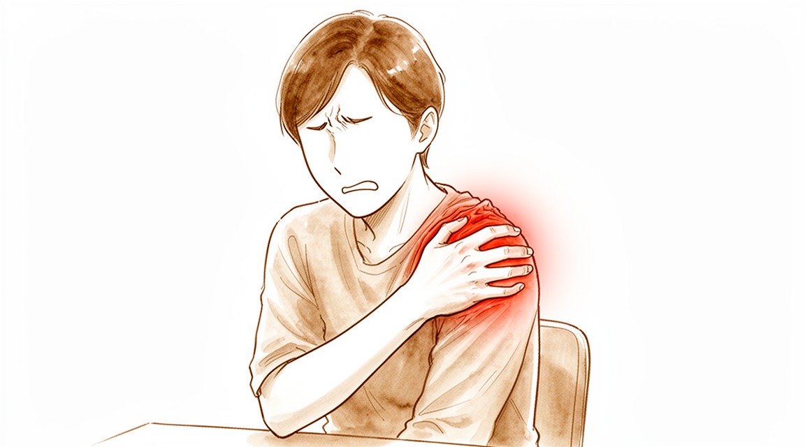
Rotator cuff disorders: common causes of shoulder pain, ranging from mild ache to debilitating injury.
您的感受
肩袖疾病很常见,且随着年龄增长,患病可能性增加。您可能会感到肩部前方或上臂外侧疼痛。这种疼痛通常源于帮助抬起手臂的肌腱。如果肱二头肌长头肌腱受累,您还可能会感到肩部前方酸痛。该肌腱沿手臂前方走行,并与肩袖协同工作。
您的症状可能表现为机械性感觉,如关节内的卡顿或研磨感。这是常见的主诉。疼痛常在夜间加剧,导致难以侧卧睡眠。您刚醒来时可能会感到僵硬。使用手臂后,酸痛可能会加重。然而,症状持续的时间长短并不能反映撕裂的严重程度。有些人即使有大撕裂也疼痛轻微,而有些人即使有小撕裂也会感到明显不适。
日常活动可能会变得困难。伸手到背后扣内衣或塞衬衫下摆可能会感到别扭或疼痛。举过头顶提物可能会引发锐痛。您可能会为了避免肩部不适而回避某些动作。主观上的不稳感或无力感也很常见。这些感觉会让您觉得肩部功能异常。
重要的是要知道,您的心理健康对疼痛程度和肩部功能恢复起着重要作用。这种关联可能比撕裂的实际大小更为重要。您的外科医生会综合评估您的整体情况,包括年龄和活动水平,以决定最佳的治疗方案。无论您选择手术治疗还是非手术治疗,两种方案均可能有效。目标是减轻您的疼痛,并帮助您恢复喜爱的活动。如果诊断不明确,您的外科医生可能会专注于直接治疗肩部疼痛,以避免不必要的操作。
实际情况
您的肩关节是一个球窝关节。肱骨头位于一个浅的关节盂内。一组称为肩袖的四条肌腱像袖子一样包裹在肱骨头周围。这些肌腱将肱骨头固定在原位,并帮助您抬起手臂。这种情况很普遍,并且随着年龄增长变得更加常见。
当肌腱撕裂时,肩关节的平衡会被打破。肱骨头可能会向上滑动,而不是保持在关节盂的中心位置。这被称为上移。这是因为肌腱无法再将肱骨头向下拉。随着时间的推移,这种错位会导致摩擦和磨损。未经治疗的慢性撕裂可能导致骨关节炎,即关节的磨损性关节炎。
您感受到的疼痛和无力源于这种不稳定性。包裹肩关节的关节囊可能会拉伸或撕裂。这会导致骨骼相互摩擦。在某些情况下,肩关节周围的肌肉会改变位置以进行代偿。这会改变您在日常活动中肩关节的运动方式。
您的外科医生会根据这些机械性变化来决定治疗方案。手术和非手术选项都可以有效。目标是恢复正常的活动并减轻疼痛。在某些情况下,手术旨在恢复肱骨头在关节盂内的自然位置。这有助于减轻关节压力。
心理健康会影响您对疼痛的感受。它可能与撕裂的大小一样重要。您的期望也会直接影响治疗效果。了解肩关节内部发生的情况有助于您为康复做好准备。
我们能做什么
您有多种选择来管理肩袖疾病,非手术和手术路径均可能有效。正确的选择取决于您的具体撕裂情况、年龄和整体健康状况。对于许多患者来说,从自我护理和物理治疗开始是一条可行的路径。在确诊后13年,约90%接受保守治疗的肩袖撕裂患者无疼痛或仅有轻微疼痛。在确诊后13年,约70%接受保守治疗的肩袖撕裂患者日常生活活动未受干扰。
物理治疗旨在增强肩部周围肌肉,以改善功能并减轻疼痛。针对非创伤性全层肩袖撕裂,特定的物理治疗方案在随访2年的患者中,对约75%的患者有效。如果您患有非创伤性冈上肌撕裂,应将保守治疗作为主要治疗方法。您的外科医生可能会建议您避免引起撞击的肩部运动,即组织在肩关节内被挤压的情况。心理健康也起着重要作用;在全层肩袖撕裂患者中,心理健康与患者报告的肩部疼痛和功能的相关性强于撕裂大小。
药物治疗侧重于缓解疼痛和减轻炎症。虽然有时会使用皮质类固醇注射,但缺乏可重复的证据支持其在管理肩袖疾病中的疗效。如果考虑这些注射,应谨慎行事,因为如果计划在接下来的6个月内进行肩袖修复,则应避免使用。目前的有限证据表明,富血小板血浆(PRP)注射在短期内对慢性肩袖疾病的非手术治疗可能无益。止痛药和抗炎药可以帮助您在接受物理治疗期间管理症状。
当保守治疗达到极限或您有未愈合的创伤性撕裂时,会考虑手术。关节镜肩袖修复是治疗肩袖撕裂症状的有效且安全的选择,其临床结果随时间推移具有持久性。该手术因改善肩部功能而受到青睐,与其他手术或关节内治疗相比,在此目标上无显著优势。对于巨大或不可修复的撕裂,部分修复可通过恢复力偶平衡来有效减轻疼痛并改善功能。在关节本身受损的情况下,如患有骨关节炎,可能会保留使用反式肩关节假体进行治疗,但类风湿关节炎患者禁用。您的外科医生在制定治疗方案时将仔细考虑您修复失败的风险因素。
预期情况
您的预后很大程度上取决于您选择手术治疗还是保守治疗。对于小到中等大小的撕裂,与单纯物理治疗相比,手术能提供更好的长期疼痛缓解和功能改善,且益处可持续长达 15 年。如果您有大撕裂或巨大撕裂,手术仍可提供令人满意的长期结果。即使需要翻修手术,您的短期结果也与初次修复相似。
如果您在不进行手术的情况下管理撕裂,病程通常较为温和。约 90% 的患者在确诊后 13 年无疼痛或仅有轻微疼痛。在同一 13 年节点,约 70% 的患者在日常活动中无干扰。然而,未经治疗的慢性撕裂最终可能导致骨关节炎,即磨损性关节炎。手术可以改变这一早期的自然病程,与非手术治疗相比,在疼痛和功能方面带来具有临床意义的差异。
如果您接受修复手术,您的康复是一个渐进的过程。您在术后 3 个月时将达到最终功能恢复的大约 60%。到 6 个月时,您将达到大约 75% 的功能恢复。术后一年并不能决定您的长期结果,因此耐心至关重要。
您的年龄和期望值会影响您的结果。如果您年龄为 50 岁或以下,您可以期望获得长期的疼痛缓解。但是,您可能不会看到活动度的显著长期改善。该年龄段的大部分患者长期结果不满意。您的心理健康也在很大程度上影响您对疼痛和功能的感知,有时甚至比撕裂本身的大小影响更大。与您的外科医生直接对齐期望有助于确保更好的结果。
何时就诊
如果肩部疼痛在休息后未见改善,请咨询您的全科医生。如果您注意到关节出现无力或不稳,请要求专科医生进行评估。如果肩部出现锁定或脱位现象,请及时就医。如果症状影响您的睡眠或工作,请联系您的医生。疼痛突然加重也是寻求医疗帮助的理由。请记住,症状持续的时间长短并不能反映撕裂的严重程度。未经治疗的慢性撕裂可能会随着时间的推移导致磨损性关节炎。早期评估有助于防止进一步损伤,并确保您获得符合您具体需求的适当治疗方案。
Evidence & references
Overview
- The majority of rotator cuff disorders are amenable to conservative treatment [1].
- Rotator cuff dysfunction may necessitate surgical treatment [1].
- The major indication for revision rotator cuff repair is the persistence of clinical symptoms despite nonsurgical management in the absence of substantial risk factors for failure [2].
- Rotator cuff injuries are most accurately diagnosed with a combination of cuff- and impingement-specific clinical tests [3].
- Deltoid complications combined with rotator cuff pathology represent a rare but devastating complication with no well-described surgical option [4].
- Treatment of chronic massive rotator cuff tears is challenging, and results are comparatively inferior to those of treating patients with smaller rotator cuff tears [17].
- Shoulder arthroscopy literature remains controversial, with conclusions often unsupported due to bias and limitations [19].
- No clinical guidelines for shoulder arthroscopy are definitive pending higher levels of evidence [19].
- No recommendations regarding suprascapular nerve release in conjunction with rotator cuff repair can be made at this time [22].
- Further research is necessary to better delineate the indications for suprascapular nerve release in conjunction with rotator cuff repair [22].
- Open approaches for rotator cuff repairs continue to have indications in certain circumstances, such as complete rotator cuff tendon avulsion and glenohumeral joint incarceration after high-velocity trauma [23].
- There is no statistically significant difference in subjective outcome after arthroscopic rotator cuff repair with or without acromioplasty at intermediate follow-up [26].
- Critical shoulder angle and acromial index do not appear to influence 24-month functional outcomes postoperatively [47].
- Critical shoulder angle and acromial index are not contraindications to arthroscopic rotator cuff repair [47].
- Predictors of pain and functional outcomes after operative treatment for rotator cuff tears can be used to select optimal candidates for operative treatment [54].
- Predictors of pain and functional outcomes after operative treatment for rotator cuff tears can assist with patient education and expectations before treatment [54].
- There were no differences of clinically relevant size between arthroscopic and open rotator cuff surgery in postoperative pain in a comparative series [71].
Anatomy & Pathophysiology
- Understanding the function and pathology surrounding the teres minor is paramount in comprehensive management of patients with shoulder pathology [6].
- A systematic approach to magnetic resonance imaging interpretation of shoulder injuries describes the normal imaging appearance of each anatomical structure, the most useful pulse sequences and imaging planes, and signs of injury [28].
- Imaging is an essential tool for evaluation of patients with shoulder pain, and understanding the extent of an injury with imaging is key to successful management [29].
- Tears of the subscapularis have greater biomechanical consequences than do tears of the infraspinatus [31].
- Dynamic superior migration of the humeral head during abduction occurs in patients with rotator cuff tears, as confirmed by in vivo 3D kinematic analysis [37].
- In massive rotator cuff tears, the pectoralis major and latissimus dorsi muscles are effective in improving glenohumeral kinematics and reducing acromiohumeral pressures [40].
- Biomechanical studies indicate that the long head of the biceps contributes to stability of the glenohumeral joint in all directions, although in vivo studies have not yet established this stabilizing effect or the physiologic load required [46].
- Increasing supraspinatus tendon loading causes a mechanical interaction between the supraspinatus and infraspinatus tendons, paralleling the increase in supraspinatus tendon strain [51].
- The physiopathology of symptomatic anterior instabilities is related to dysfunction of the anterosuperior glenohumeral capsular ligament rather than the inferior glenohumeral ligament [52].
- Additional repair of a partial subscapularis tear combined with a supraspinatus tear did not affect external rotation or glenohumeral kinematics [53].
- Shoulders with rotator cuff tears require considerable compensatory deltoid function to prevent abduction motion loss [57].
- Pain reduction from subacromial injection causes shifts in scapulohumeral rhythm, resulting in increased glenohumeral motion and reduced reliance on scapular rotation [59].
- The clinical evidence to support correction of the critical shoulder angle with lateral acromioplasty is insufficient, and further research is required to demonstrate an association between critical shoulder angle and clinical outcomes before treatment algorithms should be altered [61].
- The critical shoulder angle, posterior acromial height, and posterior acromial tilt do not change significantly over a long-term follow-up of at least 10 years, supporting the hypothesis that these scapular morphologic parameters are stable anthropometric characteristics [62].
- Simulated isolated supraspinatus cord and strap tears significantly reduced shoulder abduction force, with cord tears causing a larger decline than strap tears [63].
- The critical shoulder angle may not be responsible for rotator cuff tears; rather, patient activities throughout several decades could induce both cuff lesions and bone remodeling at the acromial level [66].
- The human scapula has two distinctive characteristics: a lateral orientation of the glenoid cavity and a narrow coraco-acromial arch [73].
- Cervical spine position may cause decreased shoulder rotation strength, meaning clinicians should assess shoulder strength in the position the patient requires to use their shoulder because weakness may be missed in standard testing positions [78].
Classification
- Rotator cuff disorders are recognized and managed conditions among patients with shoulder pain [1].
- Rotator cuff dysfunction may necessitate surgical treatment [1].
- The majority of rotator cuff conditions are amenable to conservative treatment [1].
- The major indication for revision rotator cuff repair is the persistence of clinical symptoms despite nonsurgical management in the absence of substantial risk factors for failure [2].
- Rotator cuff injuries are most accurately diagnosed with a combination of cuff- and impingement-specific clinical tests [3].
- Deltoid complications combined with rotator cuff pathology represent a rare but devastating complication with no well-described surgical option [4].
- Increasing knowledge about rotator cuff syndrome, including better imaging, has facilitated patient treatment for a stable spectrum of rotator cuff pathology [5].
- The application of endoscopic surgery has facilitated patient treatment for a stable spectrum of rotator cuff pathology [5].
- Rotator cuff disease, shoulder instability, and associated lesions are common pathologic conditions of the shoulder involving soft tissues [7].
- In the context of rotator cuff disease, the etiology of anterior shoulder pain with macroscopic changes in the biceps tendon is related to the complex interaction of the tendon and surrounding soft tissues, rather than a single entity [8].
- The Korean Shoulder Scoring System (KSS) is a useful measurement tool that combines subjective and objective evaluations for shoulder function related to rotator cuff disorders [9].
- The acromial morphology classification system is an unreliable method to assess the acromion [14].
- The acromial index shows no association with the presence of rotator cuff disease [14].
- A classification system exists to divide coracoids according to their morphology and relative risk of associated subscapularis tears [30].
- Comparing histopathological data with demographical information allows for the identification of rotator cuff tears at risk of repair failure [50].
- Gene expression in human rotator cuff muscles varied according to tendon injury severity [67].
Clinical Presentation
- Rotator cuff disorders are amenable to conservative treatment in the majority of cases [1].
- Rotator cuff dysfunction may necessitate surgical treatment [1].
- The major indication for revision rotator cuff repair is the persistence of clinical symptoms despite nonsurgical management in the absence of substantial risk factors for failure [2].
- Rotator cuff injuries are most accurately diagnosed with a combination of cuff- and impingement-specific clinical tests [3].
- Deltoid complications combined with rotator cuff pathology represent a rare but devastating complication with no well-described surgical option [4].
- Increasing knowledge about rotator cuff syndrome, including better imaging, has facilitated patient treatment for a stable spectrum of rotator cuff pathology [5].
- The application of endoscopic surgery has facilitated patient treatment for a stable spectrum of rotator cuff pathology [5].
- Understanding the function and pathology surrounding the teres minor is paramount in comprehensive management of the patient with shoulder pathology [6].
- Rotator cuff disease, shoulder instability, and associated lesions are common pathologic conditions of the shoulder involving soft tissues [7].
- In the context of rotator cuff disease, the etiology of anterior shoulder pain with macroscopic changes in the biceps tendon is related to the complex interaction of the tendon and surrounding soft tissues, rather than a single entity [8].
- The Korean Shoulder Scoring System (KSS) is a useful measurement tool that combines subjective and objective evaluations for shoulder function related to rotator cuff disorders [9].
- In one-quarter of patients with painful cuff tears, pain developed in a contralateral asymptomatic cuff tear that resulted in a measurable decline in function within 3 years [10].
- The acromial morphology classification system is an unreliable method to assess the acromion [14].
- The acromial index shows no association with the presence of rotator cuff disease [14].
- Ultrasound is an useful tool for discovering in pre-symptomatic stages subjects that may undergo shoulder symptomatic pathologies [15].
- Rotator cuff injuries in adolescents may be overlooked as a cause of disability, leading to significant delays in diagnosis [32].
- Intratendinous rotator cuff tears are difficult to diagnose preoperatively [34].
- Current physiotherapy practice in relation to rotator cuff disorders is variable [35].
- Current physiotherapy practice in relation to rotator cuff disorders is variable [36].
- The Functional Shoulder Score (FSS) is a patient-reported outcome measure that can easily be incorporated into clinical practice [38].
- The FSS provides a quick, reliable, valid and practical measure for rotator cuff problems [38].
Investigations
- Rotator cuff dysfunction may necessitate surgical treatment [1].
- Rotator cuff injuries are most accurately diagnosed with a combination of cuff- and impingement-specific clinical tests [3].
- Better imaging has facilitated patient treatment for a stable spectrum of rotator cuff pathology [5].
- Ultrasound is an useful tool for discovering subjects in pre-symptomatic stages that may undergo shoulder symptomatic pathologies [15].
- The use of MRI before a trial of conservative management in patients with atraumatic shoulder pain, minimal to no strength deficits on physical examination, and suspected cuff tendinopathy other than full-thickness tears provides negative value in the management of these patients, at both the individual and population level [24].
- A systematic approach to the interpretation of a magnetic resonance examination of the shoulder describes the normal imaging appearance of each anatomical structure, the most useful pulse sequences and imaging planes, and the signs of injury [28].
- Imaging is an essential tool for evaluation of patients with shoulder pain [29].
- Understanding the extent of an injury with imaging is key to successful management [29].
- Most abnormal MRI findings were not different in frequency between symptomatic and asymptomatic shoulders [43].
- MRI and US provide similar assessments of postoperative rotator cuff healing, although US is less sensitive [58].
- Shoulder MRI may be warranted for preoperative planning in the select population of patients with chronic calcific tendinopathy and prolonged refractory pain, although the probability of identifying additional cuff pathology requiring surgical intervention is very low [64].
- Preoperative MRI scans of the shoulder interpreted by orthopaedic surgeons with a systematic approach resulted in improved accuracy in diagnosing subscapularis tendon tears compared with previous studies [65].
- The diagnostic accuracy of US, MRI and MRA in the characterisation of full-thickness rotator cuff tears is high with overall estimates of sensitivity and specificity over 0.90 [68].
- A non-contrast shoulder MRI obtained in the community setting after non-dislocating shoulder trauma has a moderate sensitivity for most intraarticular pathologies when interpreted by musculoskeletal radiologists [70].
- The MRI tendinosis grade is associated with stiffness assessed using sonoelastography in patients with rotator cuff tendinopathy [72].
- Tendinosis severity assessed by preoperative MRI was the only factor associated with failure to heal in patients with partial-thickness and small full-thickness rotator cuff tears [75].
- A 3 T MRI protocol can be applied to evaluate morphological tendon outcomes after different treatment modalities [77].
- Shoulders with a symptomatic rotator cuff tear showed higher radioisotope uptake on bone scintigraphy than those with an asymptomatic tear [79].
- Unenhanced magnetic resonance imaging of the shoulder in asymptomatic high performance throwing athletes reveals abnormalities that may encompass a spectrum of nonclinical findings [80].
Treatment
Non-Operative Management
- The majority of rotator cuff disorders are amenable to conservative treatment [1].
- MRI use before a trial of conservative management in patients with atraumatic shoulder pain, minimal to no strength deficits, and suspected cuff tendinopathy (other than full-thickness tears) provides negative value at both individual and population levels [24].
- Nonoperative treatment is appropriate as initial therapy for partial-thickness rotator cuff tears [42].
- There is little reproducible evidence to support the efficacy of subacromial corticosteroid injection in managing rotator cuff disease [33].
- Orthobiologics offer a relatively safe management option for rotator cuff pathology, but evidence is inconclusive for or against its use [45].
- Limited evidence suggests that platelet-rich plasma (PRP) injections may not be beneficial in the short term for the nonoperative treatment of chronic rotator cuff disease [56].
- Subacromial PRP injections produce significantly worse improvement in functional outcomes in patients with partial supraspinatus tears compared to patients with isolated tendinopathy [44].
Operative Management
- Rotator cuff dysfunction may necessitate surgical treatment when conservative options are insufficient [1].
- Early operative treatment appears to be better for rotator cuff tears with a sudden onset of symptoms and poor function to achieve maximal return of shoulder function [18].
- Patients undergoing operative treatment for rotator cuff tears had significantly better pain and functional outcomes compared with patients undergoing nonoperative treatment in a prospective cohort study [76].
- Treatment results for chronic massive rotator cuff tears are comparatively inferior to those for smaller rotator cuff tears [17].
- Open approaches for rotator cuff repairs continue to have indications in certain circumstances, such as complete tendon avulsion and glenohumeral joint incarceration following high-velocity trauma [23].
- Arthroscopic revision rotator cuff repair is indicated for the persistence of clinical symptoms despite nonsurgical management in the absence of substantial risk factors for failure [2].
- Repair of partial- and full-thickness rotator cuff tears using a bioinductive implant shows safety and efficacy at 1-year follow-up [11].
- Arthroscopy is a safe and effective treatment for symptomatic calcific tendonitis of the shoulder, including or excluding patients with rotator cuff tears [48].
- For patients with intact rotator cuffs and calcific tendonitis, needling or extracorporeal shockwave therapy (ESWT) may be beneficial alternatives to arthroscopy in some cases [48].
Arthroscopic Subacromial Decompression (SAD)
- Following nonoperative treatment for at least 6 weeks, SAD is a viable and good surgical option for shoulder impingement with an intact rotator cuff [39].
- SAD without cuff repair appears to be a safe, efficacious, and sustainable procedure for patients with partial rotator cuff tears [41].
- Operative management for partial-thickness tears, including arthroscopic subacromial decompression, is considered when nonoperative treatment fails [42].
Adjunctive Procedures and Implants
- PRP does not have an effect on overall retear rates or shoulder-specific outcomes after arthroscopic rotator cuff repair [21].
- Routine arthroscopic suprascapular nerve release (SSNR) is not recommended when treating patients with rotator cuff tear [49].
- No recommendations regarding suprascapular nerve release in conjunction with rotator cuff repair can be made at this time, and further research is necessary [22].
Reverse Total Shoulder Arthroplasty (RTSA)
- Severely impaired deltoid function is a contraindication to RTSA [13].
- An isolated supraspinatus tear is a contraindication to RTSA [13].
- The presence of full active shoulder elevation with a massive rotator cuff tear and arthritis is a contraindication to RTSA [13].
Evidence Quality
- Conclusions in shoulder arthroscopy literature are often unsupported due to bias and limitations, and no clinical guidelines are definitive pending higher levels of evidence [19].
Complications
- Rotator cuff dysfunction may necessitate surgical treatment [1].
- Deltoid complications combined with rotator cuff pathology represent a rare but devastating complication with no well-described surgical option [4].
- In one-quarter of patients with painful cuff tears, pain developed in a contralateral asymptomatic cuff tear that resulted in a measurable decline in function within 3 years [10].
- Arthroscopic rotator cuff repair leads to a structural failure rate of 33% but satisfactory functional results with high patient satisfaction at midterm follow-up [27].
Recovery
- The majority of rotator cuff disorders are amenable to conservative treatment, although rotator cuff dysfunction may necessitate surgical treatment [1].
- Conservative versus surgical management for rotator cuff tears does not result in significantly improved shoulder function (evaluated by CMS) at a 2-year follow-up [12].
- Arthroscopic decompression is not recommended in the treatment of rotator cuff tendinopathy [16].
- The natural history of rotator cuff tendinopathy probably plays a significant role in long-term results [16].
- Arthroscopic acromioplasty significantly improves long-term clinical outcomes up to 2 years for chronic rotator cuff tendinopathy when used with platelet-rich plasma injection [69].
- Patients with rotator cuff disease treated without surgery experience a clinically important change in self-assessed outcome with a 2-point change in the Simple Shoulder Test (SST) score or a 12 to 17-point change in the American Shoulder and Elbow Surgeons (ASES) score [83].
- Younger age, lower BMI, more functional capacity, a shorter symptomatic period, reversible changes on MRI, and higher Constant and ASES scores at the first evaluation are good prognostic factors for the natural course of subacromial impingement syndrome [84].
- In one-quarter of patients with painful cuff tears, pain developed in a contralateral asymptomatic cuff tear that resulted in a measurable decline in function within 3 years [10].
- Early operative treatment appears to be better for rotator cuff tears with a sudden onset of symptoms and poor function to achieve maximal return of shoulder function [18].
- The major indication for revision rotator cuff repair is the persistence of clinical symptoms despite nonsurgical management in the absence of substantial risk factors for failure [2].
- Outcomes after repair of partial- and full-thickness rotator cuff tears using a bioinductive implant show safety and efficacy at 1-year follow-up [11].
- Arthroscopic rotator cuff repair leads to a structural failure rate of 33% but yields satisfactory functional results with high patient satisfaction at midterm follow-up [27].
- Increased age and longer duration of follow-up were associated with lower healing rates after double-row rotator cuff repair [20].
- The 'critical period' for healing following rotator cuff repair, during which risks of retears are high, extends to the first 6 months [85].
- Although functional status improved with time after 6 months, the structural status of repaired cuffs remained unchanged between 6 and 19 months postoperatively [55].
- Improvement in functional outcome after arthroscopic repair of a subscapularis tendon tear is maintained long-term [60].
Key Evidence
- [L4] The majority of conditions are amenable to conservative treatment, although rotator cuff dysfunction may necessitate surgical treatment. [1] (10.1002/art.20668)
- [L5] The major indication for revision rotator cuff repair is the persistence of clinical symptoms despite nonsurgical management in the absence of substantial risk factors for failure. [2] (10.5435/00124635-201111000-00002)
- [L5] Current consensus suggests rotator cuff injuries are most accurately diagnosed with a combination of cuff- and impingement-specific clinical tests. [3] (10.1016/j.arthro.2013.07.265)
- [Case_report] Deltoid complications combined with rotator cuff pathology represent a rare but devastating complication with no well-described surgical option. [4] (10.1016/j.jse.2011.09.023)
- [L3] Increasing knowledge about this syndrome, including better imaging, has facilitated patient treatment for a stable spectrum of rotator cuff pathology, as has the application of endoscopic surgery. [5] (10.1016/j.arthro.2010.02.029)
- [L5] Understanding the function and pathology surrounding the teres minor is paramount in comprehensive management of the patient with shoulder pathology. [6] (10.5435/jaaos-d-15-00258)
- [L4] In the context of rotator cuff disease, the etiology of anterior shoulder pain with macroscopic changes in the biceps tendon is related to the complex interaction of the tendon and surrounding soft tissues, rather than a single entity. [8] (10.1016/j.jse.2008.05.044)
- [L4] The KSS is a useful measurement tool that combines subjective and objective evaluations for shoulder function related to rotator cuff disorders. [9] (10.1016/j.jse.2008.11.019)
- [L2] In one-quarter of patients with painful cuff tears, pain developed in a contralateral asymptomatic cuff tear that resulted in a measurable decline in function within 3 years. [10] (10.1016/j.jse.2023.09.008)
- [L4] Outcomes after repair of partial- and full-thickness rotator cuff tears using a bioinductive implant show safety and efficacy at 1-year follow-up. [11] (10.1016/j.arthro.2019.02.019)
- [L1] At a 2-year follow-up, shoulder function evaluated in terms of CMS was not significantly improved. [12] (10.1186/s12891-020-03872-4)
- [L5] Severely impaired deltoid function, an isolated supraspinatus tear, and the presence of full active shoulder elevation with a massive rotator cuff tear and arthritis are contraindications to RTSA. [13] (10.1007/s11999-009-1188-9)
- [L3] The acromial morphology classification system is an unreliable method to assess the acromion, and the acromial index shows no association with the presence of rotator cuff disease. [14] (10.1016/j.jse.2011.09.028)
- [L3] Ultrasound is an useful tool for discovering in pre-symptomatic stages the subjects that may undergo shoulder symptomatic pathologies. [15] (10.1186/1471-2474-11-278)
- [L1] The natural history of rotator cuff tendinopathy probably plays a significant role in the results in the long-term. [16] (10.1302/0301-620x.99b6.bjj-2016-0569.r1)
- [L5] However, treatment of these patients is challenging, and results are comparatively inferior to those of treating patients with smaller rotator cuff tears. [17] (10.5435/00124635-200309000-00005)
- [L3] Early operative treatment appears to be better for rotator cuff tears with a sudden onset of symptoms and poor function to achieve maximal return of shoulder function. [18] (10.1016/j.jse.2005.07.006)
- [L5] The editorial states that shoulder arthroscopy literature remains controversial, conclusions are often unsupported due to bias and limitations, and no clinical guidelines are definitive pending higher levels of evidence. [19] (10.1016/j.arthro.2012.07.001)
- [L4] Increased age and longer duration of follow-up were associated with lower healing rates after double-row rotator cuff repair. [20] (10.1177/0363546510382835)
- [L1] PRP does not have an effect on overall retear rates or shoulder-specific outcomes after arthroscopic rotator cuff repair. [21] (10.1016/j.arthro.2012.03.007)
- [L4] No recommendations regarding suprascapular nerve release in conjunction with rotator cuff repair can be made at this time, and further research is necessary to better delineate the indications in the future. [22] (10.1016/j.jse.2011.11.033)
- [Case_report] This case highlights the importance of the initial workup after high-velocity trauma and that open approaches for rotator cuff repairs continue to have indications in certain circumstances. [23] (10.1016/j.jse.2009.07.014)
- [L4] The use of MRI before a trial of conservative management in patients with atraumatic shoulder pain, minimal to no strength deficits on physical examination, and suspected cuff tendinopathy other than full-thickness tears provides negative value in the management of these patients, at both the individual and population level. [24] (10.1016/j.jse.2019.04.003)
- [L1] On the basis of the currently available literature, there is no statistically significant difference in subjective outcome after arthroscopic rotator cuff repair with or without acromioplasty at intermediate follow-up. [26] (10.1016/j.arthro.2011.11.022)
- [L4] Arthroscopic rotator cuff repair leads to a structural failure rate of 33% but satisfactory functional results with high patient satisfaction at midterm follow-up. [27] (10.1016/j.jse.2015.05.051)
- [L5] This article provides a systematic approach to the interpretation of a magnetic resonance examination of the shoulder, describing the normal imaging appearance of each anatomical structure, the most useful pulse sequences and imaging planes, and the signs of injury. [28] (10.1177/0363546505278255)
- [L4] Imaging is an essential tool for evaluation of patients with shoulder pain; understanding the extent of an injury with imaging is key to successful management. [29] (10.1016/j.csm.2013.03.009)
- [L3] This study was the first to create a classification system to divide coracoids according to their morphology and relative risk of associated subscapularis tears. [30] (10.1016/j.jse.2020.01.074)
- [L5] Tears of the subscapularis have greater biomechanical consequences than do tears of the infraspinatus. [31] (10.1016/j.arthro.2009.09.007)
- [L4] Rotator cuff injuries in adolescents may be overlooked as a cause of disability, leading to significant delays in diagnosis. [32] (10.1177/0363546504269033)
- [L1] This systematic review of the available literature indicates that there is little reproducible evidence to support the efficacy of subacromial corticosteroid injection in managing rotator cuff disease. [33] (10.5435/00124635-200701000-00002)
- [L4] Intratendinous rotator cuff tears are difficult to diagnose preoperatively. [34] (10.1016/j.jse.2010.01.013)
- [L4] Current physiotherapy practice in relation to rotator cuff disorders is variable, which might reflect the lack of high-quality evidence available. [35] (10.1177/1758573217717103)
- [L4] Current physiotherapy practice in relation to rotator cuff disorders is variable, which might reflect the lack of high-quality evidence available. [36] (10.1111/j.1758-5740.2011.00164.x)
- [L3] This study confirms dynamic superior migration of the humeral head during abduction in patients with rotator cuff tears using in vivo 3D kinematic analysis. [37] (10.1016/j.arthro.2015.08.031)
- [L2] The FSS is a patient-reported outcome measure that can easily be incorporated into clinical practice, providing a quick, reliable, valid and practical measure for rotator cuff problems. [38] (10.1177/1758573215578589)
- [L5] Following nonoperative treatment for at least 6 weeks, SAD is a viable and good surgical option for the treatment of shoulder impingement with an intact rotator cuff. [39] (10.1016/j.arthro.2019.06.012)
- [L5] In massive rotator cuff tear, the pectoralis major and latissimus dorsi muscles are effective in improving glenohumeral kinematics and reducing acromiohumeral pressures. [40] (10.1016/j.jse.2013.11.030)
- [L4] ASD without cuff repair appears to be a safe, efficacious, and sustainable procedure for patients with partial rotator cuff tears. [41] (10.1016/j.arthro.2015.08.026)
- [L5] Nonoperative treatment is appropriate as initial therapy, while operative management including arthroscopic subacromial decompression, debridement, or repair is considered when nonoperative treatment fails. [42] (10.5435/00124635-199901000-00004)
- [L3] Most abnormal MRI findings were not different in frequency between symptomatic and asymptomatic shoulders. [43] (10.1016/j.jse.2019.04.001)
- [L2] However, improvement in symptoms and functional outcomes was significantly worse in patients who had a partial-thickness rotator cuff tear compared with patients who had an isolated tendinopathy. [44] (10.1016/j.arthro.2023.03.019)
- [L2] Orthobiologics offer a relatively safe management option with inconclusive evidence for or against its use for rotator cuff pathology. [45] (10.3233/bmr-201844)
- [L5] Biomechanical studies indicate that the long head of the biceps contributes to stability of the glenohumeral joint in all directions, though in vivo studies have yet to establish this stabilizing effect and the physiologic load required remains unknown. [46] (10.1016/j.arthro.2010.10.014)
- [L3] CSA and AI do not appear to influence 24-month functional outcomes postoperatively and hence are not contraindications to arthroscopic rotator cuff repair. [47] (10.1177/0363546517717947)
- [L5] Arthroscopy is a safe and effective treatment for symptomatic calcific tendonitis of the shoulder, excluding or including patients with rotator cuff tears, but patients with intact cuffs could benefit from needling or ESWT in some cases. [48] (10.1016/j.arthro.2015.11.003)
- [L1] Routine arthroscopic SSNR is not recommended when treating patients with rotator cuff tear. [49] (10.1007/s00167-022-07066-4)
- [L2] Comparing histopathological data with demographical information allows for the identification of rotator cuff tears at risk of repair failure. [50] (10.1007/s00167-011-1521-1)
- [L5] Increasing supraspinatus tendon loading causes a mechanical interaction between the two tendons, paralleling the increase in supraspinatus tendon strain. [51] (10.1016/j.jse.2009.10.003)
- [L3] The physiopathology is related to dysfunction of the anterosuperior glenohumeral capsular ligament rather than the inferior glenohumeral ligament. [52] (10.1016/j.jse.2022.10.005)
- [L5] Additional repair of the partial subscapularis tear with supraspinatus tear did not affect external rotation or glenohumeral kinematics. [53] (10.1016/j.jse.2013.09.015)
- [L2] These data can be used to select optimal candidates for operative treatment of rotator cuff tears and assist with patient education and expectations before treatment. [54] (10.1016/j.jse.2018.04.016)
- [L4] Although functional status improved with time after 6 months, the structural status of repaired cuffs remained unchanged between 6 and 19 months. [55] (10.1016/j.jse.2011.05.027)
- [L2] The currently limited available evidence on PRP for nonoperative treatment of chronic rotator cuff disease suggests that in the short term, PRP injections may not be beneficial. [56] (10.1016/j.arthro.2018.10.115)
- [L5] Shoulders with rotator cuff tears require considerable compensatory deltoid function to prevent abduction motion loss. [57] (10.1177/0363546518768276)
- [L3] MRI and US provide similar assessments of postoperative rotator cuff healing, although US is less sensitive. [58] (10.1016/j.otsr.2015.06.006)
- [L3] Pain reduction caused shifts in scapulohumeral rhythm resulting in an increase in glenohumeral motion and a reduced reliance on scapular rotation. [59] (10.1016/j.jse.2007.05.010)
- [L4] This study shows that improvement in functional outcome after arthroscopic repair of a subscapularis tendon tear is maintained long-term. [60] (10.1016/j.arthro.2012.02.031)
- [L5] The clinical evidence to support correction of the critical shoulder angle with lateral acromioplasty is insufficient at this time, and further research is required to demonstrate an association between critical shoulder angle and clinical outcomes before treatment algorithms should be altered. [61] (10.1016/j.arthro.2018.06.020)
- [L3] The critical shoulder angle, posterior acromial height, and posterior acromial tilt do not change significantly over a long-term follow-up of at least 10 years, supporting the hypothesis that these scapular morphologic parameters are stable anthropometric characteristics. [62] (10.1016/j.jse.2020.09.042)
- [L5] Simulated isolated supraspinatus cord and strap tears significantly reduced shoulder abduction force, with cord tears causing a larger decline than strap tears. [63] (10.1016/j.jse.2023.07.003)
- [Commentary] Shoulder MRI may be warranted for preoperative planning in the select population of patients with chronic calcific tendinopathy and prolonged refractory pain, although the probability of identifying additional cuff pathology requiring surgical intervention is very low. [64] (10.1016/j.arthro.2020.01.014)
- [L3] Preoperative MRI scans of the shoulder interpreted by orthopaedic surgeons with the described systematic approach resulted in improved accuracy in diagnosing subscapularis tendon tears compared with previous studies. [65] (10.1016/j.arthro.2012.04.142)
- [L5] The critical shoulder angle may not be responsible for rotator cuff tears; rather, patient activities throughout several decades could induce both cuff lesions and bone remodeling at the acromial level. [66] (10.1016/j.arthro.2020.04.030)
- [L4] Gene expression in human rotator cuff muscles varied according to tendon injury severity. [67] (10.2106/jbjs.m.01585)
- [L1] The diagnostic accuracy of US, MRI and MRA in the characterisation of full-thickness rotator cuff tears is high with overall estimates of sensitivity and specificity over 0.90. [68] (10.1136/bjsports-2014-094148)
- [L1] Arthroscopic acromioplasty significantly improves long-term clinical outcomes up to 2 years. [69] (10.1177/0363546515608485)
- [L4] A non-contrast shoulder MRI obtained in the community setting after non-dislocating shoulder trauma has a moderate sensitivity for most intraarticular pathologies when interpreted by musculoskeletal radiologists. [70] (10.1007/s00167-014-3102-6)
- [L2] There were no differences of clinically relevant size between arthroscopic and open rotator cuff surgery in this comparative series. [71] (10.1007/s11999-014-3715-6)
- [L3] The MRI tendinosis grade is associated with stiffness assessed using sonoelastography in patients with rotator cuff tendinopathy. [72] (10.1016/j.jse.2015.10.019)
- [L5] The study identified two distinctive characteristics of the human scapula: a lateral orientation of the glenoid cavity and a narrow coraco-acromial arch. [73] (10.1016/j.otsr.2014.09.011)
- [L3] Tendinosis severity assessed by preoperative MRI was the only factor associated with failure to heal in patients with partial-thickness and small full-thickness rotator cuff tears. [75] (10.1177/0363546514561004)
- [L3] In this prospective cohort study, patients undergoing operative treatment had significantly better pain and functional outcomes as compared with patients undergoing nonoperative treatment for rotator cuff tears. [76] (10.1177/0363546519873840)
- [L4] This rotator cuff MRI protocol can be applied to evaluate morphological tendon outcomes after different treatment modalities. [77] (10.1186/s13018-014-0128-x)
- [L3] Clinicians should assess shoulder strength in the position the patient requires to use their shoulder because cervical spine position may cause weakness that would be missed in standard testing positions. [78] (10.1097/corr.0000000000002212)
- [L3] Shoulders with a symptomatic rotator cuff tear showed higher radioisotope uptake on bone scintigraphy than those with an asymptomatic tear. [79] (10.1177/0363546513494741)
- [L4] Unenhanced magnetic resonance imaging of the shoulder in asymptomatic high performance throwing athletes reveals abnormalities that may encompass a spectrum of nonclinical findings. [80] (10.1177/03635465020300012501)
- [L2] Patients with rotator cuff disease who are treated without surgery and have a 2-point change in the SST score or a 12 to 17-point change in the ASES score experience a clinically important change in self-assessed outcome. [83] (10.2106/jbjs.h.01296)
- [L2] Younger age, lower BMI, more functional capacity, a shorter symptomatic period, reversible changes on MRI, and higher Constant and ASES scores at the first evaluation were good prognostic factors for the natural course of subacromial impingement syndrome. [84] (10.1016/j.jse.2015.06.007)
- [L3] The 'critical period' for healing following rotator cuff repair, during which risks of retears are high, extends to the first 6 months. [85] (10.1007/s00167-016-4276-x)
References
[1] Rotator cuff disorders: Recognition and management among patients with shoulder pain. Arthritis & Rheumatism. 2004. DOI: 10.1002/art.20668 [2] Arthroscopic Revision Rotator Cuff Repair. American Academy of Orthopaedic Surgeon. 2011. DOI: 10.5435/00124635-201111000-00002 [3] Management of Disorders of the Rotator Cuff: Proceedings of the ISAKOS Upper Extremity Committee Consensus Meeting. Arthroscopy. 2013. DOI: 10.1016/j.arthro.2013.07.265 [4] Rotator cuff tear arthropathy and deltoid avulsion treated with reverse total shoulder arthroplasty and latissimus dorsi transfer: case report and review of the literature. Journal of Shoulder and Elbow Surgery. 2012. DOI: 10.1016/j.jse.2011.09.023 [5] Arthroscopy and the Dramatic Increase in Frequency of Anterior Acromioplasty From 1980 to 2005: An Epidemiologic Study. Arthroscopy. 2010. DOI: 10.1016/j.arthro.2010.02.029 [6] Understanding the Importance of the Teres Minor for Shoulder Function: Functional Anatomy and Pathology. Journal of the American Academy of Orthopaedic Surgeons. 2018. DOI: 10.5435/jaaos-d-15-00258 [7] Chapter 24 Shoulder Instability, Rotator Cuff Disorders, Muscular Ruptures, Adhesive Capsulitis, Calcific Tendinitis. 2020. [8] Biceps tendinitis in chronic rotator cuff tears: A histologic perspective. Journal of Shoulder and Elbow Surgery. 2008. DOI: 10.1016/j.jse.2008.05.044 [9] The development and validation of an appraisal method for rotator cuff disorders: The Korean Shoulder Scoring System. Journal of Shoulder and Elbow Surgery. 2009. DOI: 10.1016/j.jse.2008.11.019 [10] Predictors of pain development for contralateral asymptomatic degenerative rotator cuff tears based on features of an ipsilateral painful cuff tear: a prospective longitudinal cohort study. Journal of Shoulder and Elbow Surgery. 2024. DOI: 10.1016/j.jse.2023.09.008 [11] Patient‐Reported Outcomes After Use of a Bioabsorbable Collagen Implant to Treat Partial and Full‐Thickness Rotator Cuff Tears. Arthroscopy. 2019. DOI: 10.1016/j.arthro.2019.02.019 [12] Conservative versus surgical management for patients with rotator cuff tears: a systematic review and META-analysis. BMC Musculoskeletal Disorders. 2021. DOI: 10.1186/s12891-020-03872-4 [13] Indications for Reverse Total Shoulder Arthroplasty in Rotator Cuff Disease. Clinical Orthopaedics & Related Research. 2010. DOI: 10.1007/s11999-009-1188-9 [14] Relationship of radiographic acromial characteristics and rotator cuff disease: a prospective investigation of clinical, radiographic, and sonographic findings. Journal of Shoulder and Elbow Surgery. 2012. DOI: 10.1016/j.jse.2011.09.028 [15] Sonographic evaluation of the shoulder in asymptomatic elderly subjects with diabetes. BMC Musculoskeletal Disorders. 2010. DOI: 10.1186/1471-2474-11-278 [16] Arthroscopic decompression not recommended in the treatment of rotator cuff tendinopathy. The Bone & Joint Journal. 2017. DOI: 10.1302/0301-620x.99b6.bjj-2016-0569.r1 [17] Chronic Massive Rotator Cuff Tears: Evaluation and Management. Journal of the American Academy of Orthopaedic Surgeons. 2003. DOI: 10.5435/00124635-200309000-00005 [18] Results of early operative treatment of rotator cuff tears with acute symptoms. Journal of Shoulder and Elbow Surgery. 2006. DOI: 10.1016/j.jse.2005.07.006 [19] Shoulder Arthroscopy Literature Remains Controversial. Arthroscopy. 2012. DOI: 10.1016/j.arthro.2012.07.001 [20] Factors Affecting Healing Rates after Arthroscopic Double-Row Rotator Cuff Repair. The American Journal of Sports Medicine. 2010. DOI: 10.1177/0363546510382835 [21] The Role of Platelet‐Rich Plasma in Arthroscopic Rotator Cuff Repair: A Systematic Review With Quantitative Synthesis. Arthroscopy. 2012. DOI: 10.1016/j.arthro.2012.03.007 [22] Suprascapular neuropathy: what does the literature show?. Journal of Shoulder and Elbow Surgery. 2012. DOI: 10.1016/j.jse.2011.11.033 [23] Complete rotator cuff tendon avulsion and glenohumeral joint incarceration in a young patient: A case report. Journal of Shoulder and Elbow Surgery. 2010. DOI: 10.1016/j.jse.2009.07.014 [24] A value-based care analysis of magnetic resonance imaging in patients with suspected rotator cuff tendinopathy and the implicated role of conservative management. Journal of Shoulder and Elbow Surgery. 2019. DOI: 10.1016/j.jse.2019.04.003 [26] The Role of Subacromial Decompression in Patients Undergoing Arthroscopic Repair of Full‐Thickness Tears of the Rotator Cuff: A Systematic Review and Meta‐analysis. Arthroscopy. 2012. DOI: 10.1016/j.arthro.2011.11.022 [27] Arthroscopic rotator cuff repair in the weight-bearing shoulder. Journal of Shoulder and Elbow Surgery. 2015. DOI: 10.1016/j.jse.2015.05.051 [28] A Systematic Approach to Magnetic Resonance Imaging Interpretation of Sports Medicine Injuries of the Shoulder. The American Journal of Sports Medicine. 2005. DOI: 10.1177/0363546505278255 [29] Imaging of the Shoulder with Arthroscopic Correlation. Clinics in Sports Medicine. 2013. DOI: 10.1016/j.csm.2013.03.009 [30] Coracoid morphology and humeral version as risk factors for subscapularis tears. Journal of Shoulder and Elbow Surgery. 2020. DOI: 10.1016/j.jse.2020.01.074 [31] The Effect of Posterosuperior Rotator Cuff Tears and Biceps Loading on Glenohumeral Translation. Arthroscopy. 2010. DOI: 10.1016/j.arthro.2009.09.007 [32] Rotator Cuff Tears in Adolescent Athletes. The American Journal of Sports Medicine. 2005. DOI: 10.1177/0363546504269033 [33] The Efficacy of Subacromial Corticosteroid Injection in the Treatment of Rotator Cuff Disease: A Systematic Review. Journal of the American Academy of Orthopaedic Surgeons. 2007. DOI: 10.5435/00124635-200701000-00002 [34] Surgical treatment of confirmed intratendinous rotator cuff tears: Retrospective analysis after an average of eight years of follow-up. Journal of Shoulder and Elbow Surgery. 2010. DOI: 10.1016/j.jse.2010.01.013 [35] Rotator cuff disorders: a survey of current (2016) UK physiotherapy practice. Shoulder & Elbow. 2017. DOI: 10.1177/1758573217717103 [36] Rotator Cuff Disorders: A Survey of Current Uk Physiotherapy Practice. Shoulder & Elbow. 2012. DOI: 10.1111/j.1758-5740.2011.00164.x [37] Alterations in Glenohumeral Kinematics in Patients With Rotator Cuff Tears Measured With Biplane Fluoroscopy. Arthroscopy. 2015. DOI: 10.1016/j.arthro.2015.08.031 [38] The development and validation of a questionnaire for rotator cuff disorders: The Functional Shoulder Score. Shoulder & Elbow. 2015. DOI: 10.1177/1758573215578589 [39] Indications for Arthroscopic Subacromial Decompression. A Level V Evidence Clinical Guideline. Arthroscopy. 2019. DOI: 10.1016/j.arthro.2019.06.012 [40] The role of pectoralis major and latissimus dorsi muscles in a biomechanical model of massive rotator cuff tear. Journal of Shoulder and Elbow Surgery. 2014. DOI: 10.1016/j.jse.2013.11.030 [41] Patients With Impingement Syndrome With and Without Rotator Cuff Tears Do Well 20 Years After Arthroscopic Subacromial Decompression. Arthroscopy. 2015. DOI: 10.1016/j.arthro.2015.08.026 [42] Partial-Thickness Tears of the Rotator Cuff: Evaluation and Management. Journal of the American Academy of Orthopaedic Surgeons. 1999. DOI: 10.5435/00124635-199901000-00004 [43] Bilateral magnetic resonance imaging findings in individuals with unilateral shoulder pain. Journal of Shoulder and Elbow Surgery. 2019. DOI: 10.1016/j.jse.2019.04.001 [44] Subacromial Platelet‐Rich Plasma Injections Produce Significantly Worse Improvement in Functional Outcomes in Patients With Partial Supraspinatus Tears Than in Patients With Isolated Tendinopathy. Arthroscopy. 2023. DOI: 10.1016/j.arthro.2023.03.019 [45] Non-operative orthobiologic use for rotator cuff disorders and glenohumeral osteoarthritis: A systematic review. Journal of Back and Musculoskeletal Rehabilitation. 2021. DOI: 10.3233/bmr-201844 [46] Anatomy, Function, Injuries, and Treatment of the Long Head of the Biceps Brachii Tendon. Arthroscopy. 2011. DOI: 10.1016/j.arthro.2010.10.014 [47] Critical Shoulder Angle and Acromial Index Do Not Influence 24-Month Functional Outcome After Arthroscopic Rotator Cuff Repair. The American Journal of Sports Medicine. 2017. DOI: 10.1177/0363546517717947 [48] Editorial Commentary: Options Abound for Calcific Tendonitis of the Shoulder Without a Rotator Cuff Tear. Arthroscopy. 2016. DOI: 10.1016/j.arthro.2015.11.003 [49] Suprascapular nerve release does not provide additional benefits in arthroscopic rotator cuff repair surgery: a systematic review and meta‐analysis. Knee Surgery, Sports Traumatology, Arthroscopy. 2022. DOI: 10.1007/s00167-022-07066-4 [50] Rotator cuff re‐tear or non‐healing: histopathological aspects and predictive factors. Knee Surgery, Sports Traumatology, Arthroscopy. 2011. DOI: 10.1007/s00167-011-1521-1 [51] Effect of anterior supraspinatus tendon partial-thickness tears on infraspinatus tendon strain through a range of joint rotation angles. Journal of Shoulder and Elbow Surgery. 2010. DOI: 10.1016/j.jse.2009.10.003 [52] Arthroscopic findings of the glenohumeral joint in symptomatic anterior instabilities: comparison between overhead throwing disorders and traumatic shoulder dislocation. Journal of Shoulder and Elbow Surgery. 2023. DOI: 10.1016/j.jse.2022.10.005 [53] The influence of partial subscapularis tendon tears combined with supraspinatus tendon tears. Journal of Shoulder and Elbow Surgery. 2014. DOI: 10.1016/j.jse.2013.09.015 [54] Predictors of pain and functional outcomes after operative treatment for rotator cuff tears. Journal of Shoulder and Elbow Surgery. 2018. DOI: 10.1016/j.jse.2018.04.016 [55] Serial structural and functional assessments of rotator cuff repairs: do they differ at 6 and 19 months postoperatively?. Journal of Shoulder and Elbow Surgery. 2012. DOI: 10.1016/j.jse.2011.05.027 [56] Nonoperative Treatment of Rotator Cuff Disease With Platelet‐Rich Plasma: A Systematic Review of Randomized Controlled Trials. Arthroscopy. 2019. DOI: 10.1016/j.arthro.2018.10.115 [57] Relationship Between Deltoid and Rotator Cuff Muscles During Dynamic Shoulder Abduction: A Biomechanical Study of Rotator Cuff Tear Progression. The American Journal of Sports Medicine. 2018. DOI: 10.1177/0363546518768276 [58] Evaluating postoperative rotator cuff healing: Prospective comparison of MRI and ultrasound. Orthopaedics & Traumatology: Surgery & Research. 2015. DOI: 10.1016/j.otsr.2015.06.006 [59] Shoulder kinematics in patients with full-thickness rotator cuff tears after a subacromial injection. Journal of Shoulder and Elbow Surgery. 2008. DOI: 10.1016/j.jse.2007.05.010 [60] Long‐Term Outcome of a Consecutive Series of Subscapularis Tendon Tears Repaired Arthroscopically. Arthroscopy. 2012. DOI: 10.1016/j.arthro.2012.02.031 [61] Editorial Commentary: Critical Shoulder Angle: Perhaps Not So “Critical” for Clinical Outcomes Following Rotator Cuff Repair. Arthroscopy. 2018. DOI: 10.1016/j.arthro.2018.06.020 [62] The critical shoulder angle does not change over time: a radiographic study. Journal of Shoulder and Elbow Surgery. 2021. DOI: 10.1016/j.jse.2020.09.042 [63] Relative contributions of the supraspinatus cord and strap tendons to shoulder abduction and translation. Journal of Shoulder and Elbow Surgery. 2024. DOI: 10.1016/j.jse.2023.07.003 [64] Editorial Commentary: Is Magnetic Resonance Imaging of the Shoulder Ever Appropriate in Evaluating Patients With Calcific Tendinopathy of the Rotator Cuff?. Arthroscopy: The Journal of Arthroscopic & Related Surgery. 2020. DOI: 10.1016/j.arthro.2020.01.014 [65] A Systematic Approach for Diagnosing Subscapularis Tendon Tears With Preoperative Magnetic Resonance Imaging Scans. Arthroscopy. 2012. DOI: 10.1016/j.arthro.2012.04.142 [66] The Law of Use and Disuse: Critical Shoulder Angle and Rotator Cuff Tears—Association Does Not Imply Causation. Arthroscopy. 2020. DOI: 10.1016/j.arthro.2020.04.030 [67] Muscle Gene Expression Patterns in Human Rotator Cuff Pathology. Journal of Bone and Joint Surgery. 2014. DOI: 10.2106/jbjs.m.01585 [68] Diagnostic accuracy of ultrasonography, MRI and MR arthrography in the characterisation of rotator cuff disorders: a systematic review and meta-analysis. British Journal of Sports Medicine. 2015. DOI: 10.1136/bjsports-2014-094148 [69] Platelet-Rich Plasma Injection With Arthroscopic Acromioplasty for Chronic Rotator Cuff Tendinopathy. The American Journal of Sports Medicine. 2015. DOI: 10.1177/0363546515608485 [70] Moderate value of non‐contrast magnetic resonance imaging after non‐dislocating shoulder trauma. Knee Surgery, Sports Traumatology, Arthroscopy. 2014. DOI: 10.1007/s00167-014-3102-6 [71] No Difference in Postoperative Pain After Arthroscopic versus Open Rotator Cuff Repair. Clinical Orthopaedics & Related Research. 2014. DOI: 10.1007/s11999-014-3715-6 [72] Real-time sonoelastography in the diagnosis of rotator cuff tendinopathy. Journal of Shoulder and Elbow Surgery. 2016. DOI: 10.1016/j.jse.2015.10.019 [73] The human acromion viewed from an evolutionary perspective. Orthopaedics & Traumatology: Surgery & Research. 2014. DOI: 10.1016/j.otsr.2014.09.011 [75] Arthroscopic Repair of Partial-Thickness and Small Full-Thickness Rotator Cuff Tears. The American Journal of Sports Medicine. 2014. DOI: 10.1177/0363546514561004 [76] Comparative Effectiveness of Operative Versus Nonoperative Treatment for Rotator Cuff Tears: A Propensity Score Analysis From the ROW Cohort. The American Journal of Sports Medicine. 2019. DOI: 10.1177/0363546519873840 [77] Reliability of a 3 T MRI protocol for objective grading of supraspinatus tendonosis and partial thickness tears. Journal of Orthopaedic Surgery and Research. 2014. DOI: 10.1186/s13018-014-0128-x [78] Altered Cervical Spine Position Results in Decreased Shoulder Rotation Strength. Clinical Orthopaedics & Related Research. 2022. DOI: 10.1097/corr.0000000000002212 [79] Symptomatic Rotator Cuff Tears Show Higher Radioisotope Uptake on Bone Scintigraphy Compared With Asymptomatic Tears. The American Journal of Sports Medicine. 2013. DOI: 10.1177/0363546513494741 [80] Magnetic Resonance Imaging of the Shoulder in Asymptomatic Professional Baseball Pitchers. The American Journal of Sports Medicine. 2002. DOI: 10.1177/03635465020300012501 [83] Minimal Clinically Important Differences in ASES and Simple Shoulder Test Scores After Nonoperative Treatment of Rotator Cuff Disease. The Journal of Bone & Joint Surgery. 2010. DOI: 10.2106/jbjs.h.01296 [84] Medium-term natural history of subacromial impingement syndrome. Journal of Shoulder and Elbow Surgery. 2015. DOI: 10.1016/j.jse.2015.06.007 [85] Critical period and risk factors for retear following arthroscopic repair of the rotator cuff. Knee Surgery, Sports Traumatology, Arthroscopy. 2016. DOI: 10.1007/s00167-016-4276-x


