扳机指松解术
Patients › Hand
Trigger finger release — understanding the condition, conservative treatments, and surgical options for a stuck or clicking finger.
为何建议进行此手术
扳机指松解术是一种简短的手术,旨在松解手掌中紧绷的肌腱鞘,以阻止手指出现卡顿或锁定现象。您的外科医生可能建议您进行此手术,原因是您存在持续性疼痛、手掌肿块或手指卡住的情况,特别是如果类固醇注射未能提供持久的缓解。尽管大多数人会先尝试注射治疗,但当这些治疗失败,或者您患有糖尿病且需要一个可靠的解决方案时,手术是最佳选择。
该手术总体风险较低且疗效显著。约 97% 的患者在术后症状完全缓解。主要目标是恢复平滑的活动度,并消除因手指锁定在弯曲位置而产生的疼痛。
手术前
您需要在手术前禁食数小时,并安排专人驾车送您回家。请携带您目前所有药物的清单,并穿着舒适的衣物。您的外科医生可能会安排一些简单的检查,如X光、血液检查或麻醉评估,以确保您适合接受该手术。这些检查有助于我们了解您的整体健康状况,并为您制定最佳的治疗方案。您的外科医生会告知您在手术当天之前需要停用哪些药物。该开放手术通过在手指上方做一个小切口来松解紧张的腱鞘。
手术当天
您将抵达诊所,并与麻醉医生会面讨论疼痛控制方案。该手术可在局部麻醉(仅麻醉手术区域,您保持清醒)或全身麻醉(完全入睡)下进行。大多数人选择局部麻醉:恢复更快,术后不久即可回家。如果您更倾向于全身麻醉,这也是一个合理的选择;请与您的外科医生和麻醉医生讨论。
随后,您将进入手术室,外科医生在手指上方做一个小切口以松解紧张的腱鞘。该手术时间短且安全。术后,您在恢复室苏醒,医护人员会监测您的状况,直至您准备回家。可能出现轻微问题,如瘢痕压痛或轻度僵硬,但大多数患者会立即感到弹响症状缓解。
手术过程
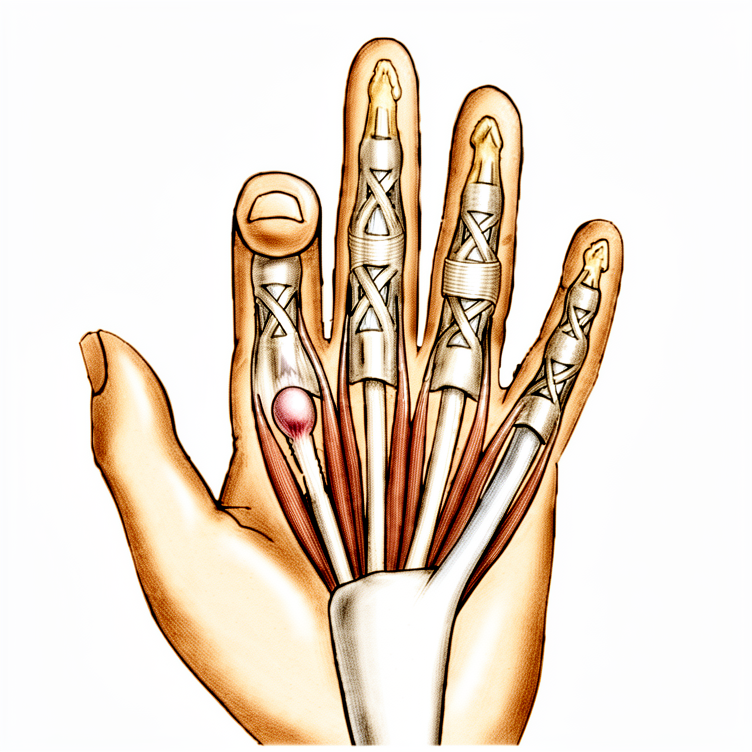
您的外科医生将在手指或拇指的掌侧做一个长约 2 厘米的切口。具体位置取决于受累的手指,通常位于手掌主横纹的下方。该切口用于暴露并进入卡压您肌腱的致密组织带,即 A1 滑车。
在手术内部,外科医生将切断该致密组织带,以释放肌腱,使其能够再次顺畅滑动。如果您患有类风湿关节炎,外科医生可能会切除一小段肌腱,以保护手指的对线。一旦组织带被松解,您将反复屈伸手指,以确认其活动自如且无卡压。
切口将用缝线缝合,缝线需在 10 至 14 天后拆除。术后会施加加压敷料,并在 48 小时后移除。敷料移除后,鼓励您正常使用手指。
术后
您将在复苏室苏醒,您的外科医生会检查您的手部。这种开放手术风险较低,但约1/20的手指可能会出现轻微、暂时性的问题。这是一个日间手术,您将在当天回家。您的手部将用敷料包裹;拇指通常不需要使用吊带或支具。疼痛通过常规药物进行管理。您应在前24小时内有人陪伴,以协助您。大多数患者会很快感觉好转,约97%的患者在术后完全恢复。
恢复
您的手在前几天会感到僵硬和疼痛。您可能会注意到手指周围有一些肿胀或轻微瘀伤。这是正常的。您的外科医生可能会建议使用冰袋来帮助缓解不适。一些患者发现轻轻活动手指有助于减轻僵硬,而不会造成伤害。
您很可能在手术当天就能活动手指。您的外科医生可能会建议您抬高患肢以减少肿胀。通常在拆除敷料后,您可以轻轻洗手。在您的外科医生确认安全之前,请避免用力抓握或提重物。简单的任务,如吃饭或打字,通常在手术后不久就可以进行。
如果您患有糖尿病,术后您的血糖水平可能会略有升高。您的外科医生会对此进行密切监测。每个人的愈合速度不同。您的外科医生和治疗师会根据您的具体进展,指导您何时可以返回工作或开车。相信您的身体,并遵循他们的建议以获得最佳效果。
可能出现的问题
大多数患者恢复良好,但偶尔也会出现一些问题。您的外科医生和医疗团队会密切监测您的情况,以便尽早发现任何异常。
术后您可能会注意到发红、肿胀或疼痛非但没有减轻,反而加重。这可能提示深部感染,特别是如果您在过去一个月内接受过类固醇注射。如果您感到深部搏动性疼痛,且普通止痛药无法缓解,或者看到伤口周围发红范围扩大,请立即致电诊所。您可能需要手术清创。
有时伤口边缘可能会轻微裂开,或者瘢痕变得非常敏感。您也可能感到手指弯曲或伸直的活动度下降。这些属于轻微问题,但如果令您困扰,请在下次复诊时提出。
在极少数情况下,拇指侧面的神经可能在松解术中受到损伤。您可能会在该区域感到麻木、刺痛或异常感觉。如果您注意到感觉或力量突然变化,请立即告知您的外科医生。
如果您患有类风湿关节炎,手术可能会导致手指关节的侧向偏移加重。您可能会发现手指比术前更偏向其他手指。请在手术前与外科医生讨论这一风险。
偶尔,术后手指可能会再次出现弹响或卡顿。这可能是由于肌腱被瘢痕组织卡住,或愈合过程中形成小结节所致。通常这种感觉会随时间自行消失,但如果持续存在,请与您的医疗团队联系。
本页的并发症表格列出了典型的发生率,如需具体数据请参阅。
何时联系我们
如果您出现发热、伤口红肿加重或渗出,请立即联系我们。若突发剧烈疼痛、新发麻木或手指无法活动,请寻求紧急医疗救助。如出现小腿肿胀或呼吸困难,请立即前往急诊室。这些症状可能提示感染、神经损伤或血栓,需立即处理。
Evidence & references
Overview
- Local anesthetic infiltration in the palm proximal to the incision site is preferred for trigger finger release [5].
- A pneumatic arm tourniquet may be helpful, although a high forearm Esmarch wrap is usually sufficient [5].
- A transverse incision about 2 cm long is made several millimeters distal to the distal palmar crease for middle, ring, and small trigger finger releases [5].
- A transverse incision about 2 cm long is made several millimeters distal to the proximal palmar crease for index trigger finger releases [5].
- Trigger thumb releases can be performed through incisions either distal or proximal to the metacarpophalangeal joint flexion crease [5].
- Alternative incisions for fingers can be made obliquely or longitudinally between the metacarpophalangeal and distal palmar creases [5].
- Alternative incisions for the thumb can be made obliquely across the thumb metacarpophalangeal flexion crease [5].
- Digital nerves on the thumb are more palmar and closer to the flexor sheath than might be anticipated [5].
- The thumb radial digital nerve is especially vulnerable during trigger finger release [5].
- Subcutaneous tissues are spread away from the underlying annular pulley system to ensure digital nerves are safely protected [5].
- Trigger thumbs require release of only the A1 pulley [5].
- Trigger digits require division of the A1 and A0, or proximal palmar pulley [5].
- Pulley division is usually accomplished with an initial opening of the pulley with a No. 15 knife blade and a pair of tenotomy scissors [5].
- For trigger thumb release, cutting too far distally should be avoided to prevent disruption of the oblique pulley [5].
- The sheath is incised from proximal to distal approximately 1 cm during trigger finger release [5].
- The patient should be asked to actively flex and extend the digit after sheath incision to reassess for triggering [5].
- Persistent triggering implies that either the A1 and palmar pulleys are incompletely released or an alternate site of triggering is present [5].
- When the distal A1 pulley edge is released, the divided pulley leaves are parallel rather than ending in a V-shaped pattern [5].
- The patient should be encouraged to actively flex and extend the digit after the tendon sheath has been released to ensure the release is complete [5].
- Other fingers can be found to trigger at the same surgical setting and can be managed at the same time [5].
- The skin is closed and a small, dry compression dressing is applied after trigger finger release [5].
- The compression dressing is removed after 48 hours postoperatively [5].
- Sutures are removed at 10 to 14 days postoperatively [5].
- Normal use of the finger or thumb is encouraged postoperatively [5].
Anatomy & Pathophysiology
- Stenosing tenosynovitis (trigger finger) involves mechanical impingement of the flexor tendons at the A1 pulley [9].
- The pathologic examination of affected pulleys demonstrates a proliferation of chondrocytes and increased type III collagen [9].
- The flexor digitorum profundus tendon often demonstrates a pathologic nodule, while the flexor digitorum superficialis is often unaffected [9].
- Trigger finger occurs in 2% to 3% of the general population [9].
- Women are more commonly affected than men [9].
- The digits are affected in the following order of decreasing prevalence: thumb, ring, long, little, and index [9].
- Middle and ring finger involvement is most common in adults [1, 2].
- Trigger finger is more common in patients with diabetes mellitus than in nondiabetic patients [4].
- When multiple digits are involved, the possibility of diabetes should be considered [4].
- Trigger finger is associated with systemic diseases including diabetes mellitus (10% to 20% lifetime incidence), hypothyroidism, sarcoidosis, rheumatoid arthritis, and septic tenosynovitis [9].
- Trigger finger is associated with gout, where monosodium urate precipitation elicits a fulminant inflammatory reaction in the tenosynovium [9].
- Trigger finger is associated with calcific tendinitis, where calcium salt deposition in the tenosynovium can resemble an infection [9].
- Trigger finger is associated with pseudogout, where calcium pyrophosphate dihydrate crystal deposition is often localized to the triangular fibrocartilage or within the carpal tunnel [9].
- Trigger finger is associated with amyloidosis, characterized by the deposition of beta-2-microglobulin in thick, plaque-like accumulations along the flexor tendons [9].
- Amyloidosis-associated trigger finger is most commonly seen in patients with renal failure undergoing peritoneal dialysis or hemodialysis [9].
- Trigger finger is associated with inflammatory arthropathy [1, 2].
- Trigger finger may be associated with repetitive grasping activities [1, 2].
- Fibrocartilaginous metaplasia occurs in the pulley and/or flexor digitorum superficialis (FDS) tendon [1, 2].
- A palpable lump or knot in the palm may represent a thickened area in the first annular pulley or a nodule/fusiform swelling of the flexor tendon just distal to it [7].
- The tendon nodule is usually just proximal to the anulus at the metacarpophalangeal joint level [7].
- In rheumatoid patients, a nodule distal to the metacarpophalangeal joint level may cause triggering [7].
- Triggering is often more pronounced in the morning than later in the day [4].
- Triggering may result from catching of the tendon on the palmar aponeurosis transverse fibers [7].
- A partially lacerated flexor tendon at the level of the A1 pulley may heal with a nodule sufficiently large to cause triggering [7].
- In adults, trigger thumb is a distinctly separate entity from congenital trigger thumb [7].
- Stenosing tenosynovitis in adults is usually seen in individuals older than 45 years of age [7].
- When associated with a collagen disease, several fingers may be involved, most often the long and ring fingers [7].
- Trigger finger is more common in patients with diabetes mellitus [9].
- Trigger finger is associated with Dupuytren's disease, with a high percentage of concurrent cases in the middle and ring finger [11].
- The percentage of patients suffering from both trigger finger and Dupuytren's disease increases with age [11].
- In Stage I Dupuytren's disease, thickening of the pulley wall leads to narrowing of the A1 pulley and synovial congestion [12].
- In more progressed stages of Dupuytren's contracture (Stages II or III), concomitant trigger finger is rarely seen [12].
- The absence of trigger finger in advanced Dupuytren's contracture may be explained by reduced range of motion reducing mechanical irritation at the A1 pulley [12].
- The absence of trigger finger in advanced Dupuytren's contracture may be explained by the tendon becoming slightly thinner distal to the chiasm of the deep and superficial flexor tendon [12].
- A variable annular pulley (fourth pulley) is found in 75% of patients and may contribute to stenosis [1, 2].
- Trigger finger is associated with carpal tunnel syndrome in 40% to 60% of patients [1, 2].
- Congenital trigger digit involves narrowing and thickening of the sheath, with occasional formation of a ganglion cyst [10].
- An intratendinous nodule may be present proximal to the first annular pulley, often referred to as Notta's nodule [10].
- Chronic inflammation is frequent in congenital trigger digits [10].
- Congenital trigger digit occurs far more commonly in the thumb [10].
- Congenital trigger digit is bilateral in about 25% of patients [10].
- Congenital trigger digit has been associated with trisomy 13 [10].
- Congenital trigger digit has been associated with mucopolysaccharidosis [10].
- Spontaneous resolution occurs in about 30% of children in whom congenital trigger digit appears within the first year of life [10].
- Spontaneous resolution occurs in about 12% of children in whom congenital trigger digit appears between 6 months and 2 years of age [10].
- Baek et al. noted spontaneous resolution in 63% of congenital trigger digits over a median of 48 months [10].
- Trigger finger is not often associated with a fixed flexion deformity in children, unlike congenital trigger thumb [10].
- Trigger finger in children may not respond to a simple A-pulley release [10].
- Surgical intervention for pediatric trigger finger may require excision of one or both slips of the flexor digitorum superficialis tendon and release of the A3 pulley [10].
- Triggering in adults is often caused by catching of the tendon on the palmar aponeurosis transverse fibers [7].
- Intraarticular disorders such as loose bodies, degenerative joint disease, and fractures can cause symptoms similar to trigger finger [7].
- Common extensor tendon subluxation can cause symptoms similar to trigger finger [7].
- A volar retinacular ganglion cyst may be present between the A1 and A2 pulleys [9].
- Fixed flexion deformity of the proximal interphalangeal (PIP) joint may be present in trigger finger [9].
- Triggering is often more pronounced in the morning than later in the day [4].
- Patients frequently state that the problem is in the proximal interphalangeal joint with trigger finger or in the proximal interphalangeal joint with trigger thumb [7].
- Pressure accentuates the apparent snapping or triggering of the more distal joints [7].
- Local tenderness may be present but is not a prominent complaint in trigger finger [7].
- Triggering is often more pronounced in the morning than later in the day [4].
- Triggering is often more pronounced in the morning than later in the day [4].
Classification
- Trigger finger demographics include women older than 50 years of age [1].
- The middle and ring fingers are the most commonly involved digits in adults with trigger finger [1].
- Trigger finger is associated with diabetes as a comorbidity [1].
- Trigger finger is associated with inflammatory arthropathy as a comorbidity [1].
- Repetitive grasping activities are possibly associated with the etiology of trigger finger [1].
- Histology of trigger finger shows fibrocartilaginous metaplasia in the pulley and/or flexor digitorum superficialis (FDS) tendon [1].
- Presentation of trigger finger includes pain and tenderness in the distal palm [1].
- Trigger finger presentation progresses from pain to mechanical catching and locking [1].
- Trigger finger may become fixed in advanced presentation [1].
- Referred pain at the dorsal metacarpophalangeal (MCP) or proximal interphalangeal (PIP) area is a common complaint in trigger finger [1].
- Concomitant trigger finger and carpal tunnel syndrome (CTS) occurs in 40% to 60% of patients [1].
- A fourth pulley, described as a variable annular pulley, is found in 75% of patients with thumb trigger finger [1].
- The presence of a fourth pulley in the thumb may contribute to stenosis [1].
- Grade I trigger finger is defined as pain and tenderness at the A1 pulley [1].
- Grade II trigger finger is defined as catching of the finger [1].
- Grade III trigger finger is defined as locking of the finger that is passively correctable [1].
- Grade IV trigger finger is defined as a fixed, locked finger [1].
Clinical Presentation
- Trigger finger is most common in women older than 50 years of age [1, 2].
- The middle and ring fingers are the most commonly involved digits in adults [1, 2].
- Trigger finger is associated with diabetes and inflammatory arthropathy [1, 2].
- Repetitive grasping activities are possibly associated with the etiology of trigger finger [1, 2].
- Histology of trigger finger demonstrates fibrocartilaginous metaplasia of the pulley and/or flexor digitorum superficialis (FDS) tendon [1, 2].
- Trigger finger occurs in 2% to 3% of the general population [9].
- The digits are affected in decreasing order of prevalence: thumb, ring, long, little, and index [9].
- Trigger finger is more common in patients with systemic diseases including diabetes mellitus (10% to 20% lifetime incidence), hypothyroidism, sarcoidosis, rheumatoid arthritis, and septic tenosynovitis [9].
- Gout can mimic infectious tenosynovitis with initial presentation of marked pain, erythema, swelling, and warmth [9].
- Calcific tendinitis can resemble infection and result in triggering, with males affected five times more frequently than females [9].
- Amyloidosis is characterized by deposition of beta-2-microglobulin along flexor tendons, most commonly seen in patients with renal failure undergoing dialysis [9].
- Clinical presentation includes pain and tenderness in the distal palm at the proximal edge of the digital A1 pulley [4].
- Patients frequently note catching or triggering of the affected finger or thumb after forceful flexion [4].
- In severe cases, the opposite hand must be used to force the finger or thumb passively into extension [4].
- In the most severe cases, the finger becomes locked in a flexed position [4].
- Triggering is often more pronounced in the morning than later in the day [4].
- Stenosing tenosynovitis is more common in diabetic patients than in nondiabetic patients [4].
- When multiple digits are involved, the possibility of diabetes should be considered [4].
- A common complaint is referred pain at the dorsal MCP or PIP area [1, 2].
- Concomitant trigger finger and carpal tunnel syndrome (CTS) occurs in 40% to 60% of patients [1, 2].
- A palpable lump or knot may be present in the palm, representing a thickened area in the first annular pulley or a nodule/fusiform swelling of the flexor tendon [7].
- The tendon nodule is usually just proximal to the anulus at the metacarpophalangeal joint level [7].
- In rheumatoid patients, a nodule distal to the metacarpophalangeal joint level may cause triggering [7].
- Local tenderness may be present but is not a prominent complaint [7].
- Pressure accentuates the apparent snapping or triggering of the more distal joints [7].
- Patients frequently state that the problem is in the proximal interphalangeal joint with trigger finger or in the proximal interphalangeal joint with trigger thumb [7].
- Physical examination may reveal a palpable triggering or pain with flexion and extension of the finger [9].
- Physical examination may reveal nodularity of the flexor tendon just proximal to the A1 pulley [9].
- A volar retinacular ganglion cyst may be present between the A1 and A2 pulleys [9].
- A fixed flexion deformity of the proximal interphalangeal (PIP) joint may be present [9].
- Green classification Grade I is defined as pain and tenderness at the A1 pulley [1, 2, 9].
- Green classification Grade II is defined as catching of the finger [1, 2, 9].
- Green classification Grade III is defined as locking of the finger that is passively correctable [1, 2, 9].
- Green classification Grade IV is defined as a fixed, locked finger [1, 2, 9].
- Triggering in adults is often seen in individuals older than 45 years of age [7].
- When associated with a collagen disease, several fingers may be involved, most often the long and ring fingers [7].
- Triggering may occur after operative release due to catching of the tendon on the palmar aponeurosis transverse fibers [7].
- Occasionally, a partially lacerated flexor tendon at this level heals with a nodule sufficiently large to cause triggering [7].
- Other conditions such as intraarticular disorders (loose bodies, degenerative joint disease, fractures) and common extensor tendon subluxation can cause similar symptoms [7].
- Pathologic examination demonstrates a proliferation of chondrocytes and increased type III collagen in the affected pulleys [9].
- The flexor digitorum profundus tendon often demonstrates a pathologic nodule, while the flexor digitorum superficialis is often unaffected [9].
- Newer evidence has found a fourth pulley (variable annular pulley) in 75% of patients, which may contribute to stenosis [1, 2].
Investigations
- Trigger finger demographics include women older than 50 years of age [1, 2].
- The middle and ring fingers are the most commonly involved digits in adults [1, 2].
- Trigger finger is associated with comorbidities including diabetes and inflammatory arthropathy [1, 2].
- Etiology is possibly associated with repetitive grasping activities [1, 2].
- Histology demonstrates fibrocartilaginous metaplasia of the pulley and/or flexor digitorum superficialis (FDS) tendon [1, 2].
- Pathologic examination of affected pulleys demonstrates a proliferation of chondrocytes and increased type III collagen [9].
- The flexor digitorum profundus tendon often demonstrates a pathologic nodule, while the flexor digitorum superficialis is often unaffected [9].
- Presentation includes pain and tenderness in the distal palm at the proximal edge of the digital A1 pulley [4].
- Patients frequently note catching or triggering of the affected finger or thumb after forceful flexion [4].
- In more severe cases, the opposite hand must be used to force the finger or thumb passively into extension [4].
- In the most severe cases, the finger becomes locked in a flexed position [4].
- Triggering is often more pronounced in the morning than later in the day [4].
- Stenosing tenosynovitis is more common in diabetic patients than in nondiabetic patients [4].
- When multiple digits are involved, the possibility of diabetes should be considered [4].
- A common complaint is referred pain at the dorsal MCP or PIP area [1, 2].
- Concomitant trigger finger and carpal tunnel syndrome (CTS) occurs in 40% to 60% of patients [1, 2].
- Trigger finger occurs in 2% to 3% of the general population [9].
- Women are more commonly affected than men [9].
- The digits are affected in the following order of decreasing prevalence: thumb, ring, long, little, and index [9].
- Trigger finger is more common in patients with systemic diseases such as diabetes mellitus (10% to 20% lifetime incidence), hypothyroidism, sarcoidosis, rheumatoid arthritis, and septic tenosynovitis [9].
- Physical examination may reveal tenderness to palpation of the flexor tendon at the level of the A1 pulley [9].
- Physical examination may reveal palpable triggering or pain with flexion and extension of the finger [9].
- Physical examination may reveal nodularity of the flexor tendon just proximal to the A1 pulley [9].
- A volar retinacular ganglion cyst may be present between the A1 and A2 pulleys [9].
- A fixed flexion deformity of the proximal interphalangeal (PIP) joint may be present [9].
- Green classification Grade I is defined as pain and tenderness at the A1 pulley [1, 2, 9].
- Green classification Grade II is defined as catching of the finger [1, 2, 9].
- Green classification Grade III is defined as locking of the finger that is passively correctable [1, 2, 9].
- Green classification Grade IV is defined as a fixed, locked finger [1, 2, 9].
- Newer evidence has found a fourth pulley (variable annular pulley) in 75% of patients with trigger thumb, which may contribute to stenosis [1, 2].
- Gout can mimic infectious tenosynovitis with initial presentation of marked pain, erythema, swelling, and warmth [9].
- Definitive diagnosis of gout is made by tenosynovial aspiration or biopsy showing negatively birefringent urate crystals under polarized light microscopy [9].
- Calcific tendinitis can resemble an infection and result in triggering [9].
- Radiographs of calcific tendinitis reveal fluffy ectopic calcification in the soft tissues [9].
- Pseudogout is characterized by calcium pyrophosphate dihydrate crystal deposition, often localized to the triangular fibrocartilage or within the carpal tunnel [9].
- Pathology of pseudogout reveals rhomboid-shaped crystals with positive birefringence [9].
- Amyloidosis is characterized by deposition of beta-2-microglobulin in thick, plaque-like accumulations along the flexor tendons [9].
- Amyloidosis is most commonly seen in patients with renal failure undergoing peritoneal dialysis or hemodialysis [9].
- Trigger finger is more common in patients with diabetes mellitus [9].
- Corticosteroid injection into the flexor tendon sheath is a nonoperative treatment option [1, 2].
- Injection is "curative" in about 60% of patients initially [1, 2].
- Diabetic patients are generally less responsive to corticosteroid injection [1, 2].
- There is no difference between soluble and insoluble corticosteroid preparations [1, 2].
- 65% to 90% of patients who do not have diabetes obtain relief of symptoms with one or two injections [9].
- Relief of symptoms after injection in patients with diabetes may depend on chronic glucose levels (hemoglobin A1c levels) [9].
- Surgical release of the A1 pulley is curative in digits refractory to steroid injection [4].
- Surgical release of the A1 pulley provides satisfactory results in greater than 90% of patients [9].
- Surgical treatment is required more often in patients with systemic diseases [9].
- In patients with rheumatoid arthritis, preference is to excise a slip of the FDS tendon rather than to release the A1 pulley [1, 2].
- In patients with rheumatoid arthritis, the entire annular pulley system should be preserved to prevent further ulnar drift of the fingers [4].
- Release of the A1 pulley in rheumatoid arthritis patients carries a chance that ulnar drift at the MCP joint can be exacerbated [1, 2].
- Percutaneous release of the A1 pulley may be accomplished with a needle on the middle and ring fingers, especially if they actively lock [4].
- Percutaneous release of the A1 pulley may be accomplished with a 25-gauge hypodermic needle with corticosteroid infiltration [3].
- The radial digital nerve is at risk of iatrogenic injury during thumb trigger finger release due to its superficial location [1, 2].
- Minor complications of surgical release include wound dehiscence, scar tenderness, and decreased range of motion [1, 2].
- Preoperative hypoglycemia increases infection risk after trigger finger injection and release [3].
Treatment
- Nonoperative treatment for trigger finger includes corticosteroid injection into the flexor tendon sheath [1].
- Corticosteroid injection is "curative" in about 60% of patients initially [1].
- Diabetic patients are generally less responsive to corticosteroid injection than non-diabetic patients [1].
- There is no difference in efficacy between soluble and insoluble corticosteroid preparations for trigger finger [1].
- Repeat corticosteroid injections provided symptomatic relief for a year or more in 50% of patients in a study of 292 injections [7].
- Corticosteroid injections may elevate serum glucose levels for 5 days or more in patients with diabetes mellitus [7].
- Patients with unstable diabetes may be better treated without corticosteroid injection [7].
- Immediate surgical release in the clinic was identified as the most cost-effective treatment strategy for trigger finger in diabetic patients [7].
- Surgical release of the A1 pulley is curative for digits refractory to steroid injection [4].
- Surgical release reliably relieves the problem for most patients, with approximately 97% having complete resolution after operative treatment [7].
- Persistence of triggering is more common than recurrence after trigger finger release [7].
- Surgical treatment options include open or percutaneous release of the A1 pulley [1].
- In patients with rheumatoid arthritis, the preference is to excise a slip of the FDS tendon rather than to release the A1 pulley [1].
- Excision of an FDS slip in rheumatoid arthritis patients is preferred because release of the A1 pulley carries a risk of exacerbating ulnar drift at the MCP joint [1].
- In patients with rheumatoid arthritis, the entire annular pulley system should be preserved to prevent further ulnar drift of the fingers [4].
- Triggering in rheumatoid arthritis patients is treated by tenosynovectomy and excision of one slip of the flexor digitorum superficialis [4].
- The radial digital nerve is at risk of iatrogenic injury during thumb trigger finger release due to its superficial location [1].
- Digital nerves on the thumb are more palmar and closer to the flexor sheath than might be anticipated, making them especially vulnerable [5].
- Minor complications of surgical release include wound dehiscence, scar tenderness, and decreased range of motion [1].
- Percutaneous release of the A1 pulley may be accomplished with a needle on the middle and ring fingers, especially if they actively lock [4].
- Percutaneous release using a needle or push knife has literature support for safety and effectiveness [7].
- Incomplete pulley release and damage to flexor tendons and digital nerves remain concerns with limited exposure techniques, especially in the index finger and thumb [7].
- For trigger thumb, the A1 pulley is released while avoiding cutting too far distally to disrupt the oblique pulley [5].
- Trigger digits require division of the A1 and A0 (proximal palmar) pulleys, whereas trigger thumbs require release of only the A1 pulley [5].
- If persistent triggering occurs after initial release, it implies either incomplete release of the A1 and palmar pulleys or an alternate site of triggering [5].
- When the distal A1 pulley edge is released, the divided pulley leaves are parallel rather than ending in a V-shaped pattern [5].
- Percutaneous release can be performed using an 18- or 19-gauge needle [8].
- During percutaneous release, the needle bevel should be oriented longitudinally parallel to the flexor tendons [8].
- A scraping or grating sensation felt during needle movement indicates the sheath is being incised during percutaneous release [8].
- Loss of the grating sensation as the pulley is cut indicates completion of percutaneous release [8].
- Injection of corticosteroid is optional during percutaneous trigger finger release [8].
- For combined trigger finger and Dupuytren's disease, an isolated opening of the pulley without touching Dupuytren tissue followed by corticosteroid application in the area of the opened pulley is a recommended strategy [13].
- Resecting Dupuytren's tissue concomitantly with trigger finger release resulted in a 58% recurrence rate with induration within 1 year [13].
- Normal use of the finger or thumb is encouraged after surgical release [5].
- Active hand and finger use with stretching exercises is encouraged after percutaneous release [8].
- Sutures are removed at 10 to 14 days following open surgical release [5].
- The compression dressing is removed after 48 hours following open surgical release [5].
- The needle entry site is covered with an adhesive bandage or light nonrestrictive dressing after percutaneous release [8].
- Triggering is often more pronounced in the morning than later in the day [4].
- In severe cases, the opposite hand must be used to force the finger or thumb passively into extension [4].
- In the most severe cases, the finger becomes locked in a flexed position [4].
- Stenosing tenosynovitis is more common in diabetic patients than in nondiabetic patients [4].
- When multiple digits are involved, the possibility of diabetes should be considered [4].
- Triggering in patients may occur after operative release due to catching of the tendon on the palmar aponeurosis transverse fibers, which usually resolves with time [7].
- Occasionally, a partially lacerated flexor tendon at the level of the pulley heals with a nodule sufficiently large to cause triggering [7].
- Local tenderness may be present but is not a prominent complaint in trigger finger [7].
- Pressure accentuates the apparent snapping or triggering of the more distal joints in trigger finger [7].
- Patients frequently state that the problem is in the proximal interphalangeal joint with trigger finger or in the proximal interphalangeal joint with trigger thumb [7].
- Some adjacent finger triggering may become obvious only after a given finger is released, and both can be released at the same surgical setting [7].
- Percutaneous release with steroid injection was more effective than steroid injection alone for trigger thumb [6].
- Preoperative hypoglycemia increases infection risk after both trigger finger injection and release [3].
- Database review found that preoperative hypoglycemia increased infection risk after both procedures [7].
Complications
- Concomitant trigger finger and carpal tunnel syndrome occurs in 40% to 60% of patients [1, 2].
- Diabetic patients are generally less responsive to corticosteroid injections than non-diabetic patients [1, 2].
- Preoperative hypoglycemia increases the risk of infection after both trigger finger injection and release procedures [3, 7].
- Corticosteroid injections may elevate serum glucose levels for 5 days or more in patients with diabetes mellitus [7].
- Patients with unstable diabetes may be better treated without corticosteroid injection [7].
- In patients with rheumatoid arthritis, release of the A1 pulley carries a risk of exacerbating ulnar drift at the MCP joint [1, 2, 4].
- The radial digital nerve is at risk of iatrogenic injury during thumb trigger finger release due to its superficial location [1, 2, 5].
- Incomplete pulley release and damage to flexor tendons and digital nerves remain concerns with limited exposure techniques [7].
- Minor complications following surgical release include wound dehiscence, scar tenderness, and decreased range of motion [1, 2].
- Persistence of triggering is more common than recurrence after trigger finger release [7].
- Triggering may occur after operative release due to catching of the tendon on the palmar aponeurosis transverse fibers [7].
- Occasionally, a partially lacerated flexor tendon at the level of the pulley may heal with a nodule sufficiently large to cause triggering [7].
References
[1] Miller S Review Of Orthopaedics. SECTION 16 PATELLAR TRACKING IN TOTAL KNEE ARTHROPLASTY > 2. Flexor tendon injury > 3. Stenosing tenosynovitis (trigger finger). [2] Miller S Review Of Orthopaedics. 2. Flexor tendon injury > 3. Stenosing tenosynovitis (trigger finger). [3] Campbell S Operative Orthopaedics 4 Volume Set. EXCISION OF NECROTIC MUSCLES COMBINED WITH NEUROLYSIS OF MEDIAN AND ULNAR NERVES FOR SEVERE CONTRACTURE > REFERENCES > TRIGGER THUMB AND TRIGGER FINGER. [4] A Lange Medical Book Current Diagnosis Treatment In Orthopedics Fifth Edition. 9Hand Surgery > 2. Flexor Tenosynovitis (Trigger Finger and Trigger Thumb). [5] Campbell S Operative Orthopaedics 4 Volume Set. EXCISION OF NECROTIC MUSCLES COMBINED WITH NEUROLYSIS OF MEDIAN AND ULNAR NERVES FOR SEVERE CONTRACTURE > TRIGGER FINGER AND THUMB > SURGICAL RELEASE OF TRIGGER FINGER. [6] Campbell S Operative Orthopaedics 4 Volume Set. EXCISION OF NECROTIC MUSCLES COMBINED WITH NEUROLYSIS OF MEDIAN AND ULNAR NERVES FOR SEVERE CONTRACTURE > TRIGGER THUMB AND TRIGGER FINGER. [7] Campbell S Operative Orthopaedics 4 Volume Set. EXCISION OF NECROTIC MUSCLES COMBINED WITH NEUROLYSIS OF MEDIAN AND ULNAR NERVES FOR SEVERE CONTRACTURE > TRIGGER FINGER AND THUMB. [8] Campbell S Operative Orthopaedics 4 Volume Set. EXCISION OF NECROTIC MUSCLES COMBINED WITH NEUROLYSIS OF MEDIAN AND ULNAR NERVES FOR SEVERE CONTRACTURE > PERCUTANEOUS RELEASE OF TRIGGER FINGER. [9] Aaos Comprehensive Orthopaedic Review 3. Tendinopathy of the Hand and Wrist* > II. Trigger Finger. [10] Campbell S Operative Orthopaedics 4 Volume Set. MULTIPLE Z-PLASTY RELEASE OF A CONGENITAL RING > TRIGGER FINGERS. [11] Dupuytren S Disease And Related Hyperproliferative Disorders. 31. The Influence of Dupuytren’s Disease on Trigger Fingers and Vice Versa > 31.5 Discussion. [12] Dupuytren S Disease And Related Hyperproliferative Disorders. 31. The Influence of Dupuytren’s Disease on Trigger Fingers and Vice Versa > 31.5 Discussion > 31.5.1 Why Is DD Concurring with TF?. [13] Dupuytren S Disease And Related Hyperproliferative Disorders. 31. The Influence of Dupuytren’s Disease on Trigger Fingers and Vice Versa > 31.5 Discussion > 31.5.3 Surgical Strategies and Recommendations.


