Trigger Finger Release Info Evidence Consent
Last reviewed
Also on YouTube.
Video transcript
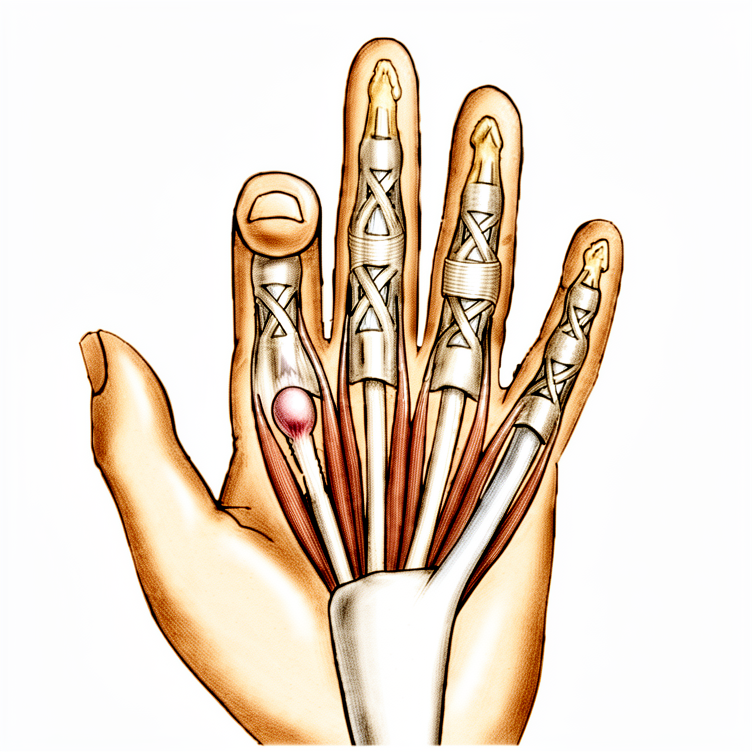
Trigger finger happens when one of the tendons that bends the finger catches as it passes through a tight pulley at the base of the finger. The finger clicks or snaps as it bends and straightens, and can lock in a bent position. It is often stiffest first thing in the morning, and there may be a tender lump in the palm at the base of the finger. Gripping can be sore, and sometimes the finger has to be eased straight with the other hand. It is common, and more so in people with diabetes. Many trigger fingers settle with simple treatment, particularly early on. Resting the finger, and easing the activities that aggravate it, can help calm things down. A splint that holds the finger straight, especially overnight, can break the catching cycle. A cortisone injection around the tendon is often very effective, and is a common first step. When the catching keeps coming back despite these measures, a small operation reliably settles it. Trigger finger release is a quick, reliable day-surgery procedure. It is done with the hand numbed, through a small incision in the palm at the base of the finger. The tight pulley that the tendon was catching on is divided, so the tendon glides freely again. You can move the finger straight away on the operating table, which confirms the catching is gone. You go home the same day, with a light dressing. You are encouraged to move the finger fully right away, to keep it gliding and prevent stiffness. The dressing stays on for a few days, and you can use the hand for light tasks soon after. The stitches can come out at around two weeks. The palm can feel tender for a couple of weeks, which settles as it heals. The catching and locking are usually gone immediately, with any residual soreness easing over the following weeks. Most people return to normal activities quickly.
Trigger finger release — understanding the condition, conservative treatments, and surgical options for a stuck or clicking finger.
Why this operation has been suggested
Trigger finger release is a short procedure that frees a tight tendon sheath in your palm to stop your finger from catching or locking. Your surgeon likely suggested this because you have persistent pain, a lump in your palm, or a finger that gets stuck, especially if steroid injections did not provide lasting relief. While most people try injections first, surgery is the best choice when those treatments fail or if you have diabetes and need a reliable solution.
This operation is generally low-risk and highly effective. About 97% of patients have complete resolution of their symptoms after the procedure. The main goal is to restore smooth movement and eliminate the pain that comes from your finger locking in a bent position.
Before the operation
You will need to fast for several hours before your surgery and arrange for someone to drive you home. Please bring a list of all your current medications and wear comfortable clothing. Your surgeon may order simple tests like X-rays, blood work, or an anaesthetic review to ensure you are safe for the procedure. These checks help us understand your overall health and plan the best care for you. Your surgeon will give you specific instructions on which medicines to stop before the day of your operation. This open surgery uses a single small cut over the finger to release the tight tendon sheath.
On the day
You will arrive at the clinic and meet your anaesthetist to discuss pain control. This operation can be done under local anaesthetic (an injection that numbs just the area of surgery, with you awake) or under general anaesthetic (fully asleep). Most people choose local — recovery is quicker and you can go home soon after. If you'd prefer to be asleep, that's also a reasonable choice — discuss it with your surgeon and anaesthetist.
You will then go to the operating theatre where your surgeon makes a single small cut over the finger to release the tight band. The procedure is short and safe. Afterward, you wake up in recovery where staff monitor you until you are ready to go home. Minor issues like scar tenderness or slight stiffness can happen, but most patients feel immediate relief from the locking.
What the operation involves
Your surgeon will make a single cut about 2 cm long on the palm side of your finger or thumb. The exact spot depends on which digit is affected, usually just below the main crease of your hand. This cut gives access to the tight band of tissue, called the A1 pulley, that is catching your tendon.
Inside, your surgeon cuts this tight band to free the tendon so it can slide smoothly again. If you have rheumatoid arthritis, your surgeon may remove a small slip of tendon instead to protect your finger alignment. Once the band is released, you will move your finger back and forth to confirm it moves freely without catching.
The cut is closed with stitches that are removed at 10 to 14 days. A compression dressing is applied and removed after 48 hours. You are encouraged to use your finger normally once the dressing is off.
After the operation
You will wake up in a recovery area where your surgeon checks your hand. This open surgery is low-risk, though about 1 in 20 fingers may have a mild, temporary issue. You will go home the same day. Your hand will be wrapped in a dressing; a sling or brace is usually not needed for the thumb. Pain is managed with standard medication. You should have someone stay with you for the first 24 hours to help you. Most patients feel better quickly, and about 97% have complete resolution after the procedure.
Recovery
Your hand will feel stiff and sore for the first few days. You may notice some swelling or mild bruising around your finger. This is normal. Your surgeon may suggest using ice packs to help ease the discomfort. Some patients find that moving their finger gently helps reduce stiffness without causing harm.
You will likely be able to move your finger on the day of surgery. Your surgeon may advise you to keep the hand elevated to reduce swelling. You can usually wash your hand gently once the dressing is removed. Avoid heavy gripping or lifting until your surgeon says it is safe. Simple tasks like eating or typing are often fine very soon after the operation.
If you have diabetes, your blood sugar levels might rise slightly after the procedure. Your surgeon will monitor this closely. Everyone heals at a different pace. Your surgeon and therapist will guide you on when to return to work or driving based on your specific progress. Trust your body and follow their advice for the best results.
What can go wrong
Most patients do well, but problems can occasionally happen. Your surgeon and the team monitor you closely to spot any issue early.
You might notice redness, swelling, or pain that gets worse instead of better after your surgery. This could signal a deep infection, especially if you had a steroid injection in the last month. If you feel a deep, throbbing pain that simple painkillers cannot ease, or if you see redness spreading from the wound, call the clinic right away. You may need surgery to clean the area.
Sometimes the wound edges can separate slightly, or the scar might become very tender. You might also feel a decrease in how easily you can bend or straighten your finger. These are minor issues, but if they bother you, bring them up at your next review.
In rare cases, the nerve on the side of your thumb could be injured during the release. You might feel numbness, tingling, or a strange sensation in that area. If you notice sudden changes in feeling or strength, tell your surgeon immediately.
If you have rheumatoid arthritis, the surgery might make a sideways drift of your finger joint worse. You might see your finger leaning more toward the other fingers than before. Discuss this risk with your surgeon before the procedure.
Occasionally, the finger might start clicking or locking again after the operation. This can happen if the tendon catches on scar tissue or heals with a small lump. Usually, this feeling goes away with time, but if it persists, mention it to your team.
The complications table on this page lists typical rates if you want the specifics.
When to call us
Call us if you develop a fever, increasing redness, or discharge from your wound. Seek urgent care for sudden severe pain, new numbness, or if you cannot move your finger. Go to the emergency room immediately if you notice calf swelling or shortness of breath. These signs could indicate an infection, nerve injury, or a blood clot that needs immediate attention.
Evidence & references
title: "Trigger Finger Release" slug: trigger-finger-release region: hand audience: patient mesh_terms: [] article_count: 0 model_used: qwen3.5-35b-a3b-q8 generated_at: '2026-05-18T13:34:04+00:00' key_articles: [] synthesis_version: "v2" verifier_status: skipped
Overview
- Local anesthetic infiltration in the palm proximal to the incision site is preferred for trigger finger release [5].
- A pneumatic arm tourniquet may be helpful, although a high forearm Esmarch wrap is usually sufficient [5].
- A transverse incision about 2 cm long is made several millimeters distal to the distal palmar crease for middle, ring, and small trigger finger releases [5].
- A transverse incision about 2 cm long is made several millimeters distal to the proximal palmar crease for index trigger finger releases [5].
- Trigger thumb releases can be performed through incisions either distal or proximal to the metacarpophalangeal joint flexion crease [5].
- Alternative incisions for fingers can be made obliquely or longitudinally between the metacarpophalangeal and distal palmar creases [5].
- Alternative incisions for the thumb can be made obliquely across the thumb metacarpophalangeal flexion crease [5].
- Digital nerves on the thumb are more palmar and closer to the flexor sheath than might be anticipated [5].
- The thumb radial digital nerve is especially vulnerable during trigger finger release [5].
- Subcutaneous tissues are spread away from the underlying annular pulley system to ensure digital nerves are safely protected [5].
- Trigger thumbs require release of only the A1 pulley [5].
- Trigger digits require division of the A1 and A0, or proximal palmar pulley [5].
- Pulley division is usually accomplished with an initial opening of the pulley with a No. 15 knife blade and a pair of tenotomy scissors [5].
- For trigger thumb release, cutting too far distally should be avoided to prevent disruption of the oblique pulley [5].
- The sheath is incised from proximal to distal approximately 1 cm during trigger finger release [5].
- The patient should be asked to actively flex and extend the digit after sheath incision to reassess for triggering [5].
- Persistent triggering implies that either the A1 and palmar pulleys are incompletely released or an alternate site of triggering is present [5].
- When the distal A1 pulley edge is released, the divided pulley leaves are parallel rather than ending in a V-shaped pattern [5].
- The patient should be encouraged to actively flex and extend the digit after the tendon sheath has been released to ensure the release is complete [5].
- Other fingers can be found to trigger at the same surgical setting and can be managed at the same time [5].
- The skin is closed and a small, dry compression dressing is applied after trigger finger release [5].
- The compression dressing is removed after 48 hours postoperatively [5].
- Sutures are removed at 10 to 14 days postoperatively [5].
- Normal use of the finger or thumb is encouraged postoperatively [5].
Anatomy & Pathophysiology
- Stenosing tenosynovitis (trigger finger) involves mechanical impingement of the flexor tendons at the A1 pulley [9].
- The pathologic examination of affected pulleys demonstrates a proliferation of chondrocytes and increased type III collagen [9].
- The flexor digitorum profundus tendon often demonstrates a pathologic nodule, while the flexor digitorum superficialis is often unaffected [9].
- Trigger finger occurs in 2% to 3% of the general population [9].
- Women are more commonly affected than men [9].
- The digits are affected in the following order of decreasing prevalence: thumb, ring, long, little, and index [9].
- Middle and ring finger involvement is most common in adults [1, 2].
- Trigger finger is more common in patients with diabetes mellitus than in nondiabetic patients [4].
- When multiple digits are involved, the possibility of diabetes should be considered [4].
- Trigger finger is associated with systemic diseases including diabetes mellitus (10% to 20% lifetime incidence), hypothyroidism, sarcoidosis, rheumatoid arthritis, and septic tenosynovitis [9].
- Trigger finger is associated with gout, where monosodium urate precipitation elicits a fulminant inflammatory reaction in the tenosynovium [9].
- Trigger finger is associated with calcific tendinitis, where calcium salt deposition in the tenosynovium can resemble an infection [9].
- Trigger finger is associated with pseudogout, where calcium pyrophosphate dihydrate crystal deposition is often localized to the triangular fibrocartilage or within the carpal tunnel [9].
- Trigger finger is associated with amyloidosis, characterized by the deposition of beta-2-microglobulin in thick, plaque-like accumulations along the flexor tendons [9].
- Amyloidosis-associated trigger finger is most commonly seen in patients with renal failure undergoing peritoneal dialysis or hemodialysis [9].
- Trigger finger is associated with inflammatory arthropathy [1, 2].
- Trigger finger may be associated with repetitive grasping activities [1, 2].
- Fibrocartilaginous metaplasia occurs in the pulley and/or flexor digitorum superficialis (FDS) tendon [1, 2].
- A palpable lump or knot in the palm may represent a thickened area in the first annular pulley or a nodule/fusiform swelling of the flexor tendon just distal to it [7].
- The tendon nodule is usually just proximal to the anulus at the metacarpophalangeal joint level [7].
- In rheumatoid patients, a nodule distal to the metacarpophalangeal joint level may cause triggering [7].
- Triggering is often more pronounced in the morning than later in the day [4].
- Triggering may result from catching of the tendon on the palmar aponeurosis transverse fibers [7].
- A partially lacerated flexor tendon at the level of the A1 pulley may heal with a nodule sufficiently large to cause triggering [7].
- In adults, trigger thumb is a distinctly separate entity from congenital trigger thumb [7].
- Stenosing tenosynovitis in adults is usually seen in individuals older than 45 years of age [7].
- When associated with a collagen disease, several fingers may be involved, most often the long and ring fingers [7].
- Trigger finger is more common in patients with diabetes mellitus [9].
- Trigger finger is associated with Dupuytren's disease, with a high percentage of concurrent cases in the middle and ring finger [11].
- The percentage of patients suffering from both trigger finger and Dupuytren's disease increases with age [11].
- In Stage I Dupuytren's disease, thickening of the pulley wall leads to narrowing of the A1 pulley and synovial congestion [12].
- In more progressed stages of Dupuytren's contracture (Stages II or III), concomitant trigger finger is rarely seen [12].
- The absence of trigger finger in advanced Dupuytren's contracture may be explained by reduced range of motion reducing mechanical irritation at the A1 pulley [12].
- The absence of trigger finger in advanced Dupuytren's contracture may be explained by the tendon becoming slightly thinner distal to the chiasm of the deep and superficial flexor tendon [12].
- A variable annular pulley (fourth pulley) is found in 75% of patients and may contribute to stenosis [1, 2].
- Trigger finger is associated with carpal tunnel syndrome in 40% to 60% of patients [1, 2].
- Congenital trigger digit involves narrowing and thickening of the sheath, with occasional formation of a ganglion cyst [10].
- An intratendinous nodule may be present proximal to the first annular pulley, often referred to as Notta's nodule [10].
- Chronic inflammation is frequent in congenital trigger digits [10].
- Congenital trigger digit occurs far more commonly in the thumb [10].
- Congenital trigger digit is bilateral in about 25% of patients [10].
- Congenital trigger digit has been associated with trisomy 13 [10].
- Congenital trigger digit has been associated with mucopolysaccharidosis [10].
- Spontaneous resolution occurs in about 30% of children in whom congenital trigger digit appears within the first year of life [10].
- Spontaneous resolution occurs in about 12% of children in whom congenital trigger digit appears between 6 months and 2 years of age [10].
- Baek et al. noted spontaneous resolution in 63% of congenital trigger digits over a median of 48 months [10].
- Trigger finger is not often associated with a fixed flexion deformity in children, unlike congenital trigger thumb [10].
- Trigger finger in children may not respond to a simple A-pulley release [10].
- Surgical intervention for pediatric trigger finger may require excision of one or both slips of the flexor digitorum superficialis tendon and release of the A3 pulley [10].
- Triggering in adults is often caused by catching of the tendon on the palmar aponeurosis transverse fibers [7].
- Intraarticular disorders such as loose bodies, degenerative joint disease, and fractures can cause symptoms similar to trigger finger [7].
- Common extensor tendon subluxation can cause symptoms similar to trigger finger [7].
- A volar retinacular ganglion cyst may be present between the A1 and A2 pulleys [9].
- Fixed flexion deformity of the proximal interphalangeal (PIP) joint may be present in trigger finger [9].
- Triggering is often more pronounced in the morning than later in the day [4].
- Patients frequently state that the problem is in the proximal interphalangeal joint with trigger finger or in the proximal interphalangeal joint with trigger thumb [7].
- Pressure accentuates the apparent snapping or triggering of the more distal joints [7].
- Local tenderness may be present but is not a prominent complaint in trigger finger [7].
- Triggering is often more pronounced in the morning than later in the day [4].
- Triggering is often more pronounced in the morning than later in the day [4].
Classification
- Trigger finger demographics include women older than 50 years of age [1].
- The middle and ring fingers are the most commonly involved digits in adults with trigger finger [1].
- Trigger finger is associated with diabetes as a comorbidity [1].
- Trigger finger is associated with inflammatory arthropathy as a comorbidity [1].
- Repetitive grasping activities are possibly associated with the etiology of trigger finger [1].
- Histology of trigger finger shows fibrocartilaginous metaplasia in the pulley and/or flexor digitorum superficialis (FDS) tendon [1].
- Presentation of trigger finger includes pain and tenderness in the distal palm [1].
- Trigger finger presentation progresses from pain to mechanical catching and locking [1].
- Trigger finger may become fixed in advanced presentation [1].
- Referred pain at the dorsal metacarpophalangeal (MCP) or proximal interphalangeal (PIP) area is a common complaint in trigger finger [1].
- Concomitant trigger finger and carpal tunnel syndrome (CTS) occurs in 40% to 60% of patients [1].
- A fourth pulley, described as a variable annular pulley, is found in 75% of patients with thumb trigger finger [1].
- The presence of a fourth pulley in the thumb may contribute to stenosis [1].
- Grade I trigger finger is defined as pain and tenderness at the A1 pulley [1].
- Grade II trigger finger is defined as catching of the finger [1].
- Grade III trigger finger is defined as locking of the finger that is passively correctable [1].
- Grade IV trigger finger is defined as a fixed, locked finger [1].
Clinical Presentation
- Trigger finger is most common in women older than 50 years of age [1, 2].
- The middle and ring fingers are the most commonly involved digits in adults [1, 2].
- Trigger finger is associated with diabetes and inflammatory arthropathy [1, 2].
- Repetitive grasping activities are possibly associated with the etiology of trigger finger [1, 2].
- Histology of trigger finger demonstrates fibrocartilaginous metaplasia of the pulley and/or flexor digitorum superficialis (FDS) tendon [1, 2].
- Trigger finger occurs in 2% to 3% of the general population [9].
- The digits are affected in decreasing order of prevalence: thumb, ring, long, little, and index [9].
- Trigger finger is more common in patients with systemic diseases including diabetes mellitus (10% to 20% lifetime incidence), hypothyroidism, sarcoidosis, rheumatoid arthritis, and septic tenosynovitis [9].
- Gout can mimic infectious tenosynovitis with initial presentation of marked pain, erythema, swelling, and warmth [9].
- Calcific tendinitis can resemble infection and result in triggering, with males affected five times more frequently than females [9].
- Amyloidosis is characterized by deposition of beta-2-microglobulin along flexor tendons, most commonly seen in patients with renal failure undergoing dialysis [9].
- Clinical presentation includes pain and tenderness in the distal palm at the proximal edge of the digital A1 pulley [4].
- Patients frequently note catching or triggering of the affected finger or thumb after forceful flexion [4].
- In severe cases, the opposite hand must be used to force the finger or thumb passively into extension [4].
- In the most severe cases, the finger becomes locked in a flexed position [4].
- Triggering is often more pronounced in the morning than later in the day [4].
- Stenosing tenosynovitis is more common in diabetic patients than in nondiabetic patients [4].
- When multiple digits are involved, the possibility of diabetes should be considered [4].
- A common complaint is referred pain at the dorsal MCP or PIP area [1, 2].
- Concomitant trigger finger and carpal tunnel syndrome (CTS) occurs in 40% to 60% of patients [1, 2].
- A palpable lump or knot may be present in the palm, representing a thickened area in the first annular pulley or a nodule/fusiform swelling of the flexor tendon [7].
- The tendon nodule is usually just proximal to the anulus at the metacarpophalangeal joint level [7].
- In rheumatoid patients, a nodule distal to the metacarpophalangeal joint level may cause triggering [7].
- Local tenderness may be present but is not a prominent complaint [7].
- Pressure accentuates the apparent snapping or triggering of the more distal joints [7].
- Patients frequently state that the problem is in the proximal interphalangeal joint with trigger finger or in the proximal interphalangeal joint with trigger thumb [7].
- Physical examination may reveal a palpable triggering or pain with flexion and extension of the finger [9].
- Physical examination may reveal nodularity of the flexor tendon just proximal to the A1 pulley [9].
- A volar retinacular ganglion cyst may be present between the A1 and A2 pulleys [9].
- A fixed flexion deformity of the proximal interphalangeal (PIP) joint may be present [9].
- Green classification Grade I is defined as pain and tenderness at the A1 pulley [1, 2, 9].
- Green classification Grade II is defined as catching of the finger [1, 2, 9].
- Green classification Grade III is defined as locking of the finger that is passively correctable [1, 2, 9].
- Green classification Grade IV is defined as a fixed, locked finger [1, 2, 9].
- Triggering in adults is often seen in individuals older than 45 years of age [7].
- When associated with a collagen disease, several fingers may be involved, most often the long and ring fingers [7].
- Triggering may occur after operative release due to catching of the tendon on the palmar aponeurosis transverse fibers [7].
- Occasionally, a partially lacerated flexor tendon at this level heals with a nodule sufficiently large to cause triggering [7].
- Other conditions such as intraarticular disorders (loose bodies, degenerative joint disease, fractures) and common extensor tendon subluxation can cause similar symptoms [7].
- Pathologic examination demonstrates a proliferation of chondrocytes and increased type III collagen in the affected pulleys [9].
- The flexor digitorum profundus tendon often demonstrates a pathologic nodule, while the flexor digitorum superficialis is often unaffected [9].
- Newer evidence has found a fourth pulley (variable annular pulley) in 75% of patients, which may contribute to stenosis [1, 2].
Investigations
- Trigger finger demographics include women older than 50 years of age [1, 2].
- The middle and ring fingers are the most commonly involved digits in adults [1, 2].
- Trigger finger is associated with comorbidities including diabetes and inflammatory arthropathy [1, 2].
- Etiology is possibly associated with repetitive grasping activities [1, 2].
- Histology demonstrates fibrocartilaginous metaplasia of the pulley and/or flexor digitorum superficialis (FDS) tendon [1, 2].
- Pathologic examination of affected pulleys demonstrates a proliferation of chondrocytes and increased type III collagen [9].
- The flexor digitorum profundus tendon often demonstrates a pathologic nodule, while the flexor digitorum superficialis is often unaffected [9].
- Presentation includes pain and tenderness in the distal palm at the proximal edge of the digital A1 pulley [4].
- Patients frequently note catching or triggering of the affected finger or thumb after forceful flexion [4].
- In more severe cases, the opposite hand must be used to force the finger or thumb passively into extension [4].
- In the most severe cases, the finger becomes locked in a flexed position [4].
- Triggering is often more pronounced in the morning than later in the day [4].
- Stenosing tenosynovitis is more common in diabetic patients than in nondiabetic patients [4].
- When multiple digits are involved, the possibility of diabetes should be considered [4].
- A common complaint is referred pain at the dorsal MCP or PIP area [1, 2].
- Concomitant trigger finger and carpal tunnel syndrome (CTS) occurs in 40% to 60% of patients [1, 2].
- Trigger finger occurs in 2% to 3% of the general population [9].
- Women are more commonly affected than men [9].
- The digits are affected in the following order of decreasing prevalence: thumb, ring, long, little, and index [9].
- Trigger finger is more common in patients with systemic diseases such as diabetes mellitus (10% to 20% lifetime incidence), hypothyroidism, sarcoidosis, rheumatoid arthritis, and septic tenosynovitis [9].
- Physical examination may reveal tenderness to palpation of the flexor tendon at the level of the A1 pulley [9].
- Physical examination may reveal palpable triggering or pain with flexion and extension of the finger [9].
- Physical examination may reveal nodularity of the flexor tendon just proximal to the A1 pulley [9].
- A volar retinacular ganglion cyst may be present between the A1 and A2 pulleys [9].
- A fixed flexion deformity of the proximal interphalangeal (PIP) joint may be present [9].
- Green classification Grade I is defined as pain and tenderness at the A1 pulley [1, 2, 9].
- Green classification Grade II is defined as catching of the finger [1, 2, 9].
- Green classification Grade III is defined as locking of the finger that is passively correctable [1, 2, 9].
- Green classification Grade IV is defined as a fixed, locked finger [1, 2, 9].
- Newer evidence has found a fourth pulley (variable annular pulley) in 75% of patients with trigger thumb, which may contribute to stenosis [1, 2].
- Gout can mimic infectious tenosynovitis with initial presentation of marked pain, erythema, swelling, and warmth [9].
- Definitive diagnosis of gout is made by tenosynovial aspiration or biopsy showing negatively birefringent urate crystals under polarized light microscopy [9].
- Calcific tendinitis can resemble an infection and result in triggering [9].
- Radiographs of calcific tendinitis reveal fluffy ectopic calcification in the soft tissues [9].
- Pseudogout is characterized by calcium pyrophosphate dihydrate crystal deposition, often localized to the triangular fibrocartilage or within the carpal tunnel [9].
- Pathology of pseudogout reveals rhomboid-shaped crystals with positive birefringence [9].
- Amyloidosis is characterized by deposition of beta-2-microglobulin in thick, plaque-like accumulations along the flexor tendons [9].
- Amyloidosis is most commonly seen in patients with renal failure undergoing peritoneal dialysis or hemodialysis [9].
- Trigger finger is more common in patients with diabetes mellitus [9].
- Corticosteroid injection into the flexor tendon sheath is a nonoperative treatment option [1, 2].
- Injection is "curative" in about 60% of patients initially [1, 2].
- Diabetic patients are generally less responsive to corticosteroid injection [1, 2].
- There is no difference between soluble and insoluble corticosteroid preparations [1, 2].
- 65% to 90% of patients who do not have diabetes obtain relief of symptoms with one or two injections [9].
- Relief of symptoms after injection in patients with diabetes may depend on chronic glucose levels (hemoglobin A1c levels) [9].
- Surgical release of the A1 pulley is curative in digits refractory to steroid injection [4].
- Surgical release of the A1 pulley provides satisfactory results in greater than 90% of patients [9].
- Surgical treatment is required more often in patients with systemic diseases [9].
- In patients with rheumatoid arthritis, preference is to excise a slip of the FDS tendon rather than to release the A1 pulley [1, 2].
- In patients with rheumatoid arthritis, the entire annular pulley system should be preserved to prevent further ulnar drift of the fingers [4].
- Release of the A1 pulley in rheumatoid arthritis patients carries a chance that ulnar drift at the MCP joint can be exacerbated [1, 2].
- Percutaneous release of the A1 pulley may be accomplished with a needle on the middle and ring fingers, especially if they actively lock [4].
- Percutaneous release of the A1 pulley may be accomplished with a 25-gauge hypodermic needle with corticosteroid infiltration [3].
- The radial digital nerve is at risk of iatrogenic injury during thumb trigger finger release due to its superficial location [1, 2].
- Minor complications of surgical release include wound dehiscence, scar tenderness, and decreased range of motion [1, 2].
- Preoperative hypoglycemia increases infection risk after trigger finger injection and release [3].
Treatment
- Nonoperative treatment for trigger finger includes corticosteroid injection into the flexor tendon sheath [1].
- Corticosteroid injection is "curative" in about 60% of patients initially [1].
- Diabetic patients are generally less responsive to corticosteroid injection than non-diabetic patients [1].
- There is no difference in efficacy between soluble and insoluble corticosteroid preparations for trigger finger [1].
- Repeat corticosteroid injections provided symptomatic relief for a year or more in 50% of patients in a study of 292 injections [7].
- Corticosteroid injections may elevate serum glucose levels for 5 days or more in patients with diabetes mellitus [7].
- Patients with unstable diabetes may be better treated without corticosteroid injection [7].
- Immediate surgical release in the clinic was identified as the most cost-effective treatment strategy for trigger finger in diabetic patients [7].
- Surgical release of the A1 pulley is curative for digits refractory to steroid injection [4].
- Surgical release reliably relieves the problem for most patients, with approximately 97% having complete resolution after operative treatment [7].
- Persistence of triggering is more common than recurrence after trigger finger release [7].
- Surgical treatment options include open or percutaneous release of the A1 pulley [1].
- In patients with rheumatoid arthritis, the preference is to excise a slip of the FDS tendon rather than to release the A1 pulley [1].
- Excision of an FDS slip in rheumatoid arthritis patients is preferred because release of the A1 pulley carries a risk of exacerbating ulnar drift at the MCP joint [1].
- In patients with rheumatoid arthritis, the entire annular pulley system should be preserved to prevent further ulnar drift of the fingers [4].
- Triggering in rheumatoid arthritis patients is treated by tenosynovectomy and excision of one slip of the flexor digitorum superficialis [4].
- The radial digital nerve is at risk of iatrogenic injury during thumb trigger finger release due to its superficial location [1].
- Digital nerves on the thumb are more palmar and closer to the flexor sheath than might be anticipated, making them especially vulnerable [5].
- Minor complications of surgical release include wound dehiscence, scar tenderness, and decreased range of motion [1].
- Percutaneous release of the A1 pulley may be accomplished with a needle on the middle and ring fingers, especially if they actively lock [4].
- Percutaneous release using a needle or push knife has literature support for safety and effectiveness [7].
- Incomplete pulley release and damage to flexor tendons and digital nerves remain concerns with limited exposure techniques, especially in the index finger and thumb [7].
- For trigger thumb, the A1 pulley is released while avoiding cutting too far distally to disrupt the oblique pulley [5].
- Trigger digits require division of the A1 and A0 (proximal palmar) pulleys, whereas trigger thumbs require release of only the A1 pulley [5].
- If persistent triggering occurs after initial release, it implies either incomplete release of the A1 and palmar pulleys or an alternate site of triggering [5].
- When the distal A1 pulley edge is released, the divided pulley leaves are parallel rather than ending in a V-shaped pattern [5].
- Percutaneous release can be performed using an 18- or 19-gauge needle [8].
- During percutaneous release, the needle bevel should be oriented longitudinally parallel to the flexor tendons [8].
- A scraping or grating sensation felt during needle movement indicates the sheath is being incised during percutaneous release [8].
- Loss of the grating sensation as the pulley is cut indicates completion of percutaneous release [8].
- Injection of corticosteroid is optional during percutaneous trigger finger release [8].
- For combined trigger finger and Dupuytren's disease, an isolated opening of the pulley without touching Dupuytren tissue followed by corticosteroid application in the area of the opened pulley is a recommended strategy [13].
- Resecting Dupuytren's tissue concomitantly with trigger finger release resulted in a 58% recurrence rate with induration within 1 year [13].
- Normal use of the finger or thumb is encouraged after surgical release [5].
- Active hand and finger use with stretching exercises is encouraged after percutaneous release [8].
- Sutures are removed at 10 to 14 days following open surgical release [5].
- The compression dressing is removed after 48 hours following open surgical release [5].
- The needle entry site is covered with an adhesive bandage or light nonrestrictive dressing after percutaneous release [8].
- Triggering is often more pronounced in the morning than later in the day [4].
- In severe cases, the opposite hand must be used to force the finger or thumb passively into extension [4].
- In the most severe cases, the finger becomes locked in a flexed position [4].
- Stenosing tenosynovitis is more common in diabetic patients than in nondiabetic patients [4].
- When multiple digits are involved, the possibility of diabetes should be considered [4].
- Triggering in patients may occur after operative release due to catching of the tendon on the palmar aponeurosis transverse fibers, which usually resolves with time [7].
- Occasionally, a partially lacerated flexor tendon at the level of the pulley heals with a nodule sufficiently large to cause triggering [7].
- Local tenderness may be present but is not a prominent complaint in trigger finger [7].
- Pressure accentuates the apparent snapping or triggering of the more distal joints in trigger finger [7].
- Patients frequently state that the problem is in the proximal interphalangeal joint with trigger finger or in the proximal interphalangeal joint with trigger thumb [7].
- Some adjacent finger triggering may become obvious only after a given finger is released, and both can be released at the same surgical setting [7].
- Percutaneous release with steroid injection was more effective than steroid injection alone for trigger thumb [6].
- Preoperative hypoglycemia increases infection risk after both trigger finger injection and release [3].
- Database review found that preoperative hypoglycemia increased infection risk after both procedures [7].
Complications
- Concomitant trigger finger and carpal tunnel syndrome occurs in 40% to 60% of patients [1, 2].
- Diabetic patients are generally less responsive to corticosteroid injections than non-diabetic patients [1, 2].
- Preoperative hypoglycemia increases the risk of infection after both trigger finger injection and release procedures [3, 7].
- Corticosteroid injections may elevate serum glucose levels for 5 days or more in patients with diabetes mellitus [7].
- Patients with unstable diabetes may be better treated without corticosteroid injection [7].
- In patients with rheumatoid arthritis, release of the A1 pulley carries a risk of exacerbating ulnar drift at the MCP joint [1, 2, 4].
- The radial digital nerve is at risk of iatrogenic injury during thumb trigger finger release due to its superficial location [1, 2, 5].
- Incomplete pulley release and damage to flexor tendons and digital nerves remain concerns with limited exposure techniques [7].
- Minor complications following surgical release include wound dehiscence, scar tenderness, and decreased range of motion [1, 2].
- Persistence of triggering is more common than recurrence after trigger finger release [7].
- Triggering may occur after operative release due to catching of the tendon on the palmar aponeurosis transverse fibers [7].
- Occasionally, a partially lacerated flexor tendon at the level of the pulley may heal with a nodule sufficiently large to cause triggering [7].
Key Evidence
References
[1] Miller S Review Of Orthopaedics. SECTION 16 PATELLAR TRACKING IN TOTAL KNEE ARTHROPLASTY > 2. Flexor tendon injury > 3. Stenosing tenosynovitis (trigger finger). [2] Miller S Review Of Orthopaedics. 2. Flexor tendon injury > 3. Stenosing tenosynovitis (trigger finger). [3] Campbell S Operative Orthopaedics 4 Volume Set. EXCISION OF NECROTIC MUSCLES COMBINED WITH NEUROLYSIS OF MEDIAN AND ULNAR NERVES FOR SEVERE CONTRACTURE > REFERENCES > TRIGGER THUMB AND TRIGGER FINGER. [4] A Lange Medical Book Current Diagnosis Treatment In Orthopedics Fifth Edition. 9Hand Surgery > 2. Flexor Tenosynovitis (Trigger Finger and Trigger Thumb). [5] Campbell S Operative Orthopaedics 4 Volume Set. EXCISION OF NECROTIC MUSCLES COMBINED WITH NEUROLYSIS OF MEDIAN AND ULNAR NERVES FOR SEVERE CONTRACTURE > TRIGGER FINGER AND THUMB > SURGICAL RELEASE OF TRIGGER FINGER. [6] Campbell S Operative Orthopaedics 4 Volume Set. EXCISION OF NECROTIC MUSCLES COMBINED WITH NEUROLYSIS OF MEDIAN AND ULNAR NERVES FOR SEVERE CONTRACTURE > TRIGGER THUMB AND TRIGGER FINGER. [7] Campbell S Operative Orthopaedics 4 Volume Set. EXCISION OF NECROTIC MUSCLES COMBINED WITH NEUROLYSIS OF MEDIAN AND ULNAR NERVES FOR SEVERE CONTRACTURE > TRIGGER FINGER AND THUMB. [8] Campbell S Operative Orthopaedics 4 Volume Set. EXCISION OF NECROTIC MUSCLES COMBINED WITH NEUROLYSIS OF MEDIAN AND ULNAR NERVES FOR SEVERE CONTRACTURE > PERCUTANEOUS RELEASE OF TRIGGER FINGER. [9] Aaos Comprehensive Orthopaedic Review 3. Tendinopathy of the Hand and Wrist* > II. Trigger Finger. [10] Campbell S Operative Orthopaedics 4 Volume Set. MULTIPLE Z-PLASTY RELEASE OF A CONGENITAL RING > TRIGGER FINGERS. [11] Dupuytren S Disease And Related Hyperproliferative Disorders. 31. The Influence of Dupuytren’s Disease on Trigger Fingers and Vice Versa > 31.5 Discussion. [12] Dupuytren S Disease And Related Hyperproliferative Disorders. 31. The Influence of Dupuytren’s Disease on Trigger Fingers and Vice Versa > 31.5 Discussion > 31.5.1 Why Is DD Concurring with TF?. [13] Dupuytren S Disease And Related Hyperproliferative Disorders. 31. The Influence of Dupuytren’s Disease on Trigger Fingers and Vice Versa > 31.5 Discussion > 31.5.3 Surgical Strategies and Recommendations.


