

反肩关节置换术 资料
该方案指导您在 Mater Private Hospital Rockhampton 接受 Kieran Hirpara 医生进行的反式肩关节置换术(reverse shoulder arthroplasty)后的康复。以下每个阶段均以通俗易懂的语言说明当前发生的情况及最关键的事项,随后是专为您的物理治疗师编写的结构化方案:请在首次物理治疗就诊时携带此页面或其 PDF 文件,以确保您的康复过程协调一致。您的物理治疗师可能会根据您的康复进展调整计划。
如果您对术后伤口有任何疑虑,请联系诊所。通常,拍摄伤口照片并通过电子邮件发送以供审阅会很有帮助。
预期情况
苏醒后,您的手臂会出现麻木感,感觉通常在 6 至 12 小时内恢复。极少数情况下,部分麻木或无力感可能持续 2 至 3 天。
手术结束后苏醒时,您将佩戴吊带,肩部覆盖一个大垫子。该垫子将在出院前移除。垫子下方是一个防水敷料,覆盖着一条医用胶水条,可保留 2 周。您的缝线是可吸收的,无需拆除,但伤口两端可能会有缝线线头,可在 2 周后齐平皮肤剪断。我们将为您预约在手术后 1–2 周由我们的护士进行伤口检查。如果您无法参加敷料检查,您可以在 2 周后自行拆除敷料。
恢复活动的预计时间范围:
- 驾驶: 6 周
- 游泳: 蛙泳:8 周;自由泳:12 周
- 高尔夫: 4–6 个月
- 提举: 轻物提举可在 6 周后开始;避免提举重物 6 个月
- 工作: 久坐工作:6 周;体力工作:由您的外科医生指导
佩戴悬吊带
您的悬吊带在早期酸痛消退期间,通过支撑肩部来提供舒适感。它的作用是承托手臂的重量。它并非用于固定修复部位。规则很简单:
- 佩戴约 2 周以获得舒适感和支撑,然后开始在白天逐步停用。如果夜间短暂佩戴悬吊带有助于您安睡,这是可以的,但并非必须。
- 在您已学会正确方法后,淋浴和进行锻炼时请取下悬吊带。
- 在家休息时,只要您感到舒适,随时可以取下悬吊带:坐着时,用枕头支撑手臂。
- 如果肩部肿胀或疼痛,尤其是锻炼后,请使用冰敷。
您的物理治疗师最初会帮助您佩戴悬吊带,并在您回家前教会您如何独立管理。正确佩戴至关重要,因为松垮的悬吊带无法为您提供适当的支撑:
- 佩戴悬吊带时,始终确保您的肘部位于悬吊带的拐角处并得到充分支撑。
- 佩戴合适的悬吊带,其末端应舒适地停在小指指关节处。如果您的手伸出悬吊带过多,它将无法为您提供足够的支撑。
- 您的悬吊带有两条魔术贴绑带:一条系在颈部周围,另一条系在腰部周围。
- 一旦您的肘部和前臂位置正确,请使用非手术侧手臂将上绑带绕至颈部前方,并通过悬吊带上的上环固定。
- 使用相同的方法将下绑带系在腰部周围,并通过悬吊带上的下环固定。
在佩戴悬吊带期间,请时刻注意您的姿势,避免让肩部处于塌陷状态。为了保持良好的姿势,请尝试保持耳朵、肩部和臀部在一条直线上;这对您的背部很重要,并有助于预防肩关节僵硬。坐着时,在后腰处放置一条卷起的毛巾,可以作为友好的提醒。
您在医院的前几天
在出院前,医院物理治疗师将指导您开始进行一套简单的练习,如下所示。了解他们将使用的三个术语会有所帮助。主动活动范围是指您自行完成的运动,无需辅助或帮助。主动辅助活动范围是指使用您的另一只手臂(或物体,如拐杖)来帮助移动手臂。被动活动范围是指手臂保持完全放松,由您的另一只手臂(或他人)完成动作。从一开始,您就可以在另一只手臂的辅助下开始活动肩部,并在舒适范围内,轻柔地依靠自身力量进行活动,通常仰卧时最容易,因为重力会起到帮助作用。请以舒适度为指引,并随着其允许的进度逐步推进。
关于最初几天的几个实用要点:
- 使用吊带以获得舒适感。如果有助于您安定下来,夜间短时间佩戴是可以的,但这是可选的。
- 如有需要,使用冰敷以缓解疼痛。
- 佩戴吊带时,放松肩部,让吊带承担手臂的重量。
- 在进行练习和物理治疗预约之前服用止痛药。
- 您可以将手臂从吊带中取出以进行练习和淋浴。
- 佩戴吊带约 2 周以获得舒适和支持,然后在白天逐渐停用。
- 除非您选择自行安排物理治疗,否则已为您安排了预约,详情见您的出院资料包。
- 如果您遇到任何问题,请联系办公室或告知您的物理治疗师。
以下是医院物理治疗师将指导您开始的练习,并将在物理治疗师的指导下在家继续。
您的医院锻炼
Kieran Hirpara 4.0
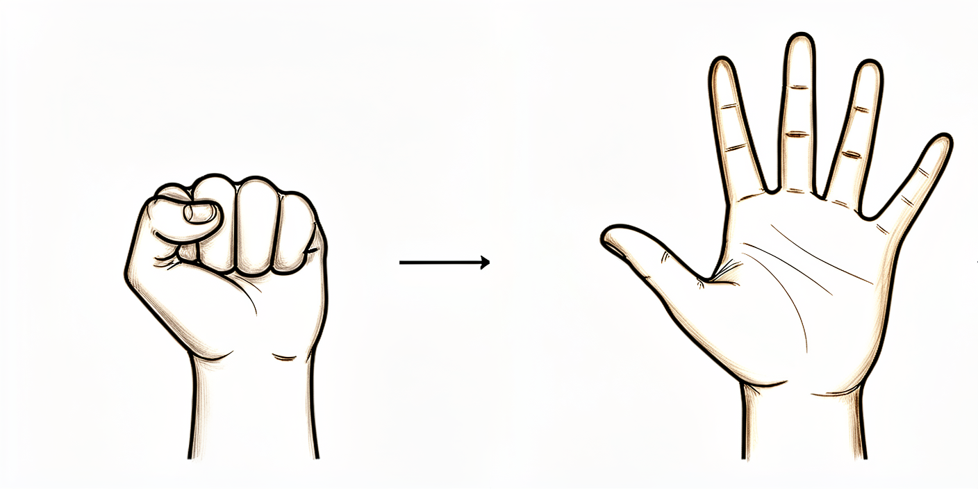
张开和握紧手
通过张开和闭合手部及手指,或挤压一个软球,保持手和手指的活动。
10次,每日3次
Kieran Hirpara 4.0
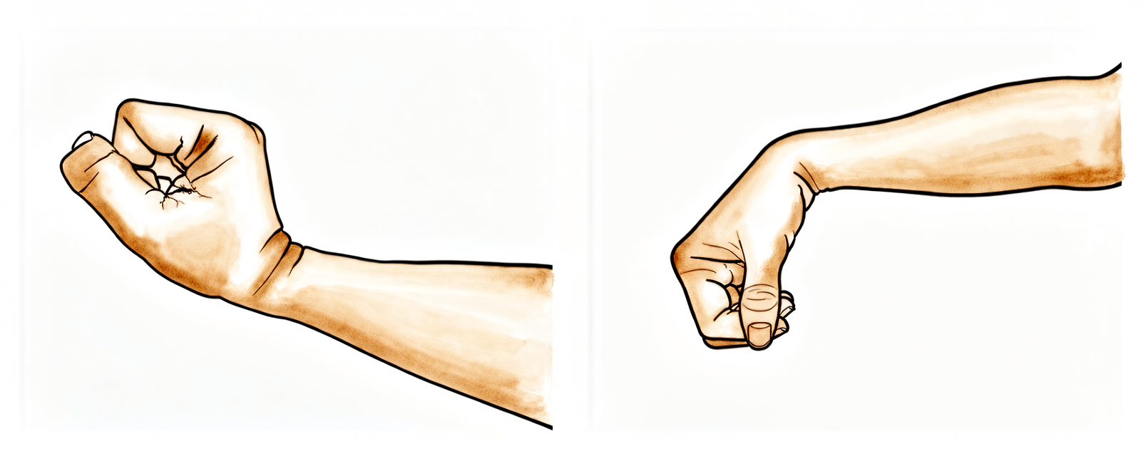
腕部运动
通过向前、向后及左右侧向弯曲手腕,保持手腕活动。
10次,每日3次
Kieran Hirpara 4.0
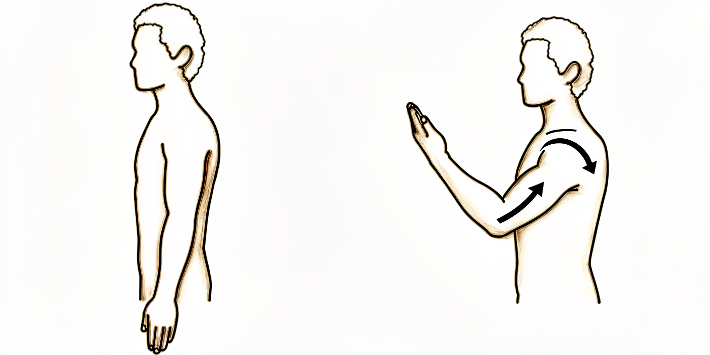
肘部
弯曲并伸直您的肘部。
10次,每日3次
Kieran Hirpara 4.0
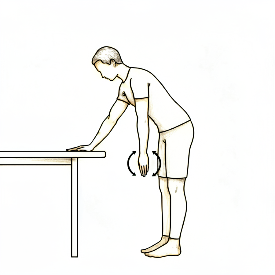
钟摆
这是一项被动练习。身体前倾,让手臂自然下垂放松。利用身体带动手臂,沿顺时针或逆时针方向轻柔转动,同时向前、向后及左右摆动。
每个方向约30秒,每天3次
Kieran Hirpara 4.0
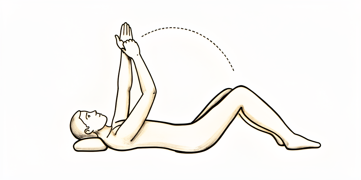
前屈
坐在椅子上并身体前倾,用另一只手臂托住手术侧手臂,轻轻将手臂在身体前方向上移动。借助未手术侧手臂的辅助将其放回原位。如果您愿意,也可以尝试仰卧在床上,并辅助手臂向上移动。
10次,每日3次
Kieran Hirpara 4.0
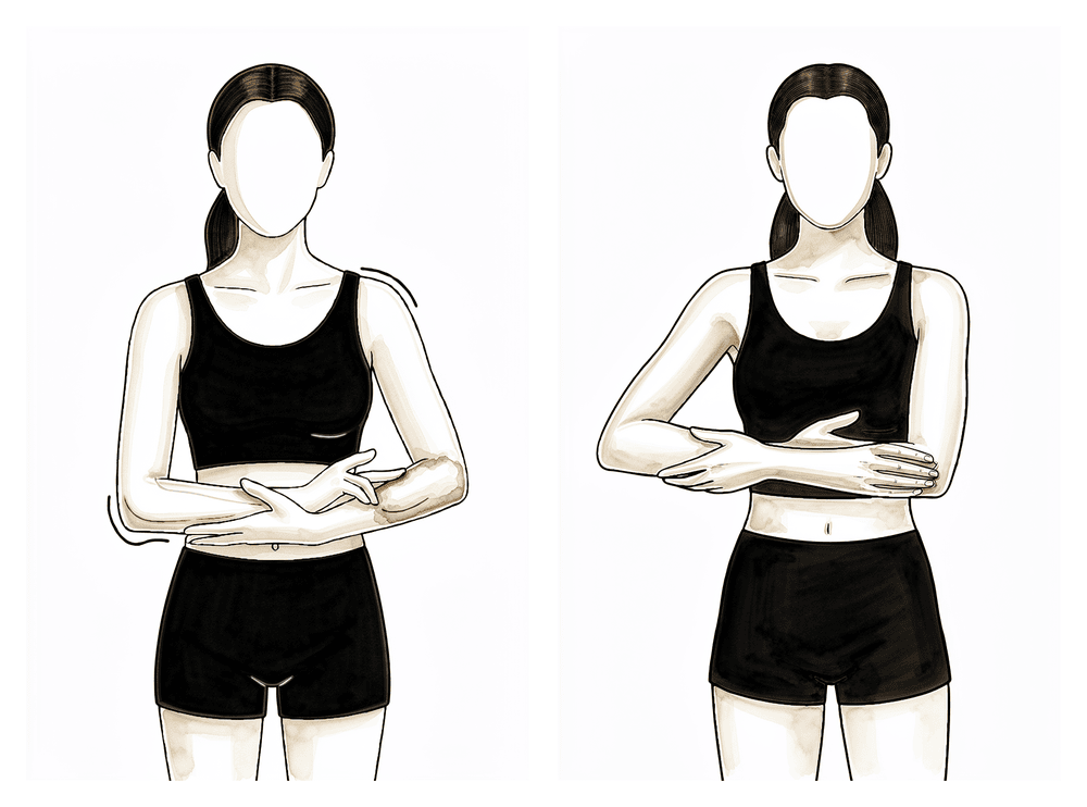
外展
坐在椅子上并向前倾身,再次托住手臂,并协助其向侧方移动(例如像摇动婴儿一样)。
10次,每日3次
Kieran Hirpara 4.0
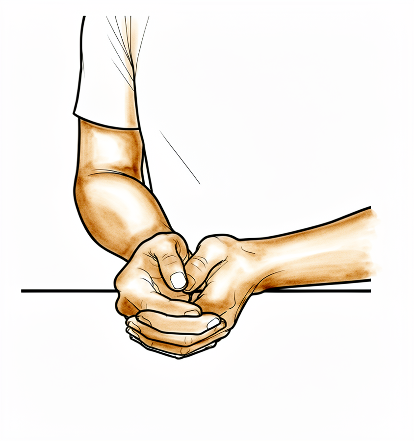
外旋
坐在椅子上,仅将手臂从吊带中的位置移动至正前方指向前方。不要向外侧移动更多。
轻柔地,10次,每日3次
Kieran Hirpara 4.0
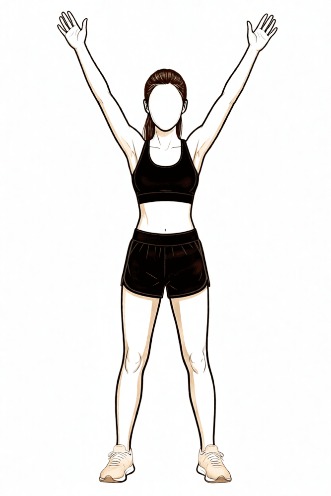
下斜方肌激活
将肩胛骨向下并向内收拢。
保持5秒,重复5次;每日重复3次
Kieran Hirpara 4.0
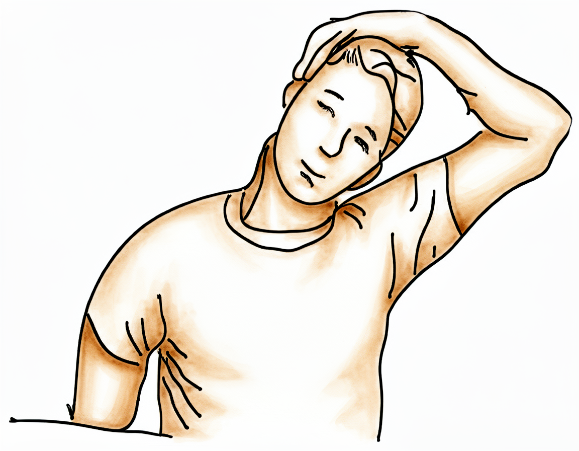
上斜方肌拉伸
用您的非手术侧手臂将耳朵向同侧肩部方向移动,远离手术侧。
保持10秒,重复3次;每天重复3次
Kieran Hirpara 4.0
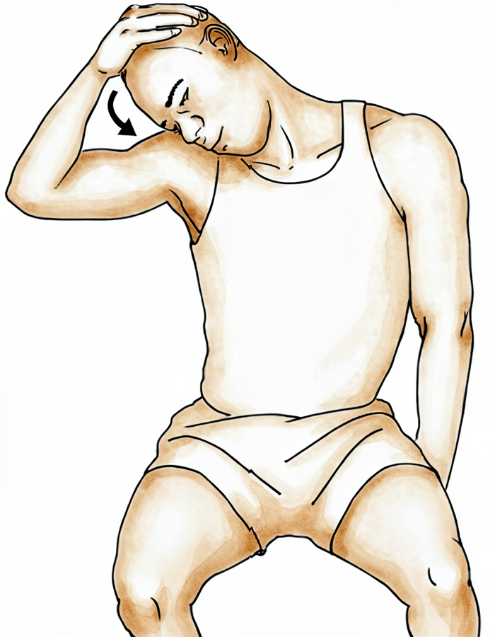
肩胛提肌拉伸
使用您的非手术侧手臂,将鼻子移向乳头或腋窝区域。
保持10秒,重复3次;每天重复3次
第一阶段 — 保护期(第 0–3 周)
Kieran Hirpara 4.0
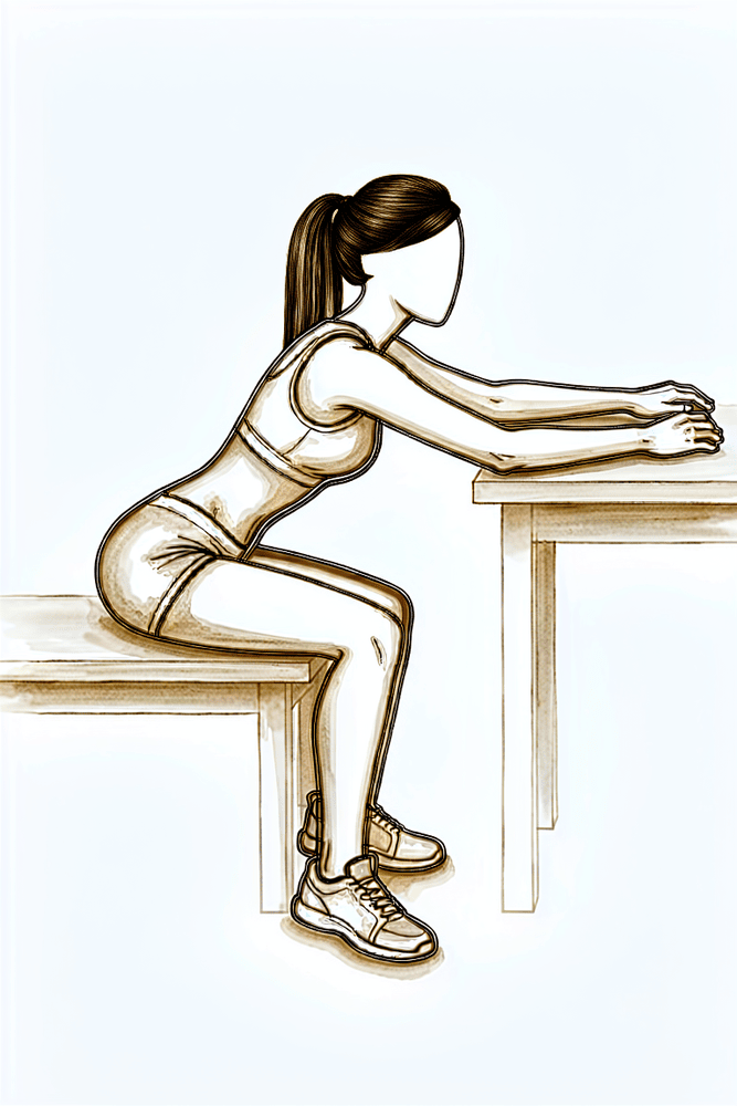
坐位桌面滑动
坐在桌前,前臂平放在桌面上。身体轻轻前倾,让肩部自然向前折叠,不要使用肩部肌肉,同时让手沿桌面向前滑动,然后向后滑动。保持动作放松且舒适——肩部本身保持被动状态。
在您的物理治疗师指导下
前三周旨在使新的肩关节稳定下来,并帮助其逐步恢复活动。您应使用吊带以获得舒适感,通过冰敷和加压控制肿胀,并保持肘部、腕部和手部的活动。从一开始,您还应开始温和的肩部活动,由另一只手臂辅助,并在舒适范围内轻柔地依靠自身力量进行,通常仰卧时最容易,因为重力会起到辅助作用。请以舒适度为指引,切勿强行活动。最重要的规则是关于保护关节以防脱位:不要向后伸手,不要将手臂向内旋转,不要提重物,也不要用手支撑身体。仰卧时,请在肘部下方放置一个小枕头或卷起的毛巾,以免肩关节向后过度伸展。
致您的物理治疗师:
目标
- 保护假体组件的稳定性(避免脱位姿势)
- 减轻肿胀,最大限度减少疼痛
- 维持上肢(UE)肘部、手部和腕部的活动范围(ROM)
- 在舒适范围内建立早期的肩部主动辅助运动和主动运动
- 最大限度减少肌肉抑制
- 患者教育
吊带
- 中立旋转位
- 用于舒适和支持;可选短暂夜间使用;从约第2周开始在白天逐渐停用
管理
- 肿胀管理:冰敷、加压
- 活动范围 / 活动度:
- 被动活动范围(PROM):在肩胛骨平面内,耐受范围内进行外旋(ER);屈曲/上举 ≤ 120度;外展(ABD)≤ 90度;坐姿盂肱(GH)屈曲桌面滑动;钟摆运动;坐姿水平桌面滑动
- 主动辅助活动范围(AAROM):从一开始:主动辅助肩部屈曲(从仰卧/重力辅助开始),在上述PROM限制范围内
- 主动活动范围(AROM):从一开始:在舒适范围内,耐受情况下进行轻柔的主动肩部运动;外加肘部、手部、腕部活动
注意事项
- 禁止肩部PROM/AROM进入内旋(IR)
- 禁止向后伸手,尤其是进入内旋位
- 避免伸展 + 内收 + 内旋(脱位)的复合姿势
- 禁止提重物
- 禁止用手支撑体重
- 仰卧时,在肘部下方放置小枕头/毛巾卷,以避免肩关节过度伸展
晋级标准
- 在舒适范围内,肩部PROM、AAROM和AROM逐渐增加
- 肩部PROM内旋为0度
- 疼痛 < 4/10
- 第一阶段无并发症
第二阶段 — 中期(第 4–6 周)
Kieran Hirpara 4.0
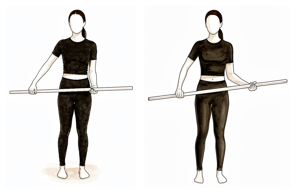
手杖外旋拉伸
仰卧,肘部置于体侧并屈曲至90度。双手握住一根拐杖或棍棒,用非手术侧手臂将手术侧手臂的手掌轻轻向外推,以舒适为度。切勿强行活动。
在您的物理治疗师指导下
Kieran Hirpara 4.0
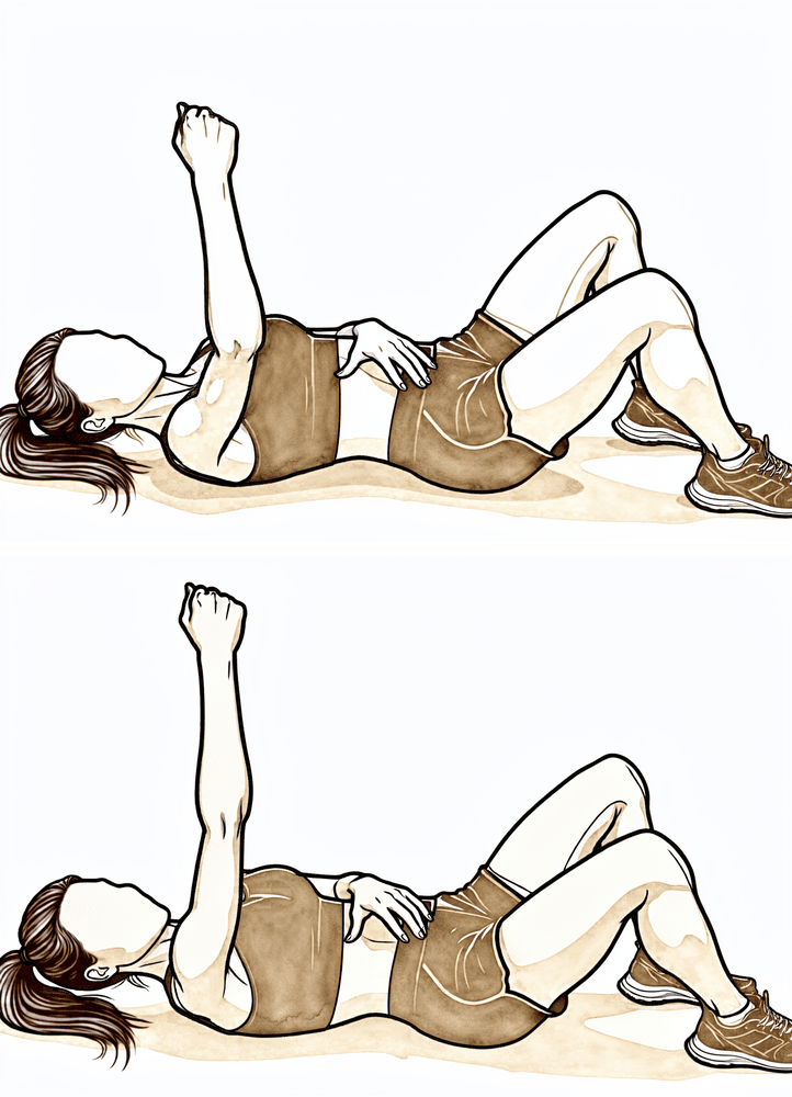
仰卧位向上推伸
仰卧,将手术侧手臂笔直向上伸向天花板。保持肘部伸直,将手进一步向天花板方向推,使肩胛骨离开床面,然后有控制地放下。
在您的物理治疗师指导下
肩部将在术后立即开始的运动基础上继续发展。关节活动度持续增加,肩胛周肌群(肩胛带肌)和三角肌的初次激活与强化训练大约从第4周开始。在此阶段,您白天应停止使用悬吊带;仅当夜间使用能增加舒适度时,方可短暂使用。保护性规则仍然适用:不要提起比咖啡杯更重的物品,不要向后伸手,不要用手支撑身体,仰卧时请在肘部下方垫枕头。
致您的物理治疗师:
目标
- 继续保护假体稳定性(避免脱位体位)
- 减轻肿胀,最小化疼痛
- 逐步增加肩关节被动活动度(PROM)
- 推进肩关节主动辅助活动度(AAROM)/主动活动度(AROM)
- 启动肩胛周肌群的激活与强化(约第4周)
- 启动三角肌的激活与强化(约第4周;激活后束三角肌时避免肩关节伸展)
- 患者教育
悬吊带
- 停止白天使用(从约第2周开始逐步停用)
- 仅在增加舒适度时可选用短暂夜间使用
管理
- 继续第一阶段干预措施
- 关节活动度 / 灵活性:
- AAROM:主动辅助肩关节屈曲、持拐杖肩关节屈曲、拐杖外旋拉伸、毛巾按压、坐姿持拐杖肩关节上举
- AROM:仰卧位屈曲、敬礼动作、仰卧位出拳
- 强化训练:
- 肩胛周肌群:肩胛骨后缩、站立位肩胛骨固定、支撑位肩胛骨固定、低位划船、下向滑动
- 三角肌:肩胛平面内的等长收缩
注意事项
- 禁止向后伸手,尤其是进入内旋体位时
- 在约第6周前,避免复合的伸展 + 内收 + 内旋(脱位)体位
- 禁止提起比咖啡杯更重的物品
- 禁止用手支撑体重
- 仰卧时在肘部下方放置小枕头/毛巾卷,以避免肩关节过度伸展
晋级标准
- 肩关节 PROM、AAROM、AROM 逐步增加
- 肩关节内旋方向 PROM 为 0 度
- 可触及肩胛肌群的肌肉收缩
- 疼痛 < 4/10
- 第二阶段无并发症
第三阶段 — 中期,续(第 7–8 周)
Kieran Hirpara 4.0
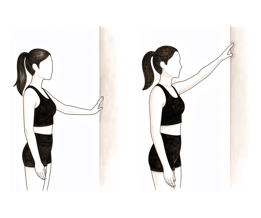
墙面攀爬
面向墙壁站立,将手术侧手臂的手指沿墙壁向上滑动,直至感到舒适为止,借助墙壁辅助手臂向上移动。受控地将手指沿墙壁向下滑动——切勿让手臂突然下落。
在您的物理治疗师指导下
Kieran Hirpara 4.0
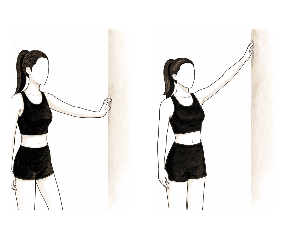
侧向爬墙练习
侧身站立面对墙壁,指尖轻触墙面。将手指沿墙壁向侧方向上滑动,将手臂抬高至舒适的最大范围,然后有控制地将手指沿墙壁向下滑动。在恢复侧方活动度时,墙壁为手臂提供支撑。
在您的物理治疗师指导下
此时吊带已完全成为过去。活动在所有方向上逐步进展,包括首次轻柔的被动内旋(将手臂向内旋转),三角肌和肩胛骨肌肉的强化训练也逐步加强,同时开始进行首批运动控制(协调)练习。仍存在的限制:不得提举比咖啡杯更重的物品,手不得伸过裤袋去够背后,不得通过手部支撑体重,并避免将手臂向后伸展至身体后方。
致您的物理治疗师:
目标
- 最小化疼痛
- 逐步推进肩关节被动活动范围(PROM);在肩胛骨平面启动肩关节被动内旋(PROM IR)
- 逐步推进肩关节主动辅助活动范围(AAROM)
- 逐步推进肩关节主动活动范围(AROM)
- 推进三角肌强化训练
- 推进肩胛周围肌肉强化训练
- 启动运动控制练习
- 患者教育
吊带
- 停止使用
管理
- 继续第一阶段和第二阶段的治疗措施
- 活动范围 / 活动度:
- 被动活动范围(PROM):各平面全范围;在肩胛骨平面逐步进行被动内旋(PROM IR)≤ 50 度
- 主动辅助活动范围(AAROM):倾斜桌面滑动、爬墙、滑轮练习、坐姿肩关节上举(使用拐杖)并主动下放
- 主动活动范围(AROM):坐姿肩胛骨平面前举(scaption)、坐姿屈曲、仰卧位前举(使用弹性阻力)至 90 度
- 强化训练:
- 肩胛周围肌肉:在健身球上进行划船动作、前锯肌出拳动作
- 三角肌:坐姿肩关节上举(使用拐杖)、坐姿肩关节上举(使用拐杖)并主动下放、墙壁球滚动
- 运动控制:
- 仰卧位,在肩胛骨平面和屈曲 90–125 度范围内进行内旋/外旋(节律性稳定)
- 拉伸:
- 侧卧位水平内收(ADD)、肱三头肌和背阔肌
注意事项
- 手不得伸过裤袋去够背后
- 不得提举比咖啡杯更重的物品
- 不得通过手部支撑体重
- 避免肩关节过度伸展
进展标准
- 活动范围目标(被动活动范围 PROM 和主动活动范围 AROM 的预期值需个体化,并取决于术后在手术室中测得的活动范围数值):
- 上举 ≤ 140 度
- 中立位外旋 ≤ 30 度
- 肩胛骨平面内旋 ≤ 50 度,或可触及后裤袋
- 肩关节主动活动范围(AROM)时最小或无代偿模式
- 疼痛 < 4/10
第四阶段 — 过渡期(第 9–11 周)
本阶段是手臂恢复正常使用的桥梁。此时被动活动度应在所有平面内达到全范围,重点转向强化三角肌和肩胛肌群,建立动态稳定性和协调性,并逐步恢复力量和耐力,最终回归完全的功能性活动。唯一的严格限制:禁止提举重物(超过 5 公斤)。
致您的物理治疗师:
目标
- 维持无痛的活动范围(ROM)
- 逐步推进肩胛周围肌群强化
- 逐步推进三角肌强化
- 逐步推进运动控制练习
- 改善肩部动态稳定性
- 逐步恢复肩部力量和耐力
- 回归完全的功能性活动
管理方案
- 继续执行第二至第三阶段的干预措施
- 活动范围 / 灵活性:
- 被动活动度(PROM):所有平面内全范围活动度
- 强化训练:
- 肩胛周围肌群:弹力带肩部伸展、弹力带坐姿划船、划船、抢劫动作、割草机动作、三脚架动作、指针动作
- 三角肌:在三角肌练习中逐渐增加阻力
- 运动控制:
- 内旋/外旋及屈曲 90–125 度(节律性稳定)
- 四肢支撑位交替等长收缩及靠墙球稳定
- 场地目标练习
- 本体感觉神经肌肉促进法(PNF):D1 对角线提升、D2 对角线提升
注意事项
- 禁止提举重物(> 5 公斤)
晋级标准
- 完成所有练习,表现出对称的肩胛骨力学机制
- 疼痛 < 2/10
第五阶段 — 高级强化训练(第 12–16 周)
Kieran Hirpara 4.0
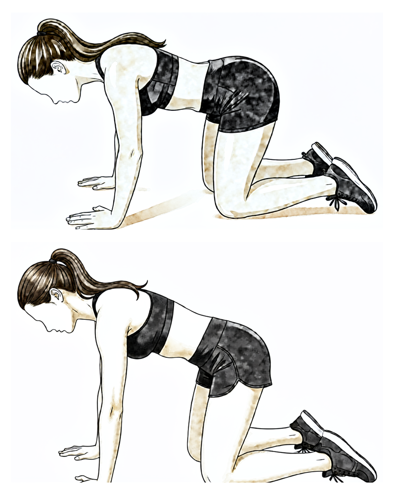
跪姿俯卧撑加强版(push-up plus)
从双膝跪地、双手置于肩下的俯卧撑起始姿势开始,保持肘部伸直,将上背部向天花板方向推起,使肩胛骨相互分离,然后有控制地缓慢下放。
在您的物理治疗师指导下
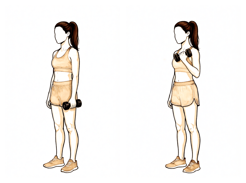
Kieran Hirpara 4.0
肱二头肌弯举
掌心向上,上臂贴于体侧,手持轻重量,屈肘将手移向肩部,随后有控制地放下。保持负重较轻——此阶段仍禁止进行重负荷提举(超过 7 公斤)。
在您的物理治疗师指导下
最终阶段旨在使肩部适应日常生活:在保持活动无疼痛的同时,建立自信使用手臂所需的肌力和耐力。如果在置换手术的同时进行了肩袖修复,则现在开始进行肩袖(RTC)强化训练。负重限制有所提高,但仍禁止搬运重物(超过 7 公斤)。本阶段及整个方案将在满足所有里程碑标准并经外科医生批准后结束。
致您的物理治疗师:
目标
- 维持无痛的活动范围(ROM)
- 在合并修复手术的情况下,启动肩袖(RTC)强化训练
- 改善肩部肌力和耐力
- 增强上肢的功能性使用
管理
- 继续实施第二至第四阶段的干预措施
- 强化训练:
- 肩胛周围肌群:跪姿俯卧撑加强版(push-up plus)、“W”字练习、弹力带 W 字练习、俯卧位肩伸展等长收缩、动态拥抱、弹力带动态拥抱、弹力带前冲拳、前冲拳、T 字和 Y 字练习、“T”字练习
- 三角肌:在功能性体位下继续逐渐增加抗阻屈曲和侧举
- 肘部:二头肌弯举、弹力带二头肌弯举以及三头肌训练
- 肩袖:内旋/外旋等长收缩、侧卧位外旋、站立位弹力带外旋、站立位弹力带内旋、内旋、外旋、侧卧位外展逐渐过渡至站立位外展
- 运动控制:
- 弹力带 PNF 模式、带阻力的 PNF D1 对角线提升、对角线上举、对角线下举、带弹力带的墙面滑动
注意事项
- 禁止搬运重物(> 7 公斤)
晋级标准
- 获得外科医生批准,且已满足所有里程碑标准
- 维持无痛的被动活动范围(PROM)和主动活动范围(AROM)
- 完成所有练习,并展示对称的肩胛骨力学机制
- QuickDASH 和 ASES 患者报告结局测量
您的方案之后
本方案与诊所的一般康复建议配合使用:请参阅术后疼痛管理和伤口护理。关于手术本身,请参阅反向肩关节置换术。
本方案背后的临床证据(已发表的康复试验、使用吊带及活动规则的依据,以及研究参考文献)详见随附的证据摘要(可下载为PDF文件)。
Evidence & references
This is the clinical evidence summary written for health professionals. It is technical, and it lists the research this page was built from. You do not need to read it to understand your treatment or to make a decision about it.
Topic scope: Post-operative rehabilitation after elective reverse total shoulder arthroplasty (RSA / rTSA) for rotator-cuff-tear arthropathy, glenohumeral arthritis with an irreparable cuff, or a massive irreparable cuff tear — not the acute proximal-humerus-fracture indication, where tuberosity healing imposes its own restrictions. The evidence base for the operation (when RSA is indicated, implant survivorship, complication profile) is extensive; the evidence base for the rehabilitation protocol is smaller but, unusually for shoulder surgery, now includes several randomised trials directly comparing immobilisation strategies.
Defining principle of this rehab: RSA rehabilitation is comparatively permissive and is increasingly run on an accelerated footing. Two features of the reconstructed joint explain why. First, the deltoid — not the rotator cuff — drives elevation after RSA; the design medialises and lowers the centre of rotation so the deltoid can elevate an arm that has no functioning cuff. Second, there is usually no subscapularis repair to protect (and many surgeons deliberately leave it unrepaired with a lateralised implant), so the external-rotation restriction that dominates anatomic-TSA rehab is far less central here. The principal early caution is therefore not tissue healing but component stability: a reverse prosthesis can dislocate, and the at-risk position is the combination of extension + adduction + internal rotation (the hand-behind-the-back / tucking-in-a-shirt movement). Early rehab accordingly protects against that position while otherwise encouraging motion. The sling is largely for comfort, support and dislocation-avoidance, and the published trial evidence shows that shortening or even omitting it does not increase the dislocation or complication rate.
Surgeon's protocol note: Dr Hirpara's protocol on this page is run on an accelerated footing, directly aligned with the randomised and cohort evidence below: a short ~2-week comfort sling (weaned off during the day; optional brief night use), active-assisted and active shoulder motion from the start (within defined limits, beginning supine/gravity-assisted), and deltoid + periscapular strengthening from ~week 4. The main early constraint is component stability, so the extension + adduction + internal-rotation (hand-behind-back) dislocation position is avoided for the first ~6 weeks while motion is otherwise encouraged. Elevation after RSA is deltoid-driven, which is why strengthening is deltoid- and periscapular-focused. Follow the protocol your surgeon has set.
The operation, in brief
In a reverse replacement the normal anatomy is inverted: a ball (glenosphere) is fixed to the shoulder blade and a cup is fixed to the top of the arm bone. This moves the joint's pivot point down and in, which lengthens and tensions the deltoid and lets that muscle lift the arm even when the rotator cuff is gone — the situation in cuff-tear arthropathy. Because the implant, not the patient's own cuff, provides stability and power, the rehabilitation logic differs fundamentally from a cuff repair (where a healing tendon must be protected) and from an anatomic replacement (where a repaired subscapularis must be protected).
Evidence by theme
1. Early / accelerated motion is safe — multiple randomised trials
This is the best-supported part of RSA rehabilitation, and it is unusually strong for a shoulder rehab question because it rests on randomised controlled trials, not just consensus:
- Hagen et al. (2020), single-blind RCT, 107 shoulders — randomised to immediate physical therapy (passive + active ROM from the start) versus 6-week delayed therapy. No difference in final ROM, patient-reported outcomes, or dislocation/complication rate (early 7.1% vs delayed 9.1%). [RAG corpus — 10.1016/j.jse.2020.11.017]
- Edwards / "two rehabilitation approaches" RCT, 61 patients (63 shoulders) — early active (submaximal isometric deltoid work from week 2) versus delayed active rehab, both with a 6-week sling. Pain and overall function were equivalent at 3, 6 and 12 months, but the early-active group had significantly better active forward flexion at 3 months (p = 0.019) — i.e. earlier functional gain without added risk. [Literature — PMC8512973]
- Lee et al. (2021), 357 shoulders — randomised to no immobilisation / early motion, 3-week sling, or 6-week sling. No difference in patient-reported outcomes, satisfaction, pain or ROM; the immediate-motion group had the lowest overall complication rate (reported as ~4% vs ~24.6% in the 6-week cohort in the pooled systematic-review re-analysis). [via Sachinis 2024 SR]
A systematic review (Sachinis et al., 2024; 3 controlled trials, ~527 shoulders) concluded that accelerated rehabilitation is safe and yields equivalent 12-month outcomes, while cautioning that protocols should still be individualised by bone quality, age, diagnosis and surgical complexity rather than applied uniformly. [Literature — PMC11034463]
Evidence: MODERATE–STRONG (several RCTs + SR), though individual trials are modest in size.
2. Sling duration can be shortened without added risk
- A large retrospective cohort (960 patients) compared 2-week vs 6-week sling immobilisation after RSA and found no difference in complication rate (12.0% vs 15.0%, p = 0.21), dislocation rate (p = 0.79), acromial stress fracture, loosening or infection; final flexion and ER were equivalent. The authors concluded shorter immobilisation does not incur additional risk. [Literature — PMC10638591]
- Published institutional protocols (e.g. Massachusetts General Brigham) have themselves moved from a 6-week to a 4-week sling standard, attributing the change to improved implant design. [Published protocol — MGH Sports Medicine]
Evidence: MODERATE (one large cohort + protocol trend; the question is now whether 6 weeks is necessary, not whether it is safe).
3. The deltoid drives recovery — and formal active PT may add little
Because elevation depends on deltoid recruitment (with documented compensatory recruitment of upper trapezius, latissimus and posterior deltoid), strengthening is deltoid- and periscapular-focused rather than cuff-focused. Notably, a multicentre RCT found that a formal active physical-therapy programme did not improve outcomes over a home/self-directed programme after RSA — many patients recover well with a structured home programme and physiotherapist supervision rather than intensive hands-on active therapy. [RAG corpus — 10.1016/j.jse.2022.12.011] This supports a pragmatic, education-and-home-exercise model.
Evidence: MODERATE (RCT).
4. Dislocation: low but real, and the early protective position is specific
Dislocation is the relevant early stability complication (as opposed to the tissue-healing concern of a cuff repair). Corpus series on dislocation following RSA identify the at-risk position as adduction + internal rotation + extension, and identify subscapularis insufficiency and implant/soft-tissue tensioning as contributors to instability. [RAG corpus — 10.1016/j.jse.2016.12.073; 10.1016/j.jse.2008.12.013] This is precisely why early protocols — accelerated or conservative — restrict reaching behind the back and forced internal rotation while otherwise allowing supported elevation. The role of routine subscapularis repair in preventing dislocation is debated and interacts with glenosphere lateralisation (a lateralised design appears less reliant on subscapularis repair for stability). [RAG corpus — 10.5435/jaaos-d-16-00781]
Evidence: MODERATE (cohort/registry); the precaution itself is universal consensus.
5. Contrast with anatomic TSA rehabilitation
The American Society of Shoulder and Elbow Therapists (ASSET) consensus on anatomic TSA rehab centres on protecting the subscapularis repair — limiting passive external rotation and active internal rotation for ~6 weeks. [RAG corpus — 10.1016/j.jse.2020.05.019] After RSA that specific constraint is usually absent or much reduced (no cuff repair to protect; subscapularis often not repaired), which is the structural reason RSA rehab is more permissive in external rotation while being more attentive to the dislocation-position combination.
Phased post-operative timeline
This table reflects Dr Hirpara's protocol on this page (the accelerated, ~2-week-sling approach). The right-hand column notes the published evidence that the protocol is grounded in.
| Phase | Window | Sling | Shoulder motion | Strengthening | Accelerated-evidence basis |
|---|---|---|---|---|---|
| I — Protection | Week 0–3 | Comfort/support; wean off during the day from ~2 weeks; optional brief night use | Active-assisted and active as tolerated from the start (begin supine/gravity-assisted; ER in scapular plane to tolerance; flexion/scaption ≤120°; abduction ≤90°); no IR, no reaching behind back, avoid the dislocation position; elbow/wrist/hand active | — | RCTs show early active/AAROM from the start carries no added dislocation/complication risk (Hagen 2020; Edwards) |
| II — Intermediate | Week 4–6 | Off during the day; optional brief night use | Progress AAROM and AROM | Periscapular + deltoid activation and strengthening initiated (~week 4) | Early active deltoid work gives earlier functional gain without added risk (Edwards) |
| III — Intermediate cont. | Week 7–8 | Discontinued | Progress AROM all planes; first gentle passive IR in scapular plane (≤50°) | Deltoid + periscapular progressed; motor control | Cohort data: 2-week sling non-inferior to 6 weeks (no extra dislocations) |
| IV — Transitional | Week 9–11 | — | Full passive ROM all planes | Resisted deltoid/periscapular; dynamic stability; PNF; no lifting >5 kg | — |
| V — Advanced strengthening | Week 12–16 | — | Maintain pain-free full ROM | Add rotator-cuff strengthening if a cuff repair was done; functional loading; no lifting >7 kg | Resisted IR / eccentric / closed-chain typically from ~12 weeks |
Return-to-activity anchors (from this protocol): driving ~6 weeks; light lifting from 6 weeks but no heavy lifting for ~6 months; swimming (breaststroke) 8 weeks / freestyle 12 weeks; golf ~3 months; sedentary work 6 weeks, manual work surgeon-guided. Published sport-return data after RSA are encouraging for low-impact activity (≈60–86% return; swimming ~84%, golf/fitness ~77%) but caution against high-impact and contact sport given prosthesis-loading and revision concerns. [Literature — PMC10043097]
Key controversies / evidence quality
-
Accelerated vs conservative rehab — and where this protocol sits. The randomised evidence shows early motion and shorter (or no) immobilisation are safe and may give earlier functional gain, with equivalent 12-month outcomes. Dr Hirpara's ~2-week comfort sling, active-assisted/active motion from the start, strengthening from ~week 4 protocol aligns with this accelerated evidence. The systematic review still endorses individualisation (bone quality, fixation, intra-operative stability, soft-tissue tensioning, any concomitant cuff repair), so the surgeon may dial the plan back for a borderline-stable construct, softer bone, or a concomitant repair. The prescribed plan is the one to follow.
-
Immobilisation duration. Trial and large-cohort data show 2–4 weeks is non-inferior to 6 weeks for dislocation and complications, which is why this protocol uses a short ~2-week comfort sling. Longer 6-week protocols persist elsewhere out of surgeon preference and construct-specific factors, with the low absolute dislocation rate making the question hard to power definitively.
-
Precautions. The extension + adduction + internal rotation (hand-behind-back) restriction is near-universal consensus early on; the disagreement is about how long, and how much external-rotation freedom to allow (more than anatomic TSA, but bounded by soft-tissue tension).
-
Subscapularis repair. Whether to repair it — and whether repair reduces dislocation — interacts with glenosphere lateralisation and remains debated; this in turn influences how restrictive early rehab needs to be.
-
Active physiotherapy intensity. At least one RCT found formal active PT did not beat a structured home programme, supporting an education-led, home-exercise model with physiotherapist oversight rather than intensive hands-on therapy.
Evidence-strength flags (summary)
- MODERATE–STRONG (RCT / SR): accelerated/early-motion rehab is safe with equivalent 12-month outcomes (Hagen 2020 RCT; Edwards RCT; Lee 2021; Sachinis 2024 SR); formal active PT not superior to home programme (multicentre RCT).
- MODERATE (large cohort): 2-week sling non-inferior to 6-week sling for dislocation/complications (960-patient cohort); deltoid-driven recovery biomechanics.
- CONSENSUS / WEAK: the dislocation-position precaution (universal but not trial-quantified for duration); the specific phase timings of this protocol (drawn from published institutional protocols + surgeon preference, not a head-to-head rehab RCT); subscapularis-repair effect on instability (conflicting cohort data).
Citations
RAG corpus (180,000+ Orthopaedic articles) — real DOIs returned by search
- Hagen MS, et al. Accelerated rehabilitation following reverse total shoulder arthroplasty. J Shoulder Elbow Surg / Semin Arthroplasty. 2021. DOI: 10.1016/j.jse.2020.11.017
- Active physical therapy does not improve outcomes after reverse total shoulder arthroplasty: a multi-center, randomized clinical trial. J Shoulder Elbow Surg. 2023. DOI: 10.1016/j.jse.2022.12.011
- Kennedy J, et al. The American Society of Shoulder and Elbow Therapists' consensus statement on rehabilitation for anatomic total shoulder arthroplasty. J Shoulder Elbow Surg. 2020. DOI: 10.1016/j.jse.2020.05.019
- Dislocation following reverse total shoulder arthroplasty. J Shoulder Elbow Surg. 2017. DOI: 10.1016/j.jse.2016.12.073
- Edwards TB, et al. Subscapularis insufficiency and the risk of shoulder dislocation after reverse shoulder arthroplasty. J Shoulder Elbow Surg. 2009. DOI: 10.1016/j.jse.2008.12.013
- Clinical Outcomes After Reverse Shoulder Arthroplasty With and Without Subscapularis Repair: The Importance of Considering Glenosphere Lateralization. J Am Acad Orthop Surg. 2018. DOI: 10.5435/jaaos-d-16-00781
Literature (URLs)
- Sachinis NP, et al. Can we accelerate rehabilitation following reverse shoulder arthroplasty? A systematic review. Shoulder Elbow. 2024. https://pmc.ncbi.nlm.nih.gov/articles/PMC11034463/
- A randomised trial comparing two rehabilitation approaches following reverse total shoulder arthroplasty (early active vs delayed active). 2021. https://pmc.ncbi.nlm.nih.gov/articles/PMC8512973/
- No difference in complications between two-week vs. six-week duration of sling immobilization after reverse total shoulder arthroplasty (960-patient cohort). 2023. https://www.ncbi.nlm.nih.gov/pmc/articles/PMC10638591/
- Optimizing Outcomes After Reverse Total Shoulder Arthroplasty: Rehabilitation, Expected Outcomes, and Maximizing Return to Activities. 2023. https://pmc.ncbi.nlm.nih.gov/articles/PMC10043097/
- Kim Y-T, et al. Four weeks of immobilisation after reverse shoulder arthroplasty yields outcomes comparable to six weeks. Clin Shoulder Elb. 2024. https://www.ncbi.nlm.nih.gov/pmc/articles/PMC11546419/
Published rehabilitation protocols (basis for the phase structure)
- Massachusetts General Brigham Sports Medicine. Rehabilitation Protocol for Reverse Shoulder Arthroplasty. Revised December 2018. https://www.massgeneral.org/assets/mgh/pdf/orthopaedics/sports-medicine/physical-therapy/rehabilitation-protocol-for-reverse-shoulder-arthroplasty.pdf
- Brigham and Women's Hospital. Reverse Total Shoulder Arthroplasty Protocol. https://www.brighamandwomens.org/assets/BWH/patients-and-families/pdfs/shoulder--reverse-total-shoulder-arthroplasty-protocol.pdf





