冻结肩
Patients › Shoulder
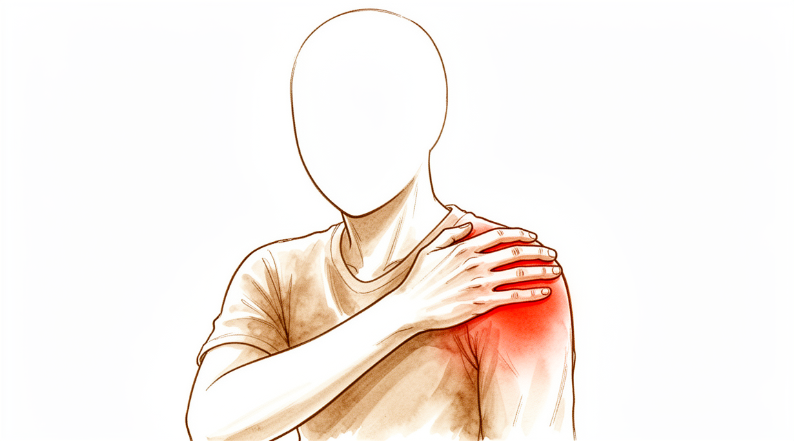
Frozen shoulder (adhesive capsulitis) causes pain and stiffness, progressing through freezing, frozen, and thawing phases.
您的感受
您可能经历逐渐出现的弥漫性肩部疼痛,疼痛在数周至数月内加重。疼痛通常在夜间更严重,并且因侧卧于患侧而加剧。随着您减少手臂的使用,疼痛会导致僵硬。您可能会通过限制活动来寻求缓解疼痛,这标志着僵硬期的开始。
原发性冻结肩的僵硬期通常持续 4 至 12 个月。随着僵硬进展,几乎全天候存在钝痛,尤其是在夜间。在运动范围达到或接近新的运动终点时,锐痛常伴随钝痛出现。您可能会描述日常生活活动困难,例如男性难以从后裤袋中取出钱包,女性难以扣上胸罩。
冻结肩的解冻期持续数周或数月。随着解冻期运动范围增加,疼痛减轻。除了良性忽视(即不进行治疗)外,大多数患者的运动功能恢复是渐进的,但可能永远不会客观地恢复正常。大多数患者在解冻期后主观感觉接近正常,这可能是由于代偿或调整日常生活活动方式所致。
实际情况
冻结肩是一种关节囊——包裹肩关节的柔性袖套——变厚、变紧并发生炎症的疾病。这种收紧限制了您的活动范围并引起剧烈疼痛。它可能在没有先前损伤或现有肩部问题的情况下单独发生。它也可能在受伤后发生,例如骨折或软组织损伤,尽管症状模式可能有所不同。
在最常见的形式中,该疾病经历三个不同的阶段。第一阶段是疼痛。您会感到逐渐扩散的酸痛,并在数周至数月内加重。这种疼痛通常在夜间更严重,并使得难以在患侧睡眠。这一疼痛阶段通常持续 10 到 36 周。
第二阶段是僵硬。您可能会自然地减少活动以避免疼痛,这会导致进一步收紧。此阶段通常持续 4 到 12 个月。在此期间,您可能会在日常任务中遇到困难,例如伸手进入后口袋或扣上胸罩。可能会一直存在钝痛,尤其是在夜间。当您尝试将手臂移动到新的有限范围附近时,您可能会感到尖锐的疼痛。
第三阶段是解冻。在数周或数月内,您的活动范围逐渐增加,疼痛减轻。如果没有特定治疗,这种自然恢复过程总共可能需要约 18 个月。虽然大多数患者在此阶段感觉接近正常,但您的活动范围可能无法完全恢复到之前的状态。
收紧是由组织本身的变化引起的。形成瘢痕组织的细胞在您肩关节周围的结构中变得过度活跃。这导致弹性丧失和僵硬增加。虽然 X 光片通常看起来正常,但这种内部增厚限制了您的活动。您的外科医生通过观察没有其他损伤或疾病能解释您特定的活动受限来诊断此病。
我们能做什么
冻结肩的病程通常分为三个阶段:疼痛期、僵硬期和缓解期。在疼痛期,您可能会感到逐渐加重的弥漫性疼痛,夜间或向患侧卧位时疼痛加剧。您的外科医生可能会建议首先采取自我管理措施和物理治疗。轻柔的活动和 supervised(监督下)的锻炼有助于维持现有的活动度并防止进一步僵硬。大多数患者通过非手术措施在相对较短的时间内即可缓解症状。然而,请注意,如果仅采取良性忽视(即不进行治疗),活动度的恢复将是渐进的,且可能永远无法在客观上恢复正常。您最终可能会感觉接近正常,这通常是通过调整日常任务的执行方式来实现的。
如果轻柔的活动无法提供足够的缓解,您的外科医生可能会建议进行药物治疗以控制疼痛和炎症。关节内类固醇注射对冻结肩有效且安全。它们可以缓解疼痛,改善功能表现,并增加您的活动范围。这些注射可以作为单次注射进行,也可以在肩关节内的多个部位进行注射。对于部分患者,还会使用水分离术(注入液体以拉伸关节囊)。如果在此过程中您无法耐受超过 20 mL 的注射量,则 41% 的病例可能需要进一步治疗。原发性冻结肩的复发率(33%)高于继发性冻结肩(16%)。您的外科医生将根据您的具体症状和病程阶段选择最佳方案。
仅当保守治疗达到极限且症状持续存在时,才会考虑手术治疗。早期手术干预可能会缩短您症状的总体持续时间。选项可能包括麻醉下手法松解,即您的外科医生轻柔地移动您的手臂以打破瘢痕组织,或关节镜下关节囊松解术,涉及仔细切割肩关节囊的紧张部分。有限的关节囊松解联合手法松解是一种安全有效的程序,可显著改善疼痛、功能和活动范围。这些干预措施适用于对其他治疗无反应的药物难治性冻结肩患者。您的外科医生将根据您的进展和僵硬程度讨论这些步骤是否适合您。
预期情况
冻结肩是一种疼痛且逐渐僵硬的疾病,通常分为三个阶段:疼痛期、僵硬期和缓解期。在第一期(疼痛期),您可能会感到逐渐加重的弥漫性酸痛,夜间或患侧卧位时疼痛加重。这种疼痛通常持续数周至数月。随着病情进入僵硬期,您可能会因避免疼痛而限制活动。此阶段通常持续4至12个月。在此期间,您可能会发现日常活动变得困难,例如伸手向后口袋或扣内衣。您可能几乎一直感到钝痛,并且在尝试将肩关节活动至极限时会出现锐痛。
最后阶段为缓解期,持续数周或数月。随着活动度逐渐增加,疼痛减轻。大多数患者在此期间主观感觉接近正常,这通常是因为他们已调整了日常活动的方式。然而,若不进行治疗,活动度的恢复缓慢。在某些情况下,活动度可能永远无法在客观上恢复正常。该疾病通常在1至3年内逐渐缓解。尽管存在这种自然缓解过程,但50%至60%的患者仍存在持续性活动受限。长期来看,59%的患者肩关节功能正常或接近正常,而41%的患者报告仍有某些持续症状。
治疗有助于管理您的症状并改善功能。早期手术干预可能会缩短症状的总持续时间,且与等待观察相比,不会导致更差的预后。麻醉下手法松解或关节镜下关节囊松解术等手术可改善活动度和功能,且长期获益得以维持。如果您患有糖尿病,您的外科医生可能会讨论特定的注意事项,因为高血糖水平是一个可能的危险因素。虽然大多数患者显著改善,但仍有高比例的患者在长期随访时仍存在一定程度的活动受限。您的外科医生将根据您的具体需求和症状持续时间,与您共同选择最佳的治疗方案。
何时就诊
若出现进行性加重的肩痛,且夜间疼痛加剧或影响睡眠,请就诊于全科医生(GP)。若关节僵硬限制了日常活动(如扣衣服或手插后裤袋),请要求专科医生评估。症状通常持续1至3年,但若未治疗,僵硬可能再持续6至12个月,随后逐渐改善。大多数患者在此阶段后感觉接近正常,但活动度可能无法完全恢复正常。若出现无力、交锁或突然加重,请及时就医。50%至60%的病例会出现持续性活动受限。早期干预可缩短症状持续时间。您的外科医生将排查糖尿病等基础疾病,糖尿病是可能的危险因素。
Evidence & references
Overview
- Frozen shoulder management guidelines provide evidence-based guidance and identify key areas for future research [2].
- Magnetic resonance imaging findings in frozen shoulder should not replace clinical judgments regarding prognosis and treatment decisions [7].
- Clinicians should monitor frozen shoulder patients with diabetes more closely and offer further treatment if pain or lack of function persist long-term [8].
- Different treatment strategies for frozen shoulder may be appropriate depending on the location [9].
- A strict definition of recalcitrant idiopathic frozen shoulder is needed to prevent unnecessary interventions, as current data should not be interpreted as a plea for surgery in patients with a mean symptom duration of 4 months [10].
- The variety of participants, methods, interventions, and outcomes across trials provides limited new evidence to inform the non-surgical management and treatment of frozen shoulder [14].
- Patients with a poor outcome or recurrent symptoms after manipulation under anaesthetic (MUA) should be offered a further MUA, with an expectation of a good outcome and a low complication rate [17].
- Early surgical intervention might shorten the overall duration of symptoms in frozen shoulder and is not associated with inferior clinical outcomes compared with late surgical intervention [19].
- There is insufficient evidence to reliably recommend a single treatment approach for frozen shoulder [32].
- Further treatment was indicated in 41% of patients who could not tolerate more than 20 mL of injection during hydrodilatation [34].
- Recurrence of frozen shoulder was more common in primary (33%) versus secondary (16%) cases [34].
- Arthroscopic capsular release is a suitable option for refractory primary frozen shoulder syndrome, leading to faster and long-lasting recovery [35].
- Manipulation under anaesthetic (MUA) and arthroscopic capsular release (ACR) are both good treatment options for primary frozen shoulder [70].
- MUA is simpler but carries risks of serious complications, whereas ACR may be safer if performed by experienced surgeons and is convenient for patients with combined rotator cuff tears [70].
Anatomy & Pathophysiology
- Primary (idiopathic) frozen shoulder consists of three phases: pain, stiffness, and thawing [3].
- Secondary frozen shoulders may not exhibit all three phases and may not follow the exact chronology of primary frozen shoulder [3].
- The pain phase of primary frozen shoulder involves a gradual onset of diffuse shoulder pain that is progressive over weeks to months [3].
- Pain in the initial phase is usually worse at night and exacerbated by lying on the affected side [3].
- Reduced arm use due to pain leads to stiffness [3].
- The stiffness phase begins when patients restrict movement to seek pain relief and usually lasts 4 to 12 months [3].
- During the stiffness phase, patients experience difficulty with activities of daily living, such as men accessing back pockets or women fastening brassieres [3].
- A dull ache is present nearly all time during the stiffness phase, especially at night, often accompanied by sharp pain during range of motion at or near new endpoints [3].
- The thawing phase lasts for weeks or months, during which motion increases and pain diminishes [3].
- Without treatment, motion return in the thawing phase is gradual and may never objectively return to normal, although most patients subjectively feel near normal [3].
- Frozen shoulder is characterized by progressive pain and stiffness that usually resolves spontaneously after about 18 months [4].
- Histological features of frozen shoulder are reminiscent of Dupuytren’s disease, with active fibroblastic and myofibroblastic proliferation in the rotator interval, anterior capsule, and coracohumeral ligament [4].
- Conditions particularly associated with frozen shoulder include diabetes, Dupuytren’s disease, hyperlipidaemia, hyperthyroidism, cardiac disease, and hemiplegia [4].
- The etiology of frozen shoulder is still not known and understanding of the pathogenesis is limited [15].
- Frozen shoulder is a poorly understood condition that typically involves substantial pain, movement restriction, and considerable morbidity [12].
- The pathophysiology of frozen shoulder is complex, involving various pathophysiologic mechanisms [62].
- A systematic review summarizes the tissue pathophysiology of primary frozen shoulder [25].
- Primary frozen shoulder is defined by total elevation restricted to 135° or less [20].
- In primary frozen shoulder, motion restriction is localized to the humero-scapular joint [20].
- Primary frozen shoulder excludes cases with post-traumatic conditions, rheumatoid arthritis, osteoarthritis, hemiplegia, or other obvious changes explaining the range of motion decrease [20].
- Secondary frozen shoulder involves decreased range of motion following a traumatic lesion, including soft tissue injury, intra- and juxtaarticular fractures, and other upper limb fractures [20].
- Symptomatic subjects with frozen shoulder demonstrate substantial kinematic deficits during humeral range of motion [44].
- Patients with frozen shoulder present with altered shoulder muscle activity and kinematics [78].
- Pathomechanics of frozen shoulder are characterized by glenohumeral motion limitations, high tension in the anteroinferior glenohumeral capsule, and altered scapular motion [75].
- The scapulohumeral rhythm (SHR) of the affected shoulder is inversely related to the severity of limitation of shoulder range of motion, suggesting a compensatory pattern [80].
- The anatomical structure of passive shoulder restraints has no impact on the difference in passive joint position sense values between external and internal rotation in frozen shoulder [59].
- The thickness of the inferior glenohumeral joint capsule in the 80° scapular plane elevated arm position is a highly reliable and valid method for assessment [79].
- Imaging is an essential tool for evaluation of patients with shoulder pain to understand the extent of injury [37].
- X-rays in frozen shoulder are normal, and their main role is to exclude other causes of pain and stiffness [4].
- Post-traumatic stiffness may persist for some months after severe shoulder injury but is maximal at the start and gradually lessens, unlike the pattern of frozen shoulder [4].
- Disuse stiffness can occur if the arm is nursed overcautiously, such as following a wrist fracture, but lacks the characteristic pain pattern of frozen shoulder [4].
- Complex regional pain syndrome may follow acute trauma or be seen in patients with myocardial infarction or stroke, presenting features similar to frozen shoulder [4].
- Rheumatoid arthritis and osteoarthritis can affect the shoulder and develop bilaterally, with diagnosis usually obvious on X-ray [4].
- Shoulder stiffness is a typical outcome after bone or soft tissue injuries around the shoulder, including contusions, subluxations, dislocations, acromioclavicular joint injuries, clavicle and scapula fractures, and proximal humerus fractures in the elderly [61].
- Repetitive, low-level trauma can cause localized contractures leading to motion loss in specific patterns [61].
- Isolated posterior capsular contracture is the most commonly described localized contracture causing motion loss [61].
- Surgical procedures such as anterior or posterior capsulorrhaphy, inferior capsular shift, and rotator cuff surgery can result in limitation of motion [61].
Classification
- Frozen shoulder is a specific, painful, and debilitating condition affecting patients mainly in middle age [6].
- Frozen shoulder is a poorly understood condition that typically involves substantial pain, movement restriction, and considerable morbidity [12].
- Most patients clinically diagnosed with primary frozen shoulder had undiagnosed systemic abnormalities and/or intra-articular pathologies [5].
- Different treatment strategies for frozen shoulder may be appropriate depending on the location [9].
- 18F-FDG PET/CT is clinically relevant in diagnostically challenging cases, such as distinguishing the first phase of frozen shoulder from subacromial impingement [11].
- Health professionals manage frozen shoulders differently for different phases of the condition [16].
- Primary frozen shoulders are defined by total elevation restricted to 135° or less [20].
- In primary frozen shoulder, the restriction of motion is localized to the humero-scapular joint [20].
- Primary frozen shoulder is diagnosed when no findings in case history, clinical examination, or radiological examination explain the decrease in range of motion [20].
- Cases with post-traumatic conditions, rheumatoid arthritis, osteoarthritis, hemiplegia, and other obvious changes are excluded from primary frozen shoulder classification [20].
- Secondary frozen shoulder is characterized by decreased range of motion following a traumatic lesion [20].
- Associated injuries in secondary frozen shoulder include soft tissue injury to the shoulder region, intra- and juxtaarticular fractures, and other fractures of the upper limb [20].
- Frozen shoulder is classified into three consecutive stages according to Reeves [20].
- Stage 1 of frozen shoulder is characterized by pain [20].
- Stage 1 of frozen shoulder has a duration of 10 to 36 weeks [20].
- In Stage 1 of frozen shoulder, there is no difference between men and women, no difference between affected dominant and nondominant shoulders, and no correlation with age [20].
- In the early stages of frozen shoulder, there is a full range of movement under anesthesia [20].
- Classification of frozen shoulder was found to be controversial [38].
- There are lower rates of agreement among Japan Shoulder Society (JSS) members than American Shoulder and Elbow Surgeons (ASES) members for the definition of primary frozen shoulder [43].
- There are lower rates of agreement among JSS members than ASES members for the classification of primary and secondary frozen shoulder [43].
- There are lower rates of agreement among JSS members than ASES members for the divisions of secondary frozen shoulder [43].
- There is disagreement among shoulder specialists regarding terminology for frozen shoulder [57].
Clinical Presentation
- Frozen shoulder is a specific, painful, and debilitating condition that primarily affects patients in middle age [6].
- Frozen shoulder is a common disease that causes significant morbidity [21].
- Frozen shoulder is characterized by severe shoulder pain and functional restriction [24].
- Frozen shoulder has considerable economic impact [28].
- Frozen shoulder typically involves substantial pain, movement restriction, and considerable morbidity [12].
- Frozen shoulder affects approximately 4% of the general population [24].
- Frozen shoulder affects up to 59% of patients with diabetes mellitus [24].
- The disease duration of frozen shoulder varies between 1 and 3 years [24].
- The clinical course of primary (idiopathic) frozen shoulder consists of three phases: pain, stiffness, and thawing [3].
- Secondary frozen shoulders may not exhibit all three phases and may not follow the exact chronology of primary frozen shoulder [3].
- Primary frozen shoulder is characterized by progressive pain and stiffness which usually resolves spontaneously after about 18 months [4].
- In Phase I (Pain), patients experience a gradual onset of diffuse shoulder pain that is progressive over weeks to months [3].
- The pain in Phase I is usually worse at night and exacerbated by lying on the affected side [3].
- In Phase II (Stiffness), patients restrict movement to seek pain relief, leading to stiffness that usually lasts 4 to 12 months [3].
- During Phase II, patients have difficulty with activities of daily living, such as men reaching back pockets or women fastening brassieres [3].
- In Phase II, a dull ache is present nearly all the time, especially at night, often accompanied by sharp pain during range of motion at or near new endpoints [3].
- In Phase III (Thawing), motion increases and pain diminishes over weeks or months [3].
- Without treatment, motion return in Phase III is gradual and may never objectively return to normal, though patients often subjectively feel near normal [3].
- The natural history of frozen shoulder involves pain that increases in severity, prevents sleeping on the affected side, and subsides after several months [4].
- As pain subsides in frozen shoulder, stiffness becomes more prominent and persists for another 6–12 months if untreated [4].
- Movement is gradually regained in frozen shoulder but may not return to normal [4].
- Patients with frozen shoulder are typically aged 40–60 years [4].
- Patients with frozen shoulder may give a history of trauma, often trivial, followed by pain [4].
- Physical examination of frozen shoulder usually reveals slight muscle wasting and tenderness [4].
- Movements in frozen shoulder are always limited, and the shoulder may be extremely stiff in severe cases [4].
- X-rays in frozen shoulder are normal; their main role is to exclude other causes of pain and stiffness [4].
- Post-traumatic stiffness is characterized by stiffness without much pain that is maximal at the start and gradually lessens, unlike the pattern of frozen shoulder [4].
- Disuse stiffness occurs if the arm is nursed overcautiously and lacks the characteristic pain pattern of frozen shoulder [4].
- Complex regional pain syndrome may follow acute trauma or occur in patients with myocardial infarction or stroke, presenting features similar to frozen shoulder [4].
- Rheumatoid arthritis and osteoarthritis can affect the shoulder bilaterally and are usually obvious on X-ray [4].
- Primary frozen shoulder is defined by total elevation restricted to 135° or less [20].
- In primary frozen shoulder, motion restriction is localized to the humero-scapular joint [20].
- Primary frozen shoulder excludes cases with post-traumatic conditions, rheumatoid arthritis, osteoarthritis, hemiplegia, or other obvious changes in history or examination [20].
- Secondary frozen shoulder involves decreased range of motion following a traumatic lesion, such as soft tissue injury or fractures of the upper limb [20].
- Stage 1 (pain) of frozen shoulder lasts 10 to 36 weeks [20].
- In Stage 1 of frozen shoulder, there is no difference in presentation between men and women, affected dominant and nondominant shoulders, or correlation with age [20].
- In the early stages of frozen shoulder, there is a full range of movement under anesthesia [20].
- Frozen shoulder following COVID-19 vaccination may present with clinical features similar to idiopathic frozen shoulder [39].
- Frozen shoulder can occur after COVID-19 vaccination [41].
- Frozen shoulder is particularly associated with diabetes, Dupuytren’s disease, hyperlipidaemia, hyperthyroidism, cardiac disease, and hemiplegia [4].
- Most patients clinically diagnosed with primary frozen shoulder had undiagnosed systemic abnormalities and/or intra-articular pathologies [5].
- MR findings in frozen shoulder should not replace clinical judgments regarding further prognosis and treatment decisions [7].
- 18F-FDG PET/CT is clinically relevant in diagnostically challenging cases, such as distinguishing the first phase of frozen shoulder from subacromial impingement [11].
- Unusual stiffness and pain in the shoulder of a young female patient suggests a wide range of disease entities, including infection [23].
- Misdiagnosing shoulder tumors as frozen shoulder syndrome is likely to cause a significant delay in making a correct diagnosis [40].
- A detailed clinical history and examination is critical in the assessment of a painful, stiff shoulder to avoid misdiagnosis of conditions like malignant shoulder girdle tumours [1].
Investigations
- A detailed clinical history and examination is critical in the assessment of a painful, stiff shoulder [1].
- Frozen shoulder is a specific, painful and debilitating condition affecting patients mainly in middle age [6].
- The term frozen shoulder should be reserved for a well-defined disorder characterized by progressive pain and stiffness which usually resolves spontaneously after about 18 months [4].
- The histological features of frozen shoulder are reminiscent of Dupuytren’s disease, with active fibroblastic and myofibroblastic proliferation in the rotator interval, anterior capsule and coracohumeral ligament [4].
- Conditions particularly associated with frozen shoulder include diabetes, Dupuytren’s disease, hyperlipidaemia, hyperthyroidism, cardiac disease, and hemiplegia [4].
- Patients with frozen shoulder are typically aged 40–60 years [4].
- Patients may give a history of trauma, often trivial, followed by pain that gradually increases in severity and often prevents sleeping on the affected side [4].
- Pain begins to subside after several months, but stiffness becomes more problematic; untreated stiffness persists for another 6–12 months before movement is gradually regained, which may not return to normal [4].
- Clinical examination usually reveals slight muscle wasting and tenderness, with always limited movements and extreme stiffness in severe cases [4].
- X-rays are normal in frozen shoulder, and their main role is to exclude other causes of pain and stiffness [4].
- Post-traumatic stiffness may persist for some months after severe shoulder injury but is maximal at the start and gradually lessens, unlike the pattern of frozen shoulder [4].
- Disuse stiffness may occur if the arm is nursed overcautiously, such as following a wrist fracture, and lacks the characteristic pain pattern of frozen shoulder [4].
- Complex regional pain syndrome may follow acute trauma or be seen in patients with myocardial infarction or stroke, and its features can be similar to those of frozen shoulder [4].
- Both rheumatoid arthritis and osteoarthritis can affect the shoulder, with diagnosis usually obvious on X-ray, and rheumatoid arthritis may present with characteristic generalized symptoms and signs [4].
- Most patients clinically diagnosed with primary frozen shoulder had undiagnosed systemic abnormalities and/or intra-articular pathologies [5].
- MR findings in frozen shoulder should not replace clinical judgments regarding further prognosis and treatment decisions [7].
- 18F-FDG PET/CT is clinically relevant in diagnostically challenging cases, such as distinguishing the first phase of frozen shoulder from subacromial impingement [11].
- Unusual stiffness and pain in the shoulder of a young female patient suggests a wide range of disease entities, from simple frozen shoulder to infection [23].
- Routine use of shoulder MRI scans in patients with frozen shoulder but without suspicion of additional pathology may not be indicated [31].
- Imaging is an essential tool for evaluation of patients with shoulder pain, and understanding the extent of an injury with imaging is key to successful management [37].
- Physicians should re-examine frozen shoulder patients with repeated plain radiographs and further imaging, especially MRI, if conservative therapy fails [56].
- MR arthrography reveals characteristic findings in patients with frozen shoulder [58].
- T2 signal hyperintensity and axillary capsule thickening are characteristic of the early stages of frozen shoulder, although MRI alone cannot completely define the disease stage [71].
- The burning sign is an abnormal finding that appears in dynamic MRI of severe frozen shoulder [73].
- Dynamic MRI semiquantitatively demonstrated a reduction in abnormal blood flow and improvement in clinical results after manipulation under cervical nerve root block (MUC) in patients with frozen shoulder [77].
- Clinical improvement in patients with frozen shoulder was associated with a decrease in the coefficient of enhancement (CE) in the glenohumeral synovium following intraarticular injections of hyaluronate [81].
- There may be a causal relationship between hypothyroidism and frozen shoulder [85].
- Patients who underwent image-guided (ultrasound) injections had statistically significant greater improvement in shoulder pain and function at 6 weeks after injection compared to blind injections [86].
- MR imaging of patients with severe frozen shoulder after MUC showed 29 capsule tears, 4 labrum tears, and 15 bone bruises of the humeral head [87].
Treatment
Non-Operative Management
- Treatment for frozen shoulder aims to improve pain and function through a shared decision-making process, utilizing a step-up approach from conservative measures to invasive treatments if symptoms persist [28].
- Health professionals manage frozen shoulders differently depending on the phase of the condition [16].
- There is insufficient evidence to reliably recommend a single treatment approach for frozen shoulder [32].
- There is limited evidence of the effectiveness of different forms of treatment used for frozen shoulder, with many studies carrying a moderate to high risk of bias and omitting details of symptom duration or condition phase [74].
- The variety of participants, methods, interventions, and outcomes across trials provides limited new evidence to inform non-surgical management [14].
- Conservative treatment is effective for the treatment of frozen shoulder regardless of the severity of symptoms [45].
- Intra-articular steroid injection is effective and safe for frozen shoulder, relieving pain, improving functional performance, and increasing range of motion [48].
- Multisite corticosteroid injection therapy is more effective than single intra-articular injection in terms of pain relief, restoration of motion, and functional status for primary frozen shoulder [46].
- Both multisite and single glenohumeral injections of corticosteroid are effective in patients with primary frozen shoulder [49].
- Ultrasound-guided administration of corticosteroid injections into the glenohumeral joint increases the likelihood of successful injection but does not improve clinical outcomes at 12 weeks compared with blind administration [52].
- Ultrasound-guided multisite injection is a nonsurgical treatment technique for frozen shoulder that emphasizes correct injection sites [72].
- Hydrodilatation has emerged as a potential nonsurgical option in the management of frozen shoulder, but its role has yet to be fully clarified [60].
- Management of frozen shoulder stage II to III using hydrodistension and a guided exercise programme by physiotherapists in primary care is an effective non-operative treatment strategy [66].
- Further treatment was indicated in 41% of patients who could not tolerate more than 20 mL of injection during hydrodilatation [34].
- Recurrence of frozen shoulder after hydrodilatation was more common in primary (33%) versus secondary (16%) frozen shoulder [34].
- A central nervous system-focused treatment approach is being evaluated in a randomized clinical trial to compare effectiveness against standard medical and physical therapy care [42].
- The comparative effectiveness of low-level laser therapy versus muscle energy technique is being evaluated in a randomized controlled trial to determine the optimal treatment approach for frozen shoulder related to diabetes [47].
Operative and Procedural Management
- Secondary frozen shoulder may be more recalcitrant to conventional conservative treatment [64].
- The study on arthroscopic capsular release timing adds information for shared decision-making but should not be a plea for surgery for patients with a mean duration of symptoms of 4 months; a strict definition of recalcitrant idiopathic frozen shoulder is needed to prevent unnecessary interventions [10].
- The arthroscopic 360° release is an effective and safe treatment modality for severe or recalcitrant frozen shoulder [53].
- A combination of limited capsular release and manipulation under anaesthesia (MUA) for primary frozen shoulder is a safe and effective procedure resulting in marked improvement in pain, function, and range of motion [55].
- The mid-term outcomes of transcatheter arterial micro embolization (TAME) for frozen shoulders resistant to conservative treatments are encouraging and warrant further evaluation [50].
- Manipulation under anesthesia versus physiotherapy treatment in stage two of a frozen shoulder is being evaluated in a randomized controlled trial to provide evidence on the best treatment strategy [51].
- Different treatment strategies for frozen shoulder may be appropriate depending on the location of pathology [9].
Complications
- Frozen shoulder is a painful and debilitating condition [6].
- Frozen shoulder causes significant morbidity [21].
- The natural history of primary frozen shoulder consists of three phases: pain, stiffness, and thawing [3].
- Primary frozen shoulder usually resolves spontaneously after about 18 months [4].
- Without treatment, motion return in primary frozen shoulder is gradual and may never objectively return to normal [3].
- In the long term, 41% of patients with frozen shoulder report some ongoing symptoms [30].
- Postoperative frozen shoulder is a serious complication after shoulder surgery with an incidence of 11% [54].
- Preoperative frozen shoulder negatively affects functional outcomes, including range of motion, at 6 months and 1 year postoperatively following arthroscopic rotator cuff repair [33].
- Preoperative frozen shoulder positively affects rotator cuff healing [33].
- Patients with diabetes should be monitored more closely for frozen shoulder, as they may experience persistent pain or lack of function long-term [8].
- An age between 46 and 60 years is a statistically significant risk factor for developing frozen shoulder after simple arthroscopic shoulder procedures [29].
- A previous history of contralateral frozen shoulder is a statistically significant risk factor for developing frozen shoulder after simple arthroscopic shoulder procedures [29].
- Most patients clinically diagnosed with primary frozen shoulder have undiagnosed systemic abnormalities and/or intra-articular pathologies [5].
- Recurrence of frozen shoulder after manipulation under anaesthetic (MUA) can occur, but late recurrence is uncommon [22].
- Patients with poor outcomes or recurrent symptoms after MUA can be offered a further MUA with an expectation of good outcome and low complication rate [17].
Recovery
- Primary (idiopathic) frozen shoulder consists of three phases: Pain, Stiffness, and Thawing [3].
- Secondary frozen shoulders may not exhibit all three phases and may not follow the exact chronology of primary frozen shoulder [3].
- Phase I (Pain) involves a gradual onset of diffuse shoulder pain that is progressive over weeks to months [3].
- Pain in Phase I is usually worse at night and exacerbated by lying on the affected side [3].
- Phase II (Stiffness) usually lasts 4 to 12 months [3].
- During Phase II, patients restrict movement to seek pain relief, leading to difficulty with activities of daily living such as reaching back pockets or fastening brassieres [3].
- Phase II is accompanied by a dull ache present nearly all the time, especially at night, and sharp pain during range of motion at or near new endpoints [3].
- Phase III (Thawing) lasts for weeks or months, during which motion increases and pain diminishes [3].
- Without treatment, motion return in Phase III is gradual in most patients [3].
- Objective motion may never return to normal, although most patients subjectively feel near normal due to compensation or adjustment in activities of daily living [3].
- Frozen shoulder affects approximately 4% of the general population and up to 59% in patients with diabetes mellitus [24].
- The disease duration of frozen shoulder varies between 1 and 3 years [24].
- In the long term, 59% of patients with frozen shoulder have normal or near normal shoulders [30].
- In the long term, 41% of patients with frozen shoulder report some ongoing symptoms [30].
- 94% of patients with spontaneous frozen shoulder recover to normal levels of function and motion without treatment [69].
- Idiopathic frozen shoulder is a self-limiting condition in which symptoms subside and full shoulder movement returns within a maximum of two years from the onset of symptoms [88].
- Idiopathic frozen shoulder is a self-limiting condition in which symptoms subside and full shoulder movement returns within a maximum of two years from the onset of symptoms [89].
- Long-term outcome after manipulation under anaesthetic (MUA) for frozen shoulder is favourable with late recurrence being uncommon [22].
- Patients with poor outcome or recurrent symptoms after MUA should be offered a further MUA with the expectation of a good outcome and a low complication rate [17].
- Arthroscopic capsular release leads to a faster and long-lasting recovery in patients with refractory primary frozen shoulder syndrome [35].
- The long-term results of arthroscopic capsular release in frozen shoulder were confirmed in 255 patients [36].
- MUA in stage 2 frozen shoulder results in a faster recovery of range of motion and improved functional outcome compared to physiotherapy alone in the short term [68].
- MUA in stage 2 frozen shoulder can be considered safe compared to physiotherapy alone in the short term [68].
- Early surgical intervention might shorten the overall duration of symptoms in frozen shoulder [19].
- Early surgical intervention is not associated with inferior clinical outcomes when compared with late surgical intervention [19].
- Timing has a significant influence on the outcome of manipulation for frozen shoulders [90].
- Diabetes is a poor prognostic factor for the conservative treatment of frozen shoulder [82].
- Male sex is a poor prognostic factor for the conservative treatment of frozen shoulder [82].
- Simultaneous bilateral involvement is a poor prognostic factor for the conservative treatment of frozen shoulder [82].
- Subsequent bilateral involvement is a poor prognostic factor for the conservative treatment of frozen shoulder [82].
- A longer duration of symptoms recorded at the first visit is a poor prognostic factor for the conservative treatment of frozen shoulder [82].
- Clinicians should monitor frozen shoulder patients with diabetes more closely and offer further treatment if pain or lack of function persist long-term [8].
- Preoperative frozen shoulder negatively affected most functional outcomes, including range of motion, at 6 months and 1 year postoperatively after arthroscopic rotator cuff repair [33].
- Preoperative frozen shoulder positively affected rotator cuff healing after arthroscopic rotator cuff repair [33].
Key Evidence
- [L4] A detailed clinical history and examination is critical in the assessment of a painful, stiff shoulder. [1] (10.1186/1477-7800-2-2)
- [L1] This updated guideline provides evidence-based guidance for managing frozen shoulder and identifies key areas for future research. [2] (10.1177/17585732251335955)
- [L4] Most patients clinically diagnosed with primary frozen shoulder had undiagnosed systemic abnormalities and/or intra-articular pathologies. [5] (10.5397/cise.2018.21.2.82)
- [Paper] Frozen shoulder is a specific, painful and debilitating condition effecting patients mainly in middle age. [6] (10.1016/j.maturitas.2014.02.009)
- [L4] MR findings in frozen shoulder should not replace clinical judgments regarding further prognosis and treatment decisions. [7] (10.1007/s00167-015-3887-y)
- [L2] If high-quality studies can confirm the findings of this review, then clinicians should monitor frozen shoulder patients with diabetes more closely and offer further treatment if pain or lack of function persist long-term. [8] (10.1016/j.arrct.2021.100141)
- [L5] Different treatment strategies for frozen shoulder may be appropriate, depending on the location. [9] (10.1016/j.jse.2018.03.010)
- [L5] The study adds information for shared decision-making but should not be a plea for surgery for patients with a mean duration of symptoms of 4 months; a strict definition of recalcitrant idiopathic frozen shoulder is needed to prevent unnecessary interventions. [10] (10.1177/2325967120903710)
- [L2] This is clinically relevant in diagnostically challenging cases, for instance in the first phase of frozen shoulder, which can be difficult to distinguish from subacromial impingement. [11] (10.1007/s00167-020-05937-2)
- [L5] Frozen shoulder is a poorly understood condition that typically involves substantial pain, movement restriction, and considerable morbidity. [12] (10.1016/j.math.2014.07.006)
- [L1] The variety of participants included/excluded in trials and the variety of methods, interventions and outcomes used across the trials provided limited new evidence to inform the non-surgical management and treatment of people with frozen shoulder. [14] (10.2340/16501977-2578)
- [L4] The etiology of frozen shoulder is still not known and our understanding of the pathogenesis is limited. [15] (10.3109/03009749009096786)
- [L4] Health professionals manage frozen shoulders differently for different phases of the condition. [16] (10.1111/j.1758-5740.2010.00073.x)
- [L4] Patients with a poor outcome or recurrent symptoms of a frozen shoulder after a MUA should be offered a further MUA with the expectation of a good outcome and a low complication rate. [17] (10.1302/0301-620x.99b6.bjj-2016-1133.r1)
- [L3] Early surgical intervention might shorten the overall duration of symptoms in frozen shoulder and is not associated with inferior clinical outcomes when compared with late surgical intervention. [19] (10.1016/j.jse.2020.07.023)
- [L4] Frozen shoulder is a common disease which causes significant morbidity. [21] (10.5312/wjo.v6.i2.263)
- [L3] Long-term outcome after MUA for frozen shoulder is favourable with late recurrence being uncommon. [22] (10.1177/17585732211070007)
- [L5] Unusual stiffness and pain in the shoulder of a young female patients suggests a wide range of disease entities, from simple frozen shoulder to (albeit rarely) infection. [23] (10.1016/j.radcr.2020.08.006)
- [L1] This systematic review presents a summary of what is currently known about the tissue pathophysiology of primary frozen shoulder. [25] (10.1186/s12891-016-1190-9)
- [L4] Frozen shoulder is a painful and debilitating condition with considerable economic impact; treatment aims to improve pain and function through a shared decision-making process, with a step-up approach from conservative measures to invasive treatments if symptoms persist. [28] (10.1177/1758573215601779)
- [L3] An age of between 46 and 60 years and a previous history of contralateral frozen shoulder were statistically significant risk factors. [29] (10.1302/0301-620x.97b7.35387)
- [L3] In the long term, 59% of patients had normal or near normal shoulders and 41% reported some ongoing symptoms. [30] (10.1016/j.jse.2007.05.009)
- [L4] Therefore, routine use of shoulder MRI scans in patients with FS but without suspicion of an additional pathology may not be indicated. [31] (10.1016/j.jseint.2022.05.009)
- [L1] There is insufficient evidence to reliably recommend a treatment approach for frozen shoulder. [32] (10.1136/bmj.i4162)
- [L3] Preoperative frozen shoulder positively affected rotator cuff healing but negatively affected most functional outcomes, including ROM, at 6 months and 1 year postoperatively. [33] (10.1177/2325967120934449)
- [L4] Further treatment was indicated in 41% of patients who could not tolerate more than 20 mL of injection, and recurrence was more common in primary (33%) versus secondary (16%) frozen shoulder. [34] (10.1177/17585732221124914)
- [Paper] In patients with refractory primary frozen shoulder syndrome, arthroscopic capsular release emerges as a suitable option that leads to a faster and long-lasting recovery. [35] (10.1016/j.eats.2015.06.004)
- [L4] The long-term results of arthroscopic capsular release in frozen shoulder were confirmed in 255 patients. [36] (10.1186/s13018-018-0758-5)
- [L4] Imaging is an essential tool for evaluation of patients with shoulder pain; understanding the extent of an injury with imaging is key to successful management. [37] (10.1016/j.csm.2013.03.009)
- [L4] However, classification of frozen shoulder was found to be controversial. [38] (10.4055/cios.2020.12.1.60)
- [L4] Frozen shoulder following COVID-19 vaccination may present with clinical features similar to those of idiopathic frozen shoulder. [39] (10.1016/j.xrrt.2023.09.013)
- [L2] Misdiagnosing shoulder tumors as frozen shoulder syndrome is likely to cause a significant delay in making a correct diagnosis. [40] (10.1016/j.jse.2009.05.010)
- [L4] Frozen shoulder can occur after COVID-19 vaccination, and musculoskeletal specialists should be aware of this diagnosis to identify and treat such patients early. [41] (10.1016/j.jseint.2022.02.013)
- [Paper] The trial aims to compare the effectiveness of a CNS-directed treatment program versus standard medical and physical therapy care on outcomes in participants with frozen shoulder. [42] (10.1186/s13063-019-3585-z)
- [L4] The survey shows lower rates of agreement among the JSS members than the ASES members for the definition of primary frozen shoulder, the classification of primary and secondary frozen shoulder, and the divisions of secondary frozen shoulder. [43] (10.1016/j.jos.2018.12.012)
- [L4] Symptomatic subjects demonstrated substantial kinematic deficits during humeral range of motion. [44] (10.1016/s0003-9993(03)00359-9)
- [L3] Conservative treatment is effective for the treatment of frozen shoulder. [45] (10.1016/j.asmr.2025.101149)
- [Commentary] Multisite corticosteroid injection therapy is more effective in terms of pain relief, restoration of motion, and functional status than single intra-articular injection for the treatment of primary frozen shoulder. [46] (10.1016/j.arthro.2021.02.028)
- [L2] The findings of the study may provide evidence on the efficacy of these interventions and most likely, the optimal treatment approach for frozen shoulder related to diabetes, which may guide clinical practice. [47] (10.1186/s13018-024-04735-7)
- [L1] Intra-articular steroid injection is effective and safe for frozen shoulder, relieving pain, improving functional performance, and increasing range of motion. [48] (10.1177/0363546516669944)
- [L1] Both treatments were effective in patients with primary frozen shoulder. [49] (10.1016/j.arthro.2021.01.069)
- [Abstract] The mid-term outcomes of TAME for frozen shoulders that were resistant to conservative treatments are encouraging and warrant further evaluation. [50] (10.1016/j.jse.2016.11.031)
- [L2] Successful completion of this trial will provide evidence on the best treatment strategy for patients with a stage two frozen shoulder. [51] (10.1186/s12891-017-1763-2)
- [L1] In patients with frozen shoulder, US-guided administration of corticosteroid injections into the glenohumeral joint increased the likelihood of successful injection but did not improve clinical outcomes at 12 weeks compared with blind administration. [52] (10.2106/jbjs.21.01007)
- [L4] The arthroscopic 360° release is an effective and safe treatment modality for severe or recalcitrant frozen shoulder. [53] (10.1016/j.jseint.2024.07.006)
- [L2] Postoperative frozen shoulder is a serious complication after shoulder surgery, with an incidence of 11%. [54] (10.1007/s00402-016-2589-3)
- [L4] A combination of limited capsular release and MUA for the treatment of primary frozen shoulder is a safe and effective procedure resulting in marked improvement in pain, function and range of motion. [55] (10.1177/1758573215578590)
- [L4] Physicians should re-examine frozen shoulder patients with repeated plain radiographs and further imaging, especially MRI, if conservative therapy fails. [56] (10.1016/j.jse.2011.07.026)
- [L4] This survey summarized the trend in prevalent practices regarding frozen shoulder among shoulder specialists and senior shoulder surgeons of SESI, revealing strong consensus on imaging and nonoperative management but disagreement on terminology and specific surgical complications. [57] (10.1177/23259671221118834)
- [L4] MR arthrography reveals characteristic findings in patients with frozen shoulder. [58] (10.1148/radiol.2332031219)
- [L3] The anatomical structure of passive shoulder restraints has no impact on the difference in passive joint position sense values between external and internal rotation. [59] (10.1186/s12891-016-0971-5)
- [L5] Hydrodilatation has emerged as a potential nonsurgical option in the management of frozen shoulder, but its role has yet to be fully clarified. [60] (10.1302/2058-5241.2.160061)
- [L5] This scoping review outlines the complexity of the pathophysiology of frozen shoulder and provides a comprehensive overview of pathophysiologic mechanisms. [62] (10.1186/s40634-020-00307-w)
- [L4] Secondary frozen shoulder may be more recalcitrant to conventional conservative treatment. [64] (10.1016/j.jor.2015.01.030)
- [L4] This service evaluation demonstrates that management of frozen shoulder stage II to III, as conducted by physiotherapists in a primary care setting utilizing hydrodistension and a guided exercise programme, represents an effective non-operative treatment strategy. [66] (10.1177/1758573217701063)
- [L1] MUA in stage 2 frozen shoulder can be considered safe and results in a faster recovery of range of motion and improved functional outcome compared to physiotherapy alone in the short term. [68] (10.1016/j.jseint.2023.11.004)
- [L4] We found 94% of patients with spontaneous frozen shoulder recovered to normal levels of function and motion without treatment. [69] (10.1007/s11999-011-2176-4)
- [L5] MUA and ACR are good treatment options for primary frozen shoulder; MUA is simpler but carries risks of serious complications, while ACR may be safer if performed by experienced surgeons and is convenient for patients with combined rotator cuff tears. [70] (10.5397/cise.2020.00311)
- [L5] T2 signal hyperintensity and axillary capsule thickening are characteristic of the early stages of frozen shoulder, although MRI alone cannot completely define the disease stage. [71] (10.1016/j.xrrt.2024.05.002)
- [L5] This technique is presented for the nonsurgical treatment of frozen shoulder, emphasizing correct injection sites. [72] (10.1016/j.eats.2022.06.020)
- [L4] The burning sign is an abnormal finding that appears in dynamic MRI of severe frozen shoulder. [73] (10.1016/j.jse.2016.06.003)
- [L2] There is limited evidence of the effectiveness of different forms of treatment used for frozen shoulder, with many studies evaluating treatment effects carrying a moderate to high risk of bias and omitting details of the duration of symptoms or the phase of the condition. [74] (10.1111/j.1758-5740.2010.00067.x)
- [L5] Pathomechanics of the frozen shoulder characterised by glenohumeral motion limitations should be considered complicated, as confirmed by high tension in the anteroinferior glenohumeral capsule and altered scapular motion. [75] (10.1016/j.jseint.2025.04.003)
- [L4] Dynamic MRI semiquantitatively demonstrated a reduction in abnormal blood flow and improvement in clinical results after MUC in patients with frozen shoulder. [77] (10.1016/j.jseint.2021.12.007)
- [L4] Patients with frozen shoulder presented with altered shoulder muscle activity and kinematics, and one-session of heat and manual muscle release showed beneficial effects on shoulder muscle performance, kinematics, mobility, and pain. [78] (10.1186/s12891-017-1867-8)
- [L3] The thickness of the inferior glenohumeral joint capsule in the 80° scapular plane elevated arm position was shown to be a highly reliable and valid method. [79] (10.1016/j.jseint.2024.06.004)
- [L4] SHR of the affected shoulder is inversely related to severity of limitation of shoulder range of motion, which suggests a compensatory pattern. [80] (10.1016/j.jbiomech.2007.09.004)
- [L4] Clinical improvement in patients with frozen shoulder was associated with a decrease in the coefficient of enhancement (CE) in the glenohumeral synovium. [81] (10.1007/s00776-004-0766-7)
- [L2] Diabetes, male sex, simultaneous bilateral involvement, subsequent bilateral involvement, and a longer duration of symptoms recorded at the first visit were identified as poor prognostic factors for the conservative treatment of frozen shoulder. [82] (10.1016/j.jos.2019.03.011)
- [L2] Our MR analysis suggests that there may be a causal relationship between hypothyroidism and frozen shoulder. [85] (10.1186/s12891-024-07826-y)
- [L1] Patients who underwent image-guided (ultrasound) injections had statistically significant greater improvement in shoulder pain and function at 6 weeks after injection compared to blind injections. [86] (10.1186/1471-2474-12-137)
- [L4] MR imaging of patients with severe frozen shoulder after MUC showed 29 capsule tears, 4 labrum tears, and 15 bone bruises of the humeral head. [87] (10.1016/j.jse.2015.06.019)
- [L4] In the great majority of patients idiopathic frozen shoulder is a self-limiting condition, in which symptoms subside and full shoulder movement returns within a maximum of two years from the onset of symptoms. [88] (10.2106/00004623-197860040-00030)
- [L4] In the great majority of patients idiopathic frozen shoulder is a self-limiting condition, in which symptoms subside and full shoulder movement returns within a maximum of two years from the onset of symptoms. [89] (10.2106/00004623-197860040-00029)
- [L4] Timing has a significant influence on the outcome of manipulation for frozen shoulders. [90] (10.1016/j.jos.2020.11.002)
References
[1] Lessons learnt from the painful shoulder; a case series of malignant shoulder girdle tumours misdiagnosed as frozen shoulder. International Seminars in Surgical Oncology. 2005. DOI: 10.1186/1477-7800-2-2 [2] British Elbow and Shoulder Society patient care pathway: Frozen shoulder. Shoulder & Elbow. 2025. DOI: 10.1177/17585732251335955 [3] Campbell S Operative Orthopaedics 4 Volume Set. ANTERIOR CRUCIATE LIGAMENT RECONSTRUCTION WITH BONE-PATELLAR TENDON-BONE GRAFT > PRIMARY FROZEN SHOULDER. [4] Apley And Solomon S Concise System Of Orthopaedics And Trauma. ADHESIVE CAPSULITIS (FROZEN SHOULDER). [5] Is the Frozen Shoulder Classification a Reliable Assessment?. Clinics in Shoulder and Elbow. 2018. DOI: 10.5397/cise.2018.21.2.82 [6] Frozen shoulder – A stiff problem that requires a flexible approach. Maturitas. 2014. DOI: 10.1016/j.maturitas.2014.02.009 [7] Correlations of magnetic resonance imaging findings with clinical symptom severity and prognosis of frozen shoulder. Knee Surgery, Sports Traumatology, Arthroscopy. 2015. DOI: 10.1007/s00167-015-3887-y [8] Diabetes as a Prognostic Factor in Frozen Shoulder: A Systematic Review. Archives of Rehabilitation Research and Clinical Translation. 2021. DOI: 10.1016/j.arrct.2021.100141 [9] Comparative proteome analysis of the capsule from patients with frozen shoulder. Journal of Shoulder and Elbow Surgery. 2018. DOI: 10.1016/j.jse.2018.03.010 [10] What Is the Right Timing for Arthroscopic Capsular Release of a Frozen Shoulder? Letter to the Editor. Orthopaedic Journal of Sports Medicine. 2020. DOI: 10.1177/2325967120903710 [11] The role of 18F‐FDG PET/CT in the diagnosis of frozen shoulder. Knee Surgery, Sports Traumatology, Arthroscopy. 2020. DOI: 10.1007/s00167-020-05937-2 [12] Frozen shoulder contracture syndrome – Aetiology, diagnosis and management. Manual Therapy. 2015. DOI: 10.1016/j.math.2014.07.006 [14] Clinical effectiveness of non-surgical interventions for primary frozen shoulder: A systematic review. Journal of Rehabilitation Medicine. 2019. DOI: 10.2340/16501977-2578 [15] Frozen Shoulder: Current Concepts. Scandinavian Journal of Rheumatology. 1990. DOI: 10.3109/03009749009096786 [16] Managing Idiopathic Frozen Shoulder: A Survey of Health Professionals' Current Practice and Research Priorities. Shoulder & Elbow. 2010. DOI: 10.1111/j.1758-5740.2010.00073.x [17] Recurrence of frozen shoulder after manipulation under anaesthetic (MUA). The Bone & Joint Journal. 2017. DOI: 10.1302/0301-620x.99b6.bjj-2016-1133.r1 [19] Does the timing of surgical intervention impact the clinical outcomes and overall duration of symptoms in frozen shoulder?. Journal of Shoulder and Elbow Surgery. 2021. DOI: 10.1016/j.jse.2020.07.023 [20] Classifications And Scores Of The Shoulder. Classifications of frozen shoulder. [21] Frozen shoulder: A systematic review of therapeutic options. World Journal of Orthopedics. 2015. DOI: 10.5312/wjo.v6.i2.263 [22] Long-Term Outcomes Following Manipulation Under Anaesthetic for Patients with Primary and Secondary Frozen Shoulder. Shoulder & Elbow. 2021. DOI: 10.1177/17585732211070007 [23] Subscapularis pyomyositis presenting as shoulder stiffness mistaken as frozen shoulder in young female: a case report. Radiology Case Reports. 2020. DOI: 10.1016/j.radcr.2020.08.006 [24] Frozen Shoulder. 2024. [25] The pathophysiology associated with primary (idiopathic) frozen shoulder: A systematic review. BMC Musculoskeletal Disorders. 2016. DOI: 10.1186/s12891-016-1190-9 [28] Frozen Shoulder. Shoulder & Elbow. 2015. DOI: 10.1177/1758573215601779 [29] Frozen shoulder after simple arthroscopic shoulder procedures. The Bone & Joint Journal. 2015. DOI: 10.1302/0301-620x.97b7.35387 [30] Long-term outcome of frozen shoulder. Journal of Shoulder and Elbow Surgery. 2008. DOI: 10.1016/j.jse.2007.05.009 [31] Is routine magnetic resonance imaging necessary in patients with clinically diagnosed frozen shoulder? Utility of magnetic resonance imaging in frozen shoulder. JSES International. 2022. DOI: 10.1016/j.jseint.2022.05.009 [32] What is the most effective treatment for frozen shoulder?. BMJ. 2016. DOI: 10.1136/bmj.i4162 [33] Effect of Preoperative Frozen Shoulder on Clinical Outcomes After Arthroscopic Rotator Cuff Repair. Orthopaedic Journal of Sports Medicine. 2020. DOI: 10.1177/2325967120934449 [34] Recurrence of the frozen shoulder after hydrodilatation, what is the true incidence?. Shoulder & Elbow. 2022. DOI: 10.1177/17585732221124914 [35] Primary Frozen Shoulder Syndrome: Arthroscopic Capsular Release. Arthroscopy Techniques. 2015. DOI: 10.1016/j.eats.2015.06.004 [36] Clinical outcome of arthroscopic capsular release for frozen shoulder: essential technical points in 255 patients. Journal of Orthopaedic Surgery and Research. 2018. DOI: 10.1186/s13018-018-0758-5 [37] Imaging of the Shoulder with Arthroscopic Correlation. Clinics in Sports Medicine. 2013. DOI: 10.1016/j.csm.2013.03.009 [38] Definition, Diagnosis, Treatment, and Prognosis of Frozen Shoulder: A Consensus Survey of Shoulder Specialists. Clinics in Orthopedic Surgery. 2020. DOI: 10.4055/cios.2020.12.1.60 [39] Frozen shoulder after COVID-19 vaccination versus idiopathic frozen shoulder: similar clinical features and functional improvement at 1-year follow-up. JSES Reviews, Reports, and Techniques. 2024. DOI: 10.1016/j.xrrt.2023.09.013 [40] Tumors masked as frozen shoulders: A retrospective analysis. Journal of Shoulder and Elbow Surgery. 2010. DOI: 10.1016/j.jse.2009.05.010 [41] Frozen shoulder after COVID-19 vaccination. JSES International. 2022. DOI: 10.1016/j.jseint.2022.02.013 [42] A central nervous system-focused treatment approach for people with frozen shoulder: protocol for a randomized clinical trial. Trials. 2019. DOI: 10.1186/s13063-019-3585-z [43] Representative survey of frozen shoulder questionnaire responses from the Japan Shoulder Society: What are the appropriate diagnostic terms for primary idiopathic frozen shoulder, stiff shoulder or frozen shoulder?. Journal of Orthopaedic Science. 2019. DOI: 10.1016/j.jos.2018.12.012 [44] Shoulder kinematics in subjects with frozen shoulder11No commercial party having a direct financial interest in the results of the research supporting this article has or will confer a benefit upon the author(s) or upon any organization with which the author(s) is/are associated.. Archives of Physical Medicine and Rehabilitation. 2003. DOI: 10.1016/s0003-9993(03)00359-9 [45] Conservative Treatment for Frozen Shoulder Is Effective Regardless of the Severity of Symptoms. Arthroscopy, Sports Medicine, and Rehabilitation. 2025. DOI: 10.1016/j.asmr.2025.101149 [46] Editorial Commentary: Corticosteroid Injections and Physical Therapy Are Effective First-Line Treatments for Frozen Shoulder. Arthroscopy: The Journal of Arthroscopic & Related Surgery. 2021. DOI: 10.1016/j.arthro.2021.02.028 [47] Comparative effectiveness of low-level laser therapy versus muscle energy technique among diabetic patients with frozen shoulder: a study protocol for a parallel group randomised controlled trial. Journal of Orthopaedic Surgery and Research. 2024. DOI: 10.1186/s13018-024-04735-7 [48] Intra-articular Steroid Injection for Frozen Shoulder: A Systematic Review and Meta-analysis of Randomized Controlled Trials With Trial Sequential Analysis. The American Journal of Sports Medicine. 2016. DOI: 10.1177/0363546516669944 [49] A Multisite Injection Is More Effective Than a Single Glenohumeral Injection of Corticosteroid in the Treatment of Primary Frozen Shoulder: A Randomized Controlled Trial. Arthroscopy: The Journal of Arthroscopic & Related Surgery. 2021. DOI: 10.1016/j.arthro.2021.01.069 [50] Mid-term outcomes of prospective clinical trial of transcatheter arterial micro embolization (TAME) for resistant frozen shoulder. Journal of Shoulder and Elbow Surgery. 2017. DOI: 10.1016/j.jse.2016.11.031 [51] Manipulation under anesthesia versus physiotherapy treatment in stage two of a frozen shoulder: a study protocol for a randomized controlled trial. BMC Musculoskeletal Disorders. 2017. DOI: 10.1186/s12891-017-1763-2 [52] In Frozen Shoulder, US-Guided Versus Blind Administration of Intra-Articular Corticosteroid Injections Increased Accuracy of Injections but Did Not Improve Clinical Outcomes at 12 Weeks. Journal of Bone and Joint Surgery. 2021. DOI: 10.2106/jbjs.21.01007 [53] The clinical course and outcomes following arthroscopic frozen shoulder 360° release. JSES International. 2024. DOI: 10.1016/j.jseint.2024.07.006 [54] Incidence and prognostic factors for postoperative frozen shoulder after shoulder surgery: a prospective cohort study. Archives of Orthopaedic and Trauma Surgery. 2017. DOI: 10.1007/s00402-016-2589-3 [55] Limited capsular release and controlled manipulation under anaesthesia for the treatment of frozen shoulder. Shoulder & Elbow. 2015. DOI: 10.1177/1758573215578590 [56] Glenohumeral joint tuberculosis that mimics frozen shoulder: a retrospective analysis. Journal of Shoulder and Elbow Surgery. 2012. DOI: 10.1016/j.jse.2011.07.026 [57] Trends in Practice Among Shoulder Specialists in the Management of Frozen Shoulder: A Consensus Survey. Orthopaedic Journal of Sports Medicine. 2022. DOI: 10.1177/23259671221118834 [58] Frozen Shoulder: MR Arthrographic Findings. Radiology. 2004. DOI: 10.1148/radiol.2332031219 [59] Shoulder proprioception – lessons we learned from idiopathic frozen shoulder. BMC Musculoskeletal Disorders. 2016. DOI: 10.1186/s12891-016-0971-5 [60] Indications for hydrodilatation for frozen shoulder. EFORT Open Reviews. 2017. DOI: 10.1302/2058-5241.2.160061 [61] Rockwood And Matsen S The Shoulder. Arthroscopic Management of Prearthritic and Arthritic Conditions of the Shoulder and the Postarthroplasty Shoulder > Secondary Frozen Shoulder. [62] The puzzling pathophysiology of frozen shoulders – a scoping review. Journal of Experimental Orthopaedics. 2020. DOI: 10.1186/s40634-020-00307-w [64] The management of secondary frozen shoulder after anterior shoulder dislocation – The results of manipulation under anaesthesia and injection. Journal of Orthopaedics. 2016. DOI: 10.1016/j.jor.2015.01.030 [66] The effectiveness of ultrasound guided hydrodistension and physiotherapy in the treatment of frozen shoulder/adhesive capsulitis in primary care: a single centre service evaluation. Shoulder & Elbow. 2017. DOI: 10.1177/1758573217701063 [68] Improved range of motion after manipulation under anesthesia versus physiotherapy for stage two frozen shoulder: a randomized controlled trial. JSES International. 2024. DOI: 10.1016/j.jseint.2023.11.004 [69] The Natural History of Idiopathic Frozen Shoulder: A 2- to 27-year Followup Study. Clinical Orthopaedics & Related Research. 2012. DOI: 10.1007/s11999-011-2176-4 [70] Arthroscopic capsular release versus manipulation under anesthesia for primary frozen shoulder. Clinics in Shoulder and Elbow. 2020. DOI: 10.5397/cise.2020.00311 [71] Can magnetic resonance imaging distinguish clinical stages of frozen shoulder? A state-of-the-art review. JSES Reviews, Reports, and Techniques. 2024. DOI: 10.1016/j.xrrt.2024.05.002 [72] Ultrasound Guıded Multisıte Injectıon Technıque in the Treatment of Frozen Shoulder. Arthroscopy Techniques. 2022. DOI: 10.1016/j.eats.2022.06.020 [73] Characteristics of dynamic magnetic resonance imaging of idiopathic severe frozen shoulder. Journal of Shoulder and Elbow Surgery. 2017. DOI: 10.1016/j.jse.2016.06.003 [74] An Overview of Factors Relevant to Undertaking Research and Reviews on the Effectiveness of Treatment for Frozen Shoulder. Shoulder & Elbow. 2010. DOI: 10.1111/j.1758-5740.2010.00067.x [75] Pathomechanics of glenohumeral capsule and scapula in idiopathic frozen shoulder: a study using a three-dimensional finite element model. JSES International. 2025. DOI: 10.1016/j.jseint.2025.04.003 [77] Reduction of abnormal blood flow in frozen shoulder after shoulder manipulation under ultrasound-guided cervical nerve root block: semiquantitative analysis using dynamic magnetic resonance imaging. JSES International. 2022. DOI: 10.1016/j.jseint.2021.12.007 [78] The immediate effect of muscle release intervention on muscle activity and shoulder kinematics in patients with frozen shoulder: a cross-sectional, exploratory study. BMC Musculoskeletal Disorders. 2017. DOI: 10.1186/s12891-017-1867-8 [79] Inferior glenohumeral joint capsule thickness in frozen shoulder via ultrasonography. JSES International. 2024. DOI: 10.1016/j.jseint.2024.06.004 [80] Three-dimensional scapular kinematics and scapulohumeral rhythm in patients with glenohumeral osteoarthritis or frozen shoulder. Journal of Biomechanics. 2008. DOI: 10.1016/j.jbiomech.2007.09.004 [81] Synovial response to intraarticular injections of hyaluronate in frozen shoulder: a quantitative assessment with dynamic magnetic resonance imaging. Journal of Orthopaedic Science. 2004. DOI: 10.1007/s00776-004-0766-7 [82] Is frozen shoulder completely resolved at 2 years after the onset of disease?. Journal of Orthopaedic Science. 2020. DOI: 10.1016/j.jos.2019.03.011 [85] Causal associations of hypothyroidism with frozen shoulder: a two-sample bidirectional Mendelian randomization study. BMC Musculoskeletal Disorders. 2024. DOI: 10.1186/s12891-024-07826-y [86] Image-guided versus blind corticosteroid injections in adults with shoulder pain: A systematic review. BMC Musculoskeletal Disorders. 2011. DOI: 10.1186/1471-2474-12-137 [87] Magnetic resonance imaging and short-term clinical results of severe frozen shoulder treated with manipulation under ultrasound-guided cervical nerve root block. Journal of Shoulder and Elbow Surgery. 2016. DOI: 10.1016/j.jse.2015.06.019 [88] Brief Note The Natural History of 'Idiopathic' Frozen Shoulder. The Journal of Bone & Joint Surgery. 1978. DOI: 10.2106/00004623-197860040-00030 [89] The natural history of 'idiopathic' frozen shoulder.. The Journal of Bone & Joint Surgery. 1978. DOI: 10.2106/00004623-197860040-00029 [90] Does the timing of shoulder manipulation under ultrasound-guided cervical nerve root block for frozen shoulder affect the clinical outcome?. Journal of Orthopaedic Science. 2022. DOI: 10.1016/j.jos.2020.11.002


