Reverse Shoulder Arthroplasty Info Evidence Consent
Last reviewed
Reverse shoulder replacement for severe rotator cuff tears and arthritis—when a traditional replacement isn’t ideal.
Why this operation has been suggested
A reverse shoulder arthroplasty, also known as a reverse joint replacement, is a surgery that changes the shape of your shoulder joint. This procedure is typically offered to patients with severe arthritis and a damaged rotator cuff, or those with complex fractures who have not improved with non-surgical care. Your surgeon may have recommended this because standard shoulder replacements often fail when the rotator cuff is torn, whereas this design uses your shoulder muscle to lift your arm instead.
The main goal of this operation is to provide stable movement and significant pain relief when other treatments have not worked. While non-operative options are tried first, surgery is chosen when you need a reliable solution to restore function. This approach allows you to raise your arm and perform daily tasks even if your rotator cuff cannot heal on its own.
Before the operation
You will need to fast before your surgery and stop certain medications as your surgeon advises. Please arrange for someone to drive you home and wear comfortable clothing. You may need X-rays, MRI scans, blood tests, or an anaesthetic review beforehand. These checks help your surgeon plan the best approach for your shoulder. Your surgeon will perform the operation through a single open incision over the shoulder. This method allows direct access to fix the problem. Bring a list of all current medications to your appointment. Your surgeon will review these to ensure your safety during the procedure.
On the day
You will arrive at the hospital and meet your surgeon and the anaesthetist. This operation is done under general anaesthetic combined with a regional nerve block. You will be fully asleep for the operation, and the block — an injection that numbs the nerves supplying the arm before you wake up — provides pain relief for the first 12 to 24 hours after surgery. The anaesthetist will meet you before the operation and talk you through both parts.
Your surgeon will perform the surgery through a single open incision over your shoulder. You will then move to the recovery area to wake up safely. Staff will monitor your pain and comfort as the numbness wears off. You will stay in recovery until you are stable and ready to return to your room.
What the operation involves
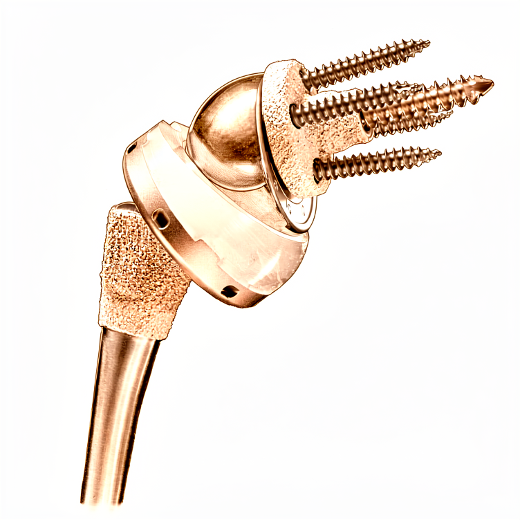
Your surgeon makes a single cut over the front of your shoulder to reach the joint. They remove the worn-out bone and cartilage, then replace them with metal and plastic parts. This new joint is designed to work even if your rotator cuff tendons are torn or damaged.
The surgeon places the metal ball on the shoulder socket and the plastic cup on the arm bone. This setup changes how the joint moves, allowing your shoulder muscle to lift your arm more easily. If you had a broken bone, your surgeon may also repair the torn bone pieces back to their normal position using small metal anchors.
Once the new parts are in place, your surgeon closes the cut with stitches. These stitches are usually dissolvable and do not need to be removed. The goal is to restore stability and help you move your shoulder again.
After the operation
You will wake up in a recovery ward where your team manages your pain. Your surgeon uses a single open incision over your shoulder. You will wear a sling and have dressings over the wound. You will begin moving your arm gently as soon as you are stable. Most patients go home the same day or stay overnight. You must have someone stay with you for the first 24 hours to help you. Early movement is safe and helps your recovery.
Recovery
Your shoulder will feel sore and swollen in the first few days. This is normal as your body heals from the open incision over your shoulder. You will wear a sling to protect the joint while you rest. Most people find that pain and stiffness ease significantly as swelling goes down.
You will begin gentle exercises early to restore movement. Your physiotherapist will guide you through simple movements to lift your arm and improve strength. You can return to daily tasks like eating or dressing once your surgeon clears you. Walking and swimming are common activities you can resume as your strength returns.
Your recovery journey is unique. While many people feel much better within two weeks, full improvement takes time. You will reach your maximum medical improvement by one year after surgery. Your surgeon and physiotherapist will tailor your plan to your specific needs. Trust the process and follow their advice to get the best result.
What can go wrong
Most patients do well, but problems can occasionally happen. Your surgeon and the team monitor you closely to spot any issue early.
If you have a wound that looks red, feels hot, or starts leaking fluid, this could be a wound complication. This risk is higher if you need blood-thinning medicine after surgery. Call your clinic immediately if you notice these signs.
You might feel sudden instability or a clicking sensation in your shoulder. This can happen if the joint slips out of place. If this occurs, stop moving the arm and contact your surgeon right away.
Deep, throbbing pain that does not ease with simple painkillers could signal a problem with the implant loosening. This is more common if you are having a second surgery on a joint that has already failed. Let your surgeon know if your pain gets worse over time.
If you are younger than 60, you may face a higher risk of surgical problems within the first 90 days. Be extra careful with your recovery and report any new symptoms to your team.
Sometimes, the bone around the shoulder blade can wear away or loosen after a revision surgery. This might feel like a grinding noise or a loss of strength. If you notice this, bring it up at your next review.
If you have had an infection before, you might need a staged procedure with antibiotic spacers. While this can help restore function, it is a complex process. Follow your surgeon's specific instructions for this path closely.
The complications table on this page lists typical rates if you want the specifics.
When to call us
Call us if you develop a fever, increasing redness, or drainage from your wound. Go to emergency if you feel sudden severe pain, new weakness, or lose the ability to move your arm. Seek immediate help for calf swelling, shortness of breath, or loss of sensation. These signs may indicate infection, blood clots, or a fracture near the shoulder blade. Contact your surgeon right away if you notice any sudden change in your recovery.
Evidence & references
title: "Reverse Shoulder Arthroplasty" slug: reverse-shoulder-arthroplasty region: shoulder audience: patient mesh_terms: [] article_count: 0 model_used: qwen3.5-35b-a3b-q8 generated_at: '2026-05-18T13:33:20+00:00' key_articles: [] synthesis_version: "v2" verifier_status: skipped
Anatomy & Pathophysiology
- Periprosthetic scapular fractures about reverse shoulder arthroplasty are universally associated with stable glenoid implants [2].
- Scapular fractures are unlikely to occur in the face of dislocation, glenosphere dissociation, or baseplate pullout at the bone–baseplate interface [2].
- Periprosthetic scapular fractures can result in new glenohumeral instability due to a change in the orientation of the glenosphere and loss of deltoid tension [2].
- The glenoid is suspended from the body of the scapula by the neck and fixed to the clavicle by the acromioclavicular and coracoclavicular ligaments [3].
- As the face of the glenoid transitions into the neck, the glenoid vault narrows [3].
- Optimal fixation for reverse shoulder arthroplasty involves implanting the central peg or screw within the glenoid vault in the axis of the body of the scapula without perforation [3].
- Peripheral locking screws enhance implant stability to allow for bony ingrowth [3].
- Optimal bony corridors for peripheral screws guide fixation within the lateral pillar of the scapula (inferior and posterior screws), the base of the coracoid (superior screw), and the scapular spine (anterior screw) [3].
- The scapular spine is subcutaneous posteriorly and widens gradually as it transitions into the base of the acromion laterally [3].
- The acromion curves anteriorly and meets the clavicle at the acromioclavicular joint and the coracoid via the coracoacromial ligament, which originates under the anterior margin of the acromion [3].
- The suprascapular nerve arises from the C4–C5 nerve roots off of the supraclavicular brachial plexus at "Erb's point" [3].
- The suprascapular nerve runs just medial to the base of the coracoid, under the transverse scapular ligament within the suprascapular notch, and gives off branches to the supraspinatus within 1 cm of the notch [3].
- The suprascapular nerve continues through the supraspinatus fossa heading laterally and distally on the under surface of the supraspinatus [3].
- The suprascapular nerve runs under the ill-defined spinoglenoid ligament around the lateral base of the scapula within the spinoglenoid notch before terminating in posterior capsular sensory branches and an infraspinatus motor branch within 1 cm of the lateral margin of the scapular spine [3].
- Cadaver studies show the suprascapular nerve is present 29 mm (23 to 35 mm) from the superior rim of the glenoid at the suprascapular notch [3].
- Cadaver studies show the suprascapular nerve is present 18 mm (14 to 24 mm) from the posterior rim at the spinoglenoid notch [3].
- Injury to the suprascapular nerve can cause pain and denervation of the supraspinatus and infraspinatus [3].
- The traditional Grammont reverse prosthesis features a glenoid component with an articulating portion shaped as a third of a sphere [4].
- In the Grammont reverse prosthesis, the center of rotation is medial to the glenoid component–bone interface [4].
- The humeral component in the Grammont reverse prosthesis is inset, resting almost completely inside the proximal humerus metaphysis [4].
- The opening angle of the polyethylene in the Grammont reverse prosthesis is relatively horizontal at 155 degrees [4].
- In the Grammont reverse prosthesis, the humerus is positioned more medially and more distally than preoperatively [4].
- The humeral component in the Grammont reverse prosthesis is recommended to be implanted in more anteversion (0 to 10 degrees of retroversion) than conventional arthroplasty [4].
- Subsequent reverse designs have modified features to place the center of rotation more lateral than the Grammont prosthesis [4].
- Later reverse designs introduced an opening angle of 135 degrees for the humeral component [4].
- Later designs introduced humeral components with an onlay humeral bearing, which lateralized the position of the humerus without changing the center of rotation of the arthroplasty [4].
- Later designs selected a 145-degree opening angle for the bearing [4].
- The RSA does not require the rotator cuff for function but is dependent on an intact deltoid neuromuscular unit [7].
- The Grammont principles of RSA include that the prosthesis must be inherently stable and perfectly concentric [7].
- The Grammont principles of RSA include that the weight-bearing part must be convex and the supported part concave (reversed) [7].
- The Grammont principles of RSA include that the center of the sphere must be at or within the glenoid neck (medialized) [7].
- The Grammont principles of RSA include that the center of rotation must be medialized and distalized [7].
- The traditional Grammont style decreases shear forces seen by the glenoid and lowers baseplate failure by medializing the center of rotation [7].
- The traditional Grammont style carries a risk of inferior scapular notching in adduction, which is associated with poorer results [7].
- Distalization in RSA doubles the lever arm of the deltoid and optimizes the length–tension curve of its sarcomeres [7].
- Distalization in RSA increases deltoid efficiency by 30% at the cost of rotational strength [7].
- Lateralized glenosphere and lateralized humerus designs have gained popularity to improve the rotational profile, deltoid function, implant stability, and decrease impingement (scapular notching) [7].
- Early reverse shoulder arthroplasty designs had a high failure rate due to the profound lever arm on the glenoid and baseplate bone [7].
- Recent reverse shoulder arthroplasty designs have a better track record but result in increased forces seen by the scapula and acromion [7].
- Postoperative periprosthetic scapular fractures occur at rates of 0.9% to 11.2% [7].
- Periprosthetic scapular fractures are a unique complication of reverse shoulder arthroplasty that occurs more commonly than periprosthetic humeral fractures [7].
- Female gender has been implicated as a risk factor for postoperative periprosthetic scapular fractures, accounting for up to 100% of some series [7].
- Postoperative periprosthetic scapular fractures typically occur in patients aged 70 to 80 years [7].
- One study of patients under age 65 undergoing reverse shoulder arthroplasty showed a 0% (0/67) postoperative periprosthetic scapular fracture rate [7].
- Osteoporosis has been implicated as a risk factor for postoperative periprosthetic scapular fractures [7].
- In one study, 75% (6/8) of acromial fractures occurred in osteoporotic hosts [7].
- A study comparing 53 postoperative scapular spine fractures with 212 matched controls identified osteoporosis as a significant risk factor (30.8% fracture patients vs. 18.4% controls; OR 1.97; p < 0.05) [7].
- Fatigue fractures of the scapula have been found to occur through already weakened acromia or those with preexisting lesions [7].
- Acromial thinning and eventual fragmentation occur at the final stages of rotator cuff-tear arthropathy [7].
Clinical Presentation
- Periprosthetic scapular fractures are universally associated with stable glenoid implants [2].
- Scapular fractures are unlikely to occur in the presence of glenohumeral dislocation, glenosphere dissociation, or baseplate pullout at the bone–baseplate interface [2].
- Periprosthetic scapular fractures can rarely result in new glenohumeral instability due to a change in glenosphere orientation and loss of deltoid tension [2].
- A case of periprosthetic scapular fracture occurred 8 months after successful reverse shoulder arthroplasty for rotator cuff tear arthropathy [2].
- Patients with periprosthetic scapular fractures may experience a profound loss of function following a fall despite substantial prior pain relief and function [2].
- Periprosthetic scapular fractures can present as minimally displaced fractures involving the scapular neck and body, confirmed via CT scanning [2].
- Diagnosis of periprosthetic fractures often requires a high index of suspicion due to subtle presentation [2].
- Workup for periprosthetic fractures should begin with a complete history and examination [2].
- History taking should elucidate the underlying diagnosis for the index surgery, subsequent surgeries, and any complications including infection [2].
- The examiner must understand the patient's shoulder function and level of disability before surgery, after surgery, and at present [2].
- The time course of functional changes should be documented during history taking [2].
- New pain at the base of the acromion may be the only finding in cases of stress reaction [2].
- Stress fractures can be more painful than when they propagate into displaced fractures [2].
- Patients with periprosthetic scapular fractures typically present in their 8th decade of life [2].
- Presentation is generally characterized by a sudden increase in pain or loss of function in an otherwise smooth postoperative course [2].
- Periprosthetic scapular fractures generally occur within 1 year but up to 2 years from surgery [2].
- Patients who develop periprosthetic scapular fractures initially outperform those who do not [2].
- Risk factors for periprosthetic scapular fractures include a history of steroid use, osteoporosis, subacromial decompression, or rotator cuff tear arthropathy [2].
- Previous operative reports, clinic notes, and imaging can help identify risk factors such as previous shoulder surgeries or history of radiation [2].
- Deformity on physical examination is concerning for dislocation, hematoma, or displaced fracture [2].
- Erythema or incisional dehiscence on physical examination is concerning for infection [2].
- Tenderness along the acromion or scapular spine raises suspicion for fracture [2].
- A complete neurovascular examination is part of the physical assessment for periprosthetic fractures [2].
- Active and passive motion should be assessed during the physical examination [2].
- Fractures can result in motion limited by pain, new weakness, or loss of function [2].
- Sudden loss of function or increase in pain is consistent with both scapular fracture and infection [2].
Investigations
- Periprosthetic scapular fractures are universally associated with stable glenoid implants [2].
- Periprosthetic scapular fractures can result in new glenohumeral instability due to a change in the orientation of the glenosphere and loss of deltoid tension [2].
- Diagnosis of periprosthetic scapular fractures often requires a high index of suspicion as identification can be subtle [2].
- Workup for periprosthetic scapular fractures should begin with a complete history and examination [2].
- New pain at the base of the acromion may be the only finding in a stress reaction and should raise suspicion for further imaging or rest [2].
- Stress fractures can be more painful than when they propagate into a displaced fracture [2].
- Patients with periprosthetic scapular fractures typically present around their 8th decade of life after a sudden increase in pain or loss of function [2].
- Periprosthetic scapular fractures generally occur within 1 year but up to 2 years from surgery [2].
- Patients who develop periprosthetic scapular fractures initially outperform those who do not [2].
- Risk factors for periprosthetic scapular fractures include a history of steroid use, osteoporosis, subacromial decompression, or rotator cuff tear arthropathy [2].
- Physical examination for periprosthetic scapular fractures should include inspection for deformity, erythema, or incisional dehiscence [2].
- Tenderness along the acromion or scapular spine raises suspicion for fracture and should be confirmed with imaging [2].
- A complete neurovascular examination and assessment of active and passive motion are required during the physical examination for periprosthetic scapular fractures [2].
- Sudden loss of function or increase in pain is consistent with both scapular fracture and infection [2].
- The glenoid is suspended from the body of the scapula by the neck and fixed to the clavicle by the acromioclavicular and coracoclavicular ligaments [3].
- The glenoid vault narrows as the face of the glenoid transitions into the neck [3].
- Optimal fixation for reverse shoulder arthroplasty involves implanting the central peg or screw within the glenoid vault in the axis of the body of the scapula without perforation [3].
- Peripheral locking screws enhance the ability to obtain implant stability long enough for bony ingrowth [3].
- Optimal bony corridors for peripheral screws guide fixation within the lateral pillar of the scapula (inferior and posterior screws), the base of the coracoid (superior screw), and scapular spine (anterior screw) [3].
- The suprascapular nerve arises from the C4–C5 nerve roots off of the supraclavicular brachial plexus at "Erb's point" [3].
- The suprascapular nerve runs just medial to the base of the coracoid, under the transverse scapular ligament within the suprascapular notch [3].
- The suprascapular nerve gives off branches to the supraspinatus within 1 cm of the notch [3].
- The suprascapular nerve runs under the spinoglenoid ligament around the lateral base of the scapula within the spinoglenoid notch before terminating in posterior capsular sensory branches and an infraspinatus motor branch [3].
- Cadaver studies show the suprascapular nerve is present 29 mm (23 to 35 mm) from the superior rim of the glenoid at the suprascapular notch [3].
- Cadaver studies show the suprascapular nerve is present 18 mm (14 to 24 mm) from the posterior rim at the spinoglenoid notch [3].
- Injury to the suprascapular nerve can cause pain and denervation of the supraspinatus and infraspinatus [3].
- It is recommended to limit superior screw length to ≤25 mm and posterior screws to ≤15 mm when possible to reduce the risk of suprascapular nerve injury [3].
- The suprascapular nerve can be injured by the fracture itself or become encased in callus [3].
- Careful assessment of preoperative radiographs and CT with three-dimensional reconstruction is extremely useful for preoperative planning in reverse shoulder arthroplasty for fracture [5].
- Preoperative planning goals include understanding the fracture pattern and anticipating the ideal height of stem implantation [5].
- Radiographs of both humeri with magnifier markers may be used to understand where the stem should be positioned in reference to the fracture line on the humeral shaft [5].
- The glenoid should be assessed in radiographs and CT to plan for component positioning, version, inclination, and rotation, as well as anticipated screw length [5].
- Rarely, there may be associated fractures of the rim of the glenoid in anterior or posterior fracture-dislocations [5].
- If a fractured glenoid rim is large enough to interfere with the stability of the glenoid baseplate, fixation with small fragment screws may be performed [5].
- Reverse shoulder arthroplasty is best performed in the beach chair position, specifically the "barber chair" position with the trunk at approximately 70 degrees [5].
- The deltopectoral approach is preferred for reverse arthroplasty for fracture due to familiarity, easier placement of the glenoid component low and with an inferior tilt, and extensibility of exposure [5].
- Management and reduction of the greater tuberosity is easier from a superior deltoid-splitting approach [5].
- Radiographic identification of periprosthetic scapular fractures can be subtle [10].
- Plane radiographs for periprosthetic scapular fractures should include AP, scapular Y, and axillary views [10].
- Radiographs should be compared with preoperative and initial postoperative images to identify subtle changes [10].
- Preoperative images can identify a missed os acromiale or insufficiency fracture which can displace after deltoid tensioning in reverse shoulder arthroplasty [10].
- Implant dissociation or loosening presents with a change in implant position on serial radiographs [10].
- Progressive downsloping of the acromion relative to the scapular spine indicates a displaced acromial fracture [10].
- Narrowing of the acromial–tuberosity interval indicates a displaced acromial fracture [10].
- The scapular Y view identifies displaced scapular spine or body fractures [10].
- The axillary view is helpful for identifying the location of the fracture, especially at the acromial base [10].
- Plain radiographs can miss more subtle fractures [10].
- Levy et al. found that plain radiographs were unreliable at detecting fracture (k = 0.05) or fracture union (k = 0.05) [10].
- In Otto's series, 32.1% (17/53) of fractures presented with pain and negative plain films [10].
- Independent reviewers were able to accurately diagnose 78.8% of periprosthetic scapular fractures with good inter-rater reliability (k = 0.782) and excellent intra-rater reliability (k = 0.862) [10].
- Patients with fractures had greater changes in acromial–tuberosity distance (p < 0.001) and acromial tilt (p < 0.001) from initial postoperative radiographs to final images [10].
- It is recommended to routinely evaluate acromial–tuberosity distance and acromial tilt to improve detection of periprosthetic scapular fractures [10].
- New pain along the scapula in the setting of normal radiographs should trigger a CT scan [10].
- In Levy et al.'s series, 39% (7/18) of fractures were associated with negative plain films and required a CT scan to diagnose nondisplaced fractures [10].
- A negative CT scan may occur in the setting of a stress reaction which may be better elicited on a bone scan [10].
Treatment
- Reverse shoulder arthroplasty is currently the replacement procedure of choice when arthroplasty is considered for proximal humeral fractures [4].
- The rate of utilization of reverse shoulder arthroplasty for proximal humeral fractures is increasing [4].
- Reverse shoulder arthroplasty was developed for the surgical management of cuff tear arthropathy [4].
- The semiconstrained nature of the reverse prosthesis provides a stable fulcrum that allows the deltoid to elevate the shoulder even in the absence of a functional rotator cuff [4].
- Tuberosity and rotator cuff-related complications are the main reason for poor functional outcomes when a humeral head replacement is implanted for management of a proximal humeral fracture [4].
- In the Grammont reverse prosthesis, the articulating portion of the glenoid component has the shape of a third of a sphere [4].
- In the Grammont reverse prosthesis, the center of rotation is medial to the glenoid component–bone interface [4].
- The intention of medializing the center of rotation in the Grammont design is to decrease shear stress and provide compressive stress to decrease the chances of glenoid loosening [4].
- In the Grammont reverse prosthesis, the humeral component is inset, resting almost completely inside the proximal humerus metaphysis [4].
- The opening angle of the polyethylene in the Grammont reverse prosthesis is relatively horizontal at 155 degrees [4].
- Once articulated in a Grammont reverse prosthesis, the humerus is positioned more medially and more distally than preoperatively [4].
- A more horizontal opening angle was selected in the Grammont design to decrease the chances of dislocation [4].
- The humeral component in the Grammont reverse prosthesis is recommended to be implanted in more anteversion (0 to 10 degrees of retroversion) than conventional arthroplasty [4].
- Subsequent reverse designs have modified features such as using a larger portion of a sphere and placing the center of rotation more lateral than the Grammont prosthesis [4].
- Later reverse designs introduced an opening angle of 135 degrees for the humeral component [4].
- Later designs introduced humeral components with an onlay humeral bearing, which lateralized the position of the humerus without changing the center of rotation of the arthroplasty [4].
- Selected later designs use a 145-degree opening angle for the bearing [4].
- There is very little published on reverse arthroplasty biomechanics in the setting of a proximal humeral fracture [4].
- Some surgeons initially elected to implant a reverse arthroplasty in proximal humeral fractures without repair or with excision of the greater and/or lesser tuberosity [4].
- Controversy remains regarding the impact of tuberosity healing on the outcome of reverse arthroplasty for fracture [4].
- Many believe that healing of at least the greater tuberosity in good position provides a higher chance of restoration of active external rotation [4].
- Active external rotation is very important for the overall functional outcome [4].
- In the treatment of proximal humeral nonunion, not performing a tuberosity repair at the time of reverse arthroplasty has been correlated with a higher rate of dislocation [4].
- Technical principles for reverse arthroplasty in cuff tear arthropathy may need to be modified to enhance tuberosity healing, such as avoiding translating the humeral shaft too lateral or too distal [4].
- The tuberosities should overlap a few millimeters with the shaft to facilitate healing [4].
- Use of a stem with fracture-dedicated features, including a proximal ingrowth surface, small cross section, and holes for suture fixation, may be beneficial [4].
- Shoulder arthroplasty is considered for proximal humeral nonunion in the presence of severe cavitation and bone loss at the humeral head and metaphysis or collapse and degenerative change of the humeral articular surface [6].
- Severe tuberosity malunion in a proximal humeral nonunion is more reliably compensated for with reverse arthroplasty than with osteotomy and internal fixation [6].
- Hemiarthroplasty is less commonly considered than reverse arthroplasty for proximal humeral nonunion [6].
- The functional outcome of hemiarthroplasty for nonunion is particularly concerning when tuberosity osteotomies need to be added [6].
- Most studies reporting on hemiarthroplasty for nonunion suggest the procedure may be effective in reducing or eliminating pain [6].
- Hemiarthroplasty for nonunion is associated with a high rate of complications that often require further surgery and disappointing functional recovery [6].
- Reverse shoulder arthroplasty may improve shoulder function in patients with nonunions associated with severe tuberosity malunions [6].
Complications
- Scapular notching is a specific complication of reverse shoulder arthroplasty that requires avoidance strategies [1, 26].
- The impact of scapular notching on clinical outcomes after reverse shoulder arthroplasty has been analyzed in a cohort of 476 shoulders [26].
- Subscapularis tendon integrity after reverse shoulder arthroplasty impacts shoulder function [1].
- Comparison of reverse total shoulder arthroplasty outcomes with and without subscapularis repair has been performed [1].
- Sonographic assessment of the subscapularis after reverse shoulder arthroplasty evaluates the impact of tendon integrity on shoulder function [1].
- Optimal screw placement for base plate fixation in reverse total shoulder arthroplasty is a critical technical consideration [1].
- The effect of component positioning on intrinsic stability of the reverse shoulder arthroplasty has been studied [1].
- Humeral component lateralization in reverse shoulder arthroplasty affects rotator cuff torque in a cadaver model [1].
- Humeral version in reverse shoulder arthroplasty affects impingement in activities of daily living [1].
- The clinical and radiographic impact of center of rotation in reverse shoulder arthroplasty has been reviewed systematically [1].
- Glenoid bone grafting is utilized in primary reverse total shoulder arthroplasty for glenoid deficiency [1].
- Structural bone grafting is used for glenoid deficiency in primary total shoulder arthroplasty [1].
- Comparison of radiographic and clinical outcomes of revision reverse total shoulder arthroplasty with structural versus nonstructural bone graft has been conducted [1].
- Posteriorly augmented glenoid components are used in anatomic total shoulder arthroplasty for primary osteoarthritis with posterior glenoid bone loss [1].
- Reverse total shoulder arthroplasty is indicated for massive irreparable rotator cuff tears in patients younger than 65 years old [1].
- Reverse total shoulder arthroplasty is indicated for massive, irreparable rotator cuff tears before the age of 60 years [1].
- Reverse total shoulder arthroplasty is indicated for the treatment of irreparable rotator cuff tear without glenohumeral arthritis [1].
- Reverse total shoulder arthroplasty improves function in cuff tear arthropathy [1].
- Functional outcomes of reverse shoulder arthroplasty compared with hemiarthroplasty for acute proximal humeral fractures have been evaluated [1].
- Comparison of hemiarthroplasty and reverse shoulder arthroplasty for the treatment of fractures in elderly patients has been performed [1].
- Reverse total shoulder arthroplasty versus hemiarthroplasty for proximal humeral fractures has been the subject of a systematic review [1].
- Three- and four-part displaced proximal humeral fractures in patients older than 70 years have been treated with reverse shoulder arthroplasty versus nonsurgical treatment [1].
- Short-stem uncemented primary reverse shoulder arthroplasty has demonstrated clinical and radiological outcomes [1].
- Reverse total shoulder arthroplasty in patients with rheumatoid arthritis has been studied [1].
- Short-term results after reverse shoulder arthroplasty in patients with rheumatoid arthritis and irreparable rotator cuff tear have been reported [1].
- Reverse total shoulder arthroplasty for primary glenohumeral osteoarthritis in patients with a biconcave glenoid has been evaluated [1].
- Long-term outcomes of reverse total shoulder arthroplasty have been followed up in a previous study [1].
Key Evidence
References
[1] Campbell S Operative Orthopaedics 4 Volume Set. RECONSTRUCTIVE PROCEDURES OF THE SHOULDER AND ELBOW IN ADULTS > REVERSE SHOULDER ARTHROPLASTY. [2] Rockwood And Green S Fractures In Adults. 29: Principles of Nonunion and Bone Defect Treatment > Injuries Associated with Periprosthetic Scapular Fractures About Reverse Shoulder Arthroplasty. [3] Rockwood And Green S Fractures In Adults. 29: Principles of Nonunion and Bone Defect Treatment > Pathoanatomy and Applied Anatomy Related to Periprosthetic Scapular Fractures About Reverse Shoulder Arthroplasty. [4] Rockwood And Green S Fractures In Adults. 29: Principles of Nonunion and Bone Defect Treatment > Reverse Shoulder Arthroplasty. [5] Rockwood And Green S Fractures In Adults. 29: Principles of Nonunion and Bone Defect Treatment > Preoperative Planning > Reverse Shoulder Arthroplasty for Fracture: Preoperative Planning Checklist. [6] Rockwood And Green S Fractures In Adults. 29: Principles of Nonunion and Bone Defect Treatment > Reverse Shoulder Arthroplasty and Hemiarthroplasty. [7] Rockwood And Green S Fractures In Adults. 29: Principles of Nonunion and Bone Defect Treatment > Periprosthetic Scapular Fractures About Reverse Shoulder Arthroplasty. [10] Rockwood And Green S Fractures In Adults. 29: Principles of Nonunion and Bone Defect Treatment > Imaging and Other Diagnostic Studies on Periprosthetic Scapular Fractures About Reverse Shoulder Arthroplasty.


