Phẫu thuật thay khớp vai ngược
Patients › Shoulder
Reverse shoulder replacement for severe rotator cuff tears and arthritis—when a traditional replacement isn’t ideal.
Lý do phẫu thuật này được đề xuất
Phẫu thuật thay khớp vai ngược, còn được gọi là thay khớp vai ngược, là một phẫu thuật làm thay đổi hình dạng khớp vai của bạn. Thủ thuật này thường được chỉ định cho bệnh nhân bị viêm khớp nặng và rách cơ chóp xoay, hoặc những người có gãy xương phức tạp không cải thiện sau điều trị nội khoa. Bác sĩ phẫu thuật của bạn có thể đã đề xuất phương pháp này vì các ca thay khớp vai thông thường thường thất bại khi cơ chóp xoay bị rách, trong khi thiết kế này sử dụng cơ vai của bạn để nâng cánh tay thay vì dựa vào cơ chóp xoay.
Mục tiêu chính của phẫu thuật này là cung cấp khả năng vận động ổn định và giảm đau đáng kể khi các phương pháp điều trị khác không hiệu quả. Mặc dù các lựa chọn điều trị nội khoa được thử nghiệm trước tiên, phẫu thuật được lựa chọn khi bạn cần một giải pháp đáng tin cậy để khôi phục chức năng. Phương pháp tiếp cận này cho phép bạn nâng cánh tay và thực hiện các công việc hàng ngày ngay cả khi cơ chóp xoay không thể tự lành.
Trước khi phẫu thuật
Bạn cần nhịn ăn trước khi phẫu thuật và ngừng một số loại thuốc theo chỉ định của bác sĩ phẫu thuật. Vui lòng sắp xếp để có người đưa bạn về nhà và mặc trang phục thoải mái. Bạn có thể cần chụp X-quang, chụp cộng hưởng từ (MRI), xét nghiệm máu hoặc đánh giá gây mê trước đó. Các kiểm tra này giúp bác sĩ phẫu thuật lập kế hoạch phương pháp tiếp cận tốt nhất cho khớp vai của bạn. Bác sĩ phẫu thuật sẽ thực hiện cuộc phẫu thuật thông qua một vết rạch mở duy nhất trên vùng vai. Phương pháp này cho phép tiếp cận trực tiếp để sửa chữa vấn đề. Hãy mang theo danh sách tất cả các loại thuốc hiện tại bạn đang dùng đến cuộc hẹn. Bác sĩ phẫu thuật sẽ xem xét những loại thuốc này để đảm bảo an toàn cho bạn trong suốt quá trình phẫu thuật.
Vào ngày phẫu thuật
Bạn sẽ đến bệnh viện và gặp bác sĩ phẫu thuật cùng bác sĩ gây mê. Phẫu thuật này được thực hiện dưới gây mê toàn thân kết hợp với phong bế thần kinh vùng. Bạn sẽ hoàn toàn ngủ trong suốt quá trình phẫu thuật, và khối phong bế (một mũi tiêm gây tê các thần kinh chi phối cánh tay trước khi bạn tỉnh dậy) giúp giảm đau trong 12 đến 24 giờ đầu sau phẫu thuật. Bác sĩ gây mê sẽ gặp bạn trước khi phẫu thuật và giải thích chi tiết về cả hai phần của phương pháp gây mê này.
Bác sĩ phẫu thuật sẽ thực hiện ca mổ thông qua một vết rạch mở duy nhất ở vùng vai của bạn. Sau đó, bạn sẽ được chuyển đến khu vực hồi tỉnh để tỉnh dậy một cách an toàn. Nhân viên y tế sẽ theo dõi mức độ đau và sự thoải mái của bạn khi hiệu quả tê dần hết. Bạn sẽ ở lại khu vực hồi tỉnh cho đến khi tình trạng ổn định và sẵn sàng trở về phòng.
Quy trình phẫu thuật
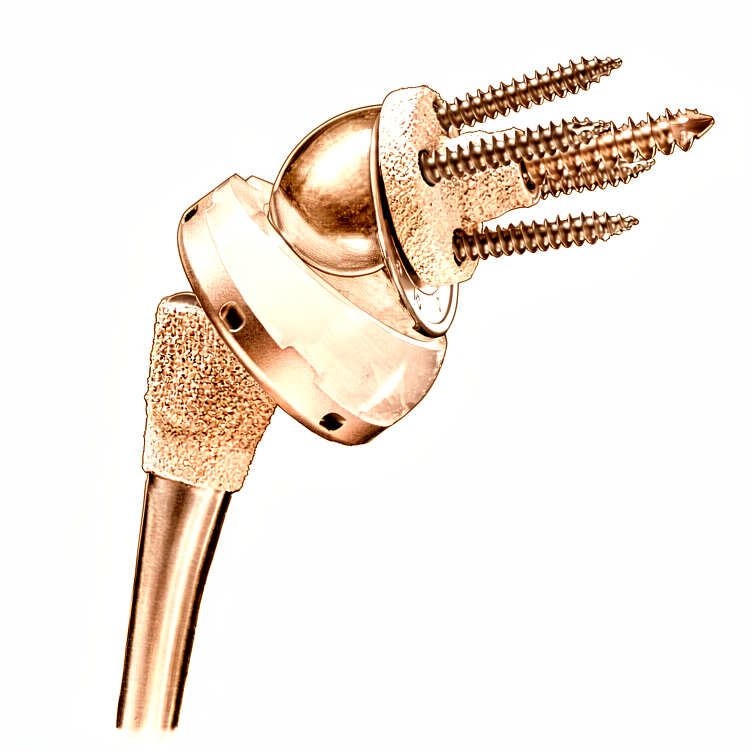
Bác sĩ phẫu thuật của bạn sẽ thực hiện một vết rạch duy nhất ở phía trước vai để tiếp cận khớp. Họ sẽ loại bỏ xương và sụn bị mòn, sau đó thay thế chúng bằng các bộ phận bằng kim loại và nhựa. Khớp mới này được thiết kế để hoạt động ngay cả khi các gân của nhóm cơ xoay cuff (rotator cuff) bị rách hoặc tổn thương.
Bác sĩ phẫu thuật đặt quả bóng kim loại vào hốc khớp vai và đặt cốc nhựa vào xương cánh tay. Cấu hình này thay đổi cách khớp vận động, cho phép cơ vai của bạn nâng cánh tay dễ dàng hơn. Nếu bạn bị gãy xương, bác sĩ phẫu thuật cũng có thể sửa chữa các mảnh xương bị rách trở lại vị trí bình thường bằng các neo kim loại nhỏ.
Sau khi các bộ phận mới được đặt vào vị trí, bác sĩ phẫu thuật của bạn sẽ khâu kín vết rạch. Các mũi khâu này thường là loại tự tiêu và không cần phải tháo bỏ. Mục tiêu là khôi phục sự ổn định và giúp bạn vận động lại khớp vai.
Sau phẫu thuật
Bạn sẽ tỉnh lại tại khoa hồi sức, nơi nhóm y tế sẽ kiểm soát cơn đau của bạn. Bác sĩ phẫu thuật sử dụng một vết rạch mở duy nhất ở vùng vai của bạn. Bạn sẽ đeo nạng và có băng gạc che phủ vết mổ. Bạn sẽ bắt đầu cử động cánh tay một cách nhẹ nhàng ngay khi tình trạng của bạn ổn định. Hầu hết bệnh nhân nằm lại bệnh viện qua đêm sau ca phẫu thuật này, mặc dù một số có thể về nhà cùng ngày. Bạn cần có người ở lại bên cạnh trong 24 giờ đầu để hỗ trợ bạn. Việc vận động sớm là an toàn và giúp quá trình hồi phục của bạn diễn ra tốt hơn.
Phục hồi
Vai của bạn sẽ cảm thấy đau và sưng trong vài ngày đầu. Đây là điều bình thường khi cơ thể bạn đang lành vết mổ mở trên vai. Bạn sẽ đeo nạng để bảo vệ khớp trong khi nghỉ ngơi. Hầu hết mọi người nhận thấy rằng cơn đau và cứng khớp giảm đáng kể khi tình trạng sưng giảm.
Bạn sẽ bắt đầu các bài tập nhẹ nhàng sớm để khôi phục tầm vận động. Chuyên viên vật lý trị liệu sẽ hướng dẫn bạn thực hiện các chuyển động đơn giản để nâng cánh tay và cải thiện sức mạnh. Bạn có thể quay trở lại các hoạt động hàng ngày như ăn uống hoặc mặc quần áo khi bác sĩ phẫu thuật cho phép. Đi bộ và bơi lội là những hoạt động phổ biến mà bạn có thể bắt đầu lại khi sức mạnh của bạn phục hồi.
Hành trình phục hồi của bạn là duy nhất. Trong khi nhiều người cảm thấy tốt hơn đáng kể trong vòng hai tuần, việc cải thiện hoàn toàn cần có thời gian. Bạn sẽ đạt được mức cải thiện y khoa tối đa sau một năm kể từ ngày phẫu thuật. Bác sĩ phẫu thuật và chuyên viên vật lý trị liệu sẽ điều chỉnh kế hoạch của bạn cho phù hợp với nhu cầu cụ thể của bạn. Hãy tin tưởng vào quá trình và tuân theo lời khuyên của họ để đạt được kết quả tốt nhất.
Những rủi ro có thể xảy ra
Hầu hết bệnh nhân đều hồi phục tốt, nhưng đôi khi có thể xảy ra các biến chứng. Bác sĩ phẫu thuật và đội ngũ y tế sẽ theo dõi sát sao bạn để phát hiện sớm bất kỳ vấn đề nào.
Nếu vết mổ có dấu hiệu đỏ, nóng hoặc bắt đầu chảy dịch, đây có thể là biến chứng về vết mổ. Nguy cơ này cao hơn nếu bạn cần dùng thuốc làm loãng máu sau phẫu thuật. Hãy gọi ngay cho phòng khám của bạn nếu nhận thấy các dấu hiệu này.
Bạn có thể cảm thấy mất ổn định đột ngột hoặc nghe thấy tiếng lách cách ở vai. Điều này có thể xảy ra nếu khớp bị trật vị trí. Nếu điều này xảy ra, hãy ngừng cử động cánh tay và liên hệ ngay với bác sĩ phẫu thuật của bạn.
Cơn đau dữ dội, nhói từng cơn không giảm với thuốc giảm đau thông thường có thể là dấu hiệu của vấn đề lỏng lẻo ở implant. Tình trạng này phổ biến hơn nếu bạn đang trải qua phẫu thuật lần thứ hai trên một khớp đã từng thất bại. Hãy thông báo cho bác sĩ phẫu thuật nếu cơn đau của bạn ngày càng nghiêm trọng theo thời gian.
Nếu bạn dưới 60 tuổi, bạn có thể đối mặt với nguy cơ cao hơn về các biến chứng phẫu thuật trong 90 ngày đầu tiên. Hãy cẩn thận hơn trong quá trình hồi phục và báo cáo bất kỳ triệu chứng mới nào cho đội ngũ y tế của bạn.
Đôi khi, xương xung quanh xương bả vai có thể bị mòn hoặc lỏng lẻo sau phẫu thuật sửa chữa. Điều này có thể cảm thấy như có tiếng ma sát hoặc mất sức mạnh. Nếu bạn nhận thấy điều này, hãy đề cập đến nó trong lần tái khám tiếp theo của bạn.
Nếu bạn đã từng bị nhiễm trùng trước đây, bạn có thể cần một quy trình từng giai đoạn với các spacer kháng sinh. Mặc dù điều này có thể giúp khôi phục chức năng, nhưng đây là một quy trình phức tạp. Hãy tuân thủ chặt chẽ các hướng dẫn cụ thể của bác sĩ phẫu thuật cho lộ trình này.
Bảng biến chứng trên trang này liệt kê các tỷ lệ điển hình nếu bạn muốn biết chi tiết.
Khi nào nên gọi cho chúng tôi
Gọi cho chúng tôi nếu bạn bị sốt, đỏ tăng dần hoặc có dịch chảy ra từ vết thương. Đến phòng cấp cứu nếu bạn cảm thấy đau dữ dội đột ngột, yếu mới xuất hiện hoặc mất khả năng cử động cánh tay. Tìm kiếm sự giúp đỡ ngay lập tức nếu bạn bị sưng bắp chân, khó thở hoặc mất cảm giác. Những dấu hiệu này có thể cho thấy nhiễm trùng, huyết khối hoặc gãy xương gần xương bả vai. Liên hệ với bác sĩ phẫu thuật của bạn ngay lập tức nếu bạn nhận thấy bất kỳ thay đổi đột ngột nào trong quá trình hồi phục của mình.
Evidence & references
Anatomy & Pathophysiology
- Periprosthetic scapular fractures about reverse shoulder arthroplasty are universally associated with stable glenoid implants [2].
- Scapular fractures are unlikely to occur in the face of dislocation, glenosphere dissociation, or baseplate pullout at the bone–baseplate interface [2].
- Periprosthetic scapular fractures can result in new glenohumeral instability due to a change in the orientation of the glenosphere and loss of deltoid tension [2].
- The glenoid is suspended from the body of the scapula by the neck and fixed to the clavicle by the acromioclavicular and coracoclavicular ligaments [3].
- As the face of the glenoid transitions into the neck, the glenoid vault narrows [3].
- Optimal fixation for reverse shoulder arthroplasty involves implanting the central peg or screw within the glenoid vault in the axis of the body of the scapula without perforation [3].
- Peripheral locking screws enhance implant stability to allow for bony ingrowth [3].
- Optimal bony corridors for peripheral screws guide fixation within the lateral pillar of the scapula (inferior and posterior screws), the base of the coracoid (superior screw), and the scapular spine (anterior screw) [3].
- The scapular spine is subcutaneous posteriorly and widens gradually as it transitions into the base of the acromion laterally [3].
- The acromion curves anteriorly and meets the clavicle at the acromioclavicular joint and the coracoid via the coracoacromial ligament, which originates under the anterior margin of the acromion [3].
- The suprascapular nerve arises from the C4–C5 nerve roots off of the supraclavicular brachial plexus at "Erb's point" [3].
- The suprascapular nerve runs just medial to the base of the coracoid, under the transverse scapular ligament within the suprascapular notch, and gives off branches to the supraspinatus within 1 cm of the notch [3].
- The suprascapular nerve continues through the supraspinatus fossa heading laterally and distally on the under surface of the supraspinatus [3].
- The suprascapular nerve runs under the ill-defined spinoglenoid ligament around the lateral base of the scapula within the spinoglenoid notch before terminating in posterior capsular sensory branches and an infraspinatus motor branch within 1 cm of the lateral margin of the scapular spine [3].
- Cadaver studies show the suprascapular nerve is present 29 mm (23 to 35 mm) from the superior rim of the glenoid at the suprascapular notch [3].
- Cadaver studies show the suprascapular nerve is present 18 mm (14 to 24 mm) from the posterior rim at the spinoglenoid notch [3].
- Injury to the suprascapular nerve can cause pain and denervation of the supraspinatus and infraspinatus [3].
- The traditional Grammont reverse prosthesis features a glenoid component with an articulating portion shaped as a third of a sphere [4].
- In the Grammont reverse prosthesis, the center of rotation is medial to the glenoid component–bone interface [4].
- The humeral component in the Grammont reverse prosthesis is inset, resting almost completely inside the proximal humerus metaphysis [4].
- The opening angle of the polyethylene in the Grammont reverse prosthesis is relatively horizontal at 155 degrees [4].
- In the Grammont reverse prosthesis, the humerus is positioned more medially and more distally than preoperatively [4].
- The humeral component in the Grammont reverse prosthesis is recommended to be implanted in more anteversion (0 to 10 degrees of retroversion) than conventional arthroplasty [4].
- Subsequent reverse designs have modified features to place the center of rotation more lateral than the Grammont prosthesis [4].
- Later reverse designs introduced an opening angle of 135 degrees for the humeral component [4].
- Later designs introduced humeral components with an onlay humeral bearing, which lateralized the position of the humerus without changing the center of rotation of the arthroplasty [4].
- Later designs selected a 145-degree opening angle for the bearing [4].
- The RSA does not require the rotator cuff for function but is dependent on an intact deltoid neuromuscular unit [7].
- The Grammont principles of RSA include that the prosthesis must be inherently stable and perfectly concentric [7].
- The Grammont principles of RSA include that the weight-bearing part must be convex and the supported part concave (reversed) [7].
- The Grammont principles of RSA include that the center of the sphere must be at or within the glenoid neck (medialized) [7].
- The Grammont principles of RSA include that the center of rotation must be medialized and distalized [7].
- The traditional Grammont style decreases shear forces seen by the glenoid and lowers baseplate failure by medializing the center of rotation [7].
- The traditional Grammont style carries a risk of inferior scapular notching in adduction, which is associated with poorer results [7].
- Distalization in RSA doubles the lever arm of the deltoid and optimizes the length–tension curve of its sarcomeres [7].
- Distalization in RSA increases deltoid efficiency by 30% at the cost of rotational strength [7].
- Lateralized glenosphere and lateralized humerus designs have gained popularity to improve the rotational profile, deltoid function, implant stability, and decrease impingement (scapular notching) [7].
- Early reverse shoulder arthroplasty designs had a high failure rate due to the profound lever arm on the glenoid and baseplate bone [7].
- Recent reverse shoulder arthroplasty designs have a better track record but result in increased forces seen by the scapula and acromion [7].
- Postoperative periprosthetic scapular fractures occur at rates of 0.9% to 11.2% [7].
- Periprosthetic scapular fractures are a unique complication of reverse shoulder arthroplasty that occurs more commonly than periprosthetic humeral fractures [7].
- Female gender has been implicated as a risk factor for postoperative periprosthetic scapular fractures, accounting for up to 100% of some series [7].
- Postoperative periprosthetic scapular fractures typically occur in patients aged 70 to 80 years [7].
- One study of patients under age 65 undergoing reverse shoulder arthroplasty showed a 0% (0/67) postoperative periprosthetic scapular fracture rate [7].
- Osteoporosis has been implicated as a risk factor for postoperative periprosthetic scapular fractures [7].
- In one study, 75% (6/8) of acromial fractures occurred in osteoporotic hosts [7].
- A study comparing 53 postoperative scapular spine fractures with 212 matched controls identified osteoporosis as a significant risk factor (30.8% fracture patients vs. 18.4% controls; OR 1.97; p < 0.05) [7].
- Fatigue fractures of the scapula have been found to occur through already weakened acromia or those with preexisting lesions [7].
- Acromial thinning and eventual fragmentation occur at the final stages of rotator cuff-tear arthropathy [7].
Clinical Presentation
- Periprosthetic scapular fractures are universally associated with stable glenoid implants [2].
- Scapular fractures are unlikely to occur in the presence of glenohumeral dislocation, glenosphere dissociation, or baseplate pullout at the bone–baseplate interface [2].
- Periprosthetic scapular fractures can rarely result in new glenohumeral instability due to a change in glenosphere orientation and loss of deltoid tension [2].
- A case of periprosthetic scapular fracture occurred 8 months after successful reverse shoulder arthroplasty for rotator cuff tear arthropathy [2].
- Patients with periprosthetic scapular fractures may experience a profound loss of function following a fall despite substantial prior pain relief and function [2].
- Periprosthetic scapular fractures can present as minimally displaced fractures involving the scapular neck and body, confirmed via CT scanning [2].
- Diagnosis of periprosthetic fractures often requires a high index of suspicion due to subtle presentation [2].
- Workup for periprosthetic fractures should begin with a complete history and examination [2].
- History taking should elucidate the underlying diagnosis for the index surgery, subsequent surgeries, and any complications including infection [2].
- The examiner must understand the patient's shoulder function and level of disability before surgery, after surgery, and at present [2].
- The time course of functional changes should be documented during history taking [2].
- New pain at the base of the acromion may be the only finding in cases of stress reaction [2].
- Stress fractures can be more painful than when they propagate into displaced fractures [2].
- Patients with periprosthetic scapular fractures typically present in their 8th decade of life [2].
- Presentation is generally characterized by a sudden increase in pain or loss of function in an otherwise smooth postoperative course [2].
- Periprosthetic scapular fractures generally occur within 1 year but up to 2 years from surgery [2].
- Patients who develop periprosthetic scapular fractures initially outperform those who do not [2].
- Risk factors for periprosthetic scapular fractures include a history of steroid use, osteoporosis, subacromial decompression, or rotator cuff tear arthropathy [2].
- Previous operative reports, clinic notes, and imaging can help identify risk factors such as previous shoulder surgeries or history of radiation [2].
- Deformity on physical examination is concerning for dislocation, hematoma, or displaced fracture [2].
- Erythema or incisional dehiscence on physical examination is concerning for infection [2].
- Tenderness along the acromion or scapular spine raises suspicion for fracture [2].
- A complete neurovascular examination is part of the physical assessment for periprosthetic fractures [2].
- Active and passive motion should be assessed during the physical examination [2].
- Fractures can result in motion limited by pain, new weakness, or loss of function [2].
- Sudden loss of function or increase in pain is consistent with both scapular fracture and infection [2].
Investigations
- Periprosthetic scapular fractures are universally associated with stable glenoid implants [2].
- Periprosthetic scapular fractures can result in new glenohumeral instability due to a change in the orientation of the glenosphere and loss of deltoid tension [2].
- Diagnosis of periprosthetic scapular fractures often requires a high index of suspicion as identification can be subtle [2].
- Workup for periprosthetic scapular fractures should begin with a complete history and examination [2].
- New pain at the base of the acromion may be the only finding in a stress reaction and should raise suspicion for further imaging or rest [2].
- Stress fractures can be more painful than when they propagate into a displaced fracture [2].
- Patients with periprosthetic scapular fractures typically present around their 8th decade of life after a sudden increase in pain or loss of function [2].
- Periprosthetic scapular fractures generally occur within 1 year but up to 2 years from surgery [2].
- Patients who develop periprosthetic scapular fractures initially outperform those who do not [2].
- Risk factors for periprosthetic scapular fractures include a history of steroid use, osteoporosis, subacromial decompression, or rotator cuff tear arthropathy [2].
- Physical examination for periprosthetic scapular fractures should include inspection for deformity, erythema, or incisional dehiscence [2].
- Tenderness along the acromion or scapular spine raises suspicion for fracture and should be confirmed with imaging [2].
- A complete neurovascular examination and assessment of active and passive motion are required during the physical examination for periprosthetic scapular fractures [2].
- Sudden loss of function or increase in pain is consistent with both scapular fracture and infection [2].
- The glenoid is suspended from the body of the scapula by the neck and fixed to the clavicle by the acromioclavicular and coracoclavicular ligaments [3].
- The glenoid vault narrows as the face of the glenoid transitions into the neck [3].
- Optimal fixation for reverse shoulder arthroplasty involves implanting the central peg or screw within the glenoid vault in the axis of the body of the scapula without perforation [3].
- Peripheral locking screws enhance the ability to obtain implant stability long enough for bony ingrowth [3].
- Optimal bony corridors for peripheral screws guide fixation within the lateral pillar of the scapula (inferior and posterior screws), the base of the coracoid (superior screw), and scapular spine (anterior screw) [3].
- The suprascapular nerve arises from the C4–C5 nerve roots off of the supraclavicular brachial plexus at "Erb's point" [3].
- The suprascapular nerve runs just medial to the base of the coracoid, under the transverse scapular ligament within the suprascapular notch [3].
- The suprascapular nerve gives off branches to the supraspinatus within 1 cm of the notch [3].
- The suprascapular nerve runs under the spinoglenoid ligament around the lateral base of the scapula within the spinoglenoid notch before terminating in posterior capsular sensory branches and an infraspinatus motor branch [3].
- Cadaver studies show the suprascapular nerve is present 29 mm (23 to 35 mm) from the superior rim of the glenoid at the suprascapular notch [3].
- Cadaver studies show the suprascapular nerve is present 18 mm (14 to 24 mm) from the posterior rim at the spinoglenoid notch [3].
- Injury to the suprascapular nerve can cause pain and denervation of the supraspinatus and infraspinatus [3].
- It is recommended to limit superior screw length to ≤25 mm and posterior screws to ≤15 mm when possible to reduce the risk of suprascapular nerve injury [3].
- The suprascapular nerve can be injured by the fracture itself or become encased in callus [3].
- Careful assessment of preoperative radiographs and CT with three-dimensional reconstruction is extremely useful for preoperative planning in reverse shoulder arthroplasty for fracture [5].
- Preoperative planning goals include understanding the fracture pattern and anticipating the ideal height of stem implantation [5].
- Radiographs of both humeri with magnifier markers may be used to understand where the stem should be positioned in reference to the fracture line on the humeral shaft [5].
- The glenoid should be assessed in radiographs and CT to plan for component positioning, version, inclination, and rotation, as well as anticipated screw length [5].
- Rarely, there may be associated fractures of the rim of the glenoid in anterior or posterior fracture-dislocations [5].
- If a fractured glenoid rim is large enough to interfere with the stability of the glenoid baseplate, fixation with small fragment screws may be performed [5].
- Reverse shoulder arthroplasty is best performed in the beach chair position, specifically the "barber chair" position with the trunk at approximately 70 degrees [5].
- The deltopectoral approach is preferred for reverse arthroplasty for fracture due to familiarity, easier placement of the glenoid component low and with an inferior tilt, and extensibility of exposure [5].
- Management and reduction of the greater tuberosity is easier from a superior deltoid-splitting approach [5].
- Radiographic identification of periprosthetic scapular fractures can be subtle [10].
- Plane radiographs for periprosthetic scapular fractures should include AP, scapular Y, and axillary views [10].
- Radiographs should be compared with preoperative and initial postoperative images to identify subtle changes [10].
- Preoperative images can identify a missed os acromiale or insufficiency fracture which can displace after deltoid tensioning in reverse shoulder arthroplasty [10].
- Implant dissociation or loosening presents with a change in implant position on serial radiographs [10].
- Progressive downsloping of the acromion relative to the scapular spine indicates a displaced acromial fracture [10].
- Narrowing of the acromial–tuberosity interval indicates a displaced acromial fracture [10].
- The scapular Y view identifies displaced scapular spine or body fractures [10].
- The axillary view is helpful for identifying the location of the fracture, especially at the acromial base [10].
- Plain radiographs can miss more subtle fractures [10].
- Levy et al. found that plain radiographs were unreliable at detecting fracture (k = 0.05) or fracture union (k = 0.05) [10].
- In Otto's series, 32.1% (17/53) of fractures presented with pain and negative plain films [10].
- Independent reviewers were able to accurately diagnose 78.8% of periprosthetic scapular fractures with good inter-rater reliability (k = 0.782) and excellent intra-rater reliability (k = 0.862) [10].
- Patients with fractures had greater changes in acromial–tuberosity distance (p < 0.001) and acromial tilt (p < 0.001) from initial postoperative radiographs to final images [10].
- It is recommended to routinely evaluate acromial–tuberosity distance and acromial tilt to improve detection of periprosthetic scapular fractures [10].
- New pain along the scapula in the setting of normal radiographs should trigger a CT scan [10].
- In Levy et al.'s series, 39% (7/18) of fractures were associated with negative plain films and required a CT scan to diagnose nondisplaced fractures [10].
- A negative CT scan may occur in the setting of a stress reaction which may be better elicited on a bone scan [10].
Treatment
- Reverse shoulder arthroplasty is currently the replacement procedure of choice when arthroplasty is considered for proximal humeral fractures [4].
- The rate of utilization of reverse shoulder arthroplasty for proximal humeral fractures is increasing [4].
- Reverse shoulder arthroplasty was developed for the surgical management of cuff tear arthropathy [4].
- The semiconstrained nature of the reverse prosthesis provides a stable fulcrum that allows the deltoid to elevate the shoulder even in the absence of a functional rotator cuff [4].
- Tuberosity and rotator cuff-related complications are the main reason for poor functional outcomes when a humeral head replacement is implanted for management of a proximal humeral fracture [4].
- In the Grammont reverse prosthesis, the articulating portion of the glenoid component has the shape of a third of a sphere [4].
- In the Grammont reverse prosthesis, the center of rotation is medial to the glenoid component–bone interface [4].
- The intention of medializing the center of rotation in the Grammont design is to decrease shear stress and provide compressive stress to decrease the chances of glenoid loosening [4].
- In the Grammont reverse prosthesis, the humeral component is inset, resting almost completely inside the proximal humerus metaphysis [4].
- The opening angle of the polyethylene in the Grammont reverse prosthesis is relatively horizontal at 155 degrees [4].
- Once articulated in a Grammont reverse prosthesis, the humerus is positioned more medially and more distally than preoperatively [4].
- A more horizontal opening angle was selected in the Grammont design to decrease the chances of dislocation [4].
- The humeral component in the Grammont reverse prosthesis is recommended to be implanted in more anteversion (0 to 10 degrees of retroversion) than conventional arthroplasty [4].
- Subsequent reverse designs have modified features such as using a larger portion of a sphere and placing the center of rotation more lateral than the Grammont prosthesis [4].
- Later reverse designs introduced an opening angle of 135 degrees for the humeral component [4].
- Later designs introduced humeral components with an onlay humeral bearing, which lateralized the position of the humerus without changing the center of rotation of the arthroplasty [4].
- Selected later designs use a 145-degree opening angle for the bearing [4].
- There is very little published on reverse arthroplasty biomechanics in the setting of a proximal humeral fracture [4].
- Some surgeons initially elected to implant a reverse arthroplasty in proximal humeral fractures without repair or with excision of the greater and/or lesser tuberosity [4].
- Controversy remains regarding the impact of tuberosity healing on the outcome of reverse arthroplasty for fracture [4].
- Many believe that healing of at least the greater tuberosity in good position provides a higher chance of restoration of active external rotation [4].
- Active external rotation is very important for the overall functional outcome [4].
- In the treatment of proximal humeral nonunion, not performing a tuberosity repair at the time of reverse arthroplasty has been correlated with a higher rate of dislocation [4].
- Technical principles for reverse arthroplasty in cuff tear arthropathy may need to be modified to enhance tuberosity healing, such as avoiding translating the humeral shaft too lateral or too distal [4].
- The tuberosities should overlap a few millimeters with the shaft to facilitate healing [4].
- Use of a stem with fracture-dedicated features, including a proximal ingrowth surface, small cross section, and holes for suture fixation, may be beneficial [4].
- Shoulder arthroplasty is considered for proximal humeral nonunion in the presence of severe cavitation and bone loss at the humeral head and metaphysis or collapse and degenerative change of the humeral articular surface [6].
- Severe tuberosity malunion in a proximal humeral nonunion is more reliably compensated for with reverse arthroplasty than with osteotomy and internal fixation [6].
- Hemiarthroplasty is less commonly considered than reverse arthroplasty for proximal humeral nonunion [6].
- The functional outcome of hemiarthroplasty for nonunion is particularly concerning when tuberosity osteotomies need to be added [6].
- Most studies reporting on hemiarthroplasty for nonunion suggest the procedure may be effective in reducing or eliminating pain [6].
- Hemiarthroplasty for nonunion is associated with a high rate of complications that often require further surgery and disappointing functional recovery [6].
- Reverse shoulder arthroplasty may improve shoulder function in patients with nonunions associated with severe tuberosity malunions [6].
Complications
- Scapular notching is a specific complication of reverse shoulder arthroplasty that requires avoidance strategies [1, 26].
- The impact of scapular notching on clinical outcomes after reverse shoulder arthroplasty has been analyzed in a cohort of 476 shoulders [26].
- Subscapularis tendon integrity after reverse shoulder arthroplasty impacts shoulder function [1].
- Comparison of reverse total shoulder arthroplasty outcomes with and without subscapularis repair has been performed [1].
- Sonographic assessment of the subscapularis after reverse shoulder arthroplasty evaluates the impact of tendon integrity on shoulder function [1].
- Optimal screw placement for base plate fixation in reverse total shoulder arthroplasty is a critical technical consideration [1].
- The effect of component positioning on intrinsic stability of the reverse shoulder arthroplasty has been studied [1].
- Humeral component lateralization in reverse shoulder arthroplasty affects rotator cuff torque in a cadaver model [1].
- Humeral version in reverse shoulder arthroplasty affects impingement in activities of daily living [1].
- The clinical and radiographic impact of center of rotation in reverse shoulder arthroplasty has been reviewed systematically [1].
- Glenoid bone grafting is utilized in primary reverse total shoulder arthroplasty for glenoid deficiency [1].
- Structural bone grafting is used for glenoid deficiency in primary total shoulder arthroplasty [1].
- Comparison of radiographic and clinical outcomes of revision reverse total shoulder arthroplasty with structural versus nonstructural bone graft has been conducted [1].
- Posteriorly augmented glenoid components are used in anatomic total shoulder arthroplasty for primary osteoarthritis with posterior glenoid bone loss [1].
- Reverse total shoulder arthroplasty is indicated for massive irreparable rotator cuff tears in patients younger than 65 years old [1].
- Reverse total shoulder arthroplasty is indicated for massive, irreparable rotator cuff tears before the age of 60 years [1].
- Reverse total shoulder arthroplasty is indicated for the treatment of irreparable rotator cuff tear without glenohumeral arthritis [1].
- Reverse total shoulder arthroplasty improves function in cuff tear arthropathy [1].
- Functional outcomes of reverse shoulder arthroplasty compared with hemiarthroplasty for acute proximal humeral fractures have been evaluated [1].
- Comparison of hemiarthroplasty and reverse shoulder arthroplasty for the treatment of fractures in elderly patients has been performed [1].
- Reverse total shoulder arthroplasty versus hemiarthroplasty for proximal humeral fractures has been the subject of a systematic review [1].
- Three- and four-part displaced proximal humeral fractures in patients older than 70 years have been treated with reverse shoulder arthroplasty versus nonsurgical treatment [1].
- Short-stem uncemented primary reverse shoulder arthroplasty has demonstrated clinical and radiological outcomes [1].
- Reverse total shoulder arthroplasty in patients with rheumatoid arthritis has been studied [1].
- Short-term results after reverse shoulder arthroplasty in patients with rheumatoid arthritis and irreparable rotator cuff tear have been reported [1].
- Reverse total shoulder arthroplasty for primary glenohumeral osteoarthritis in patients with a biconcave glenoid has been evaluated [1].
- Long-term outcomes of reverse total shoulder arthroplasty have been followed up in a previous study [1].
References
[1] Campbell S Operative Orthopaedics 4 Volume Set. RECONSTRUCTIVE PROCEDURES OF THE SHOULDER AND ELBOW IN ADULTS > REVERSE SHOULDER ARTHROPLASTY. [2] Rockwood And Green S Fractures In Adults. 29: Principles of Nonunion and Bone Defect Treatment > Injuries Associated with Periprosthetic Scapular Fractures About Reverse Shoulder Arthroplasty. [3] Rockwood And Green S Fractures In Adults. 29: Principles of Nonunion and Bone Defect Treatment > Pathoanatomy and Applied Anatomy Related to Periprosthetic Scapular Fractures About Reverse Shoulder Arthroplasty. [4] Rockwood And Green S Fractures In Adults. 29: Principles of Nonunion and Bone Defect Treatment > Reverse Shoulder Arthroplasty. [5] Rockwood And Green S Fractures In Adults. 29: Principles of Nonunion and Bone Defect Treatment > Preoperative Planning > Reverse Shoulder Arthroplasty for Fracture: Preoperative Planning Checklist. [6] Rockwood And Green S Fractures In Adults. 29: Principles of Nonunion and Bone Defect Treatment > Reverse Shoulder Arthroplasty and Hemiarthroplasty. [7] Rockwood And Green S Fractures In Adults. 29: Principles of Nonunion and Bone Defect Treatment > Periprosthetic Scapular Fractures About Reverse Shoulder Arthroplasty. [10] Rockwood And Green S Fractures In Adults. 29: Principles of Nonunion and Bone Defect Treatment > Imaging and Other Diagnostic Studies on Periprosthetic Scapular Fractures About Reverse Shoulder Arthroplasty.


