反式肩关节置换术
Patients › Shoulder
Reverse shoulder replacement for severe rotator cuff tears and arthritis—when a traditional replacement isn’t ideal.
为何建议进行此手术
反向肩关节置换术(也称为反向关节置换术)是一种改变肩关节形态的手术。该手术通常适用于患有严重关节炎和肩袖损伤的患者,或那些经非手术治疗后未见改善的复杂骨折患者。您的外科医生可能已推荐此手术,因为当肩袖撕裂时,标准肩关节置换术往往失败,而反向置换的设计是利用您的肩部肌肉来抬起手臂。
此手术的主要目标是在其他治疗无效时,提供稳定的活动度和显著的疼痛缓解。虽然通常会先尝试非手术方案,但当您需要一种可靠的解决方案以恢复功能时,则会选择手术。即使您的肩袖无法自行愈合,这种方法也能帮助您抬起手臂并执行日常任务。
手术前
您需要在手术前禁食,并遵医嘱停用某些药物。请安排专人驾车送您回家,并穿着舒适的衣物。您可能需要在术前接受X线检查、磁共振成像(MRI)扫描、血液检查或麻醉评估。这些检查有助于您的外科医生为肩部手术制定最佳方案。您的外科医生将通过肩部上方的单个开放切口进行手术。该方法可直接暴露病灶以进行处理。请将您目前服用的所有药物清单带给您的就诊医生。您的外科医生将审查这些药物,以确保手术过程中的安全。
手术当天
您将抵达医院,并与您的外科医生和麻醉师会面。本手术将在全身麻醉联合区域神经阻滞下进行。您在手术过程中将完全处于睡眠状态,而神经阻滞(在苏醒前注射以麻木支配手臂的神经)可为术后最初12至24小时提供镇痛效果。麻醉师将在手术前与您见面,并向您详细说明这两个部分。
您的外科医生将通过肩部上方的一个开放切口进行手术。随后,您将移至复苏区安全苏醒。在麻木感消退期间,医护人员将监测您的疼痛和舒适度。您将在复苏区留观,直至生命体征稳定并准备返回病房。
手术过程
您的外科医生会在肩部前方做一个切口以进入关节。他们移除磨损的骨头和软骨,然后用金属和塑料部件替换。这种新关节的设计旨在即使您的肩袖肌腱撕裂或受损时也能正常工作。
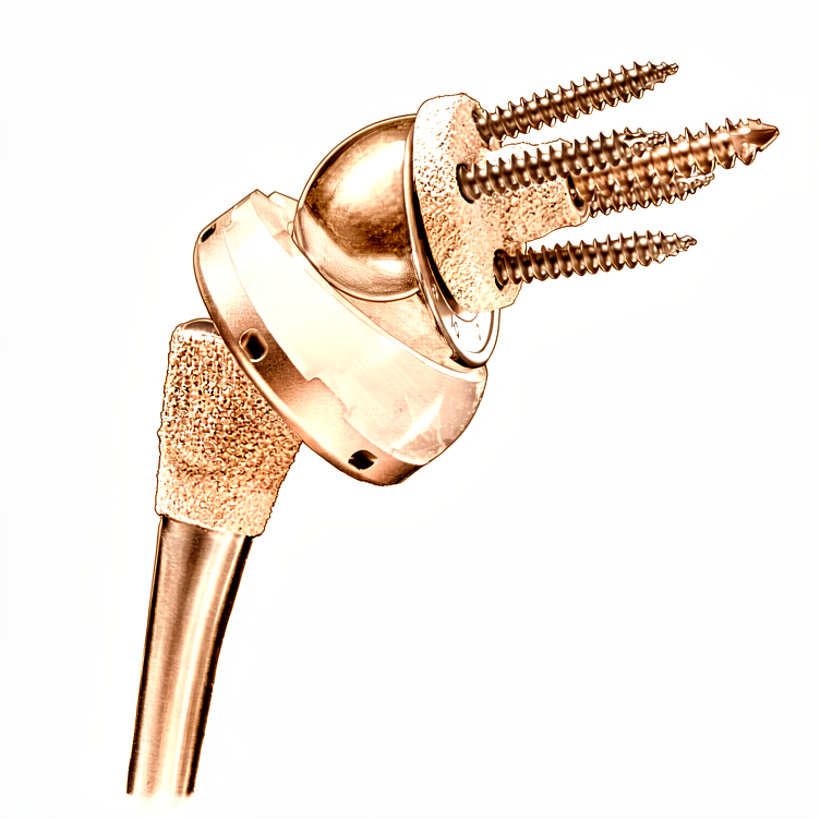
外科医生将金属球置于肩胛骨盂(肩关节窝),将塑料杯置于肱骨(上臂骨)。这种配置改变了关节的运动方式,使您的肩部肌肉能够更轻松地抬起手臂。如果您伴有骨折,您的外科医生还可能使用小型金属锚钉将撕裂的骨块复位至正常位置。
一旦新部件安装到位,您的外科医生会用缝线缝合切口。这些缝线通常是可吸收的,无需拆除。手术的目的是恢复稳定性并帮助您重新活动肩部。
术后
您将在恢复室苏醒,您的医疗团队将为您管理疼痛。您的外科医生会在您的肩部做一个单一的开放切口。您需要佩戴悬吊带,并且伤口上会有敷料。一旦您情况稳定,您将开始轻柔地活动手臂。大多数患者在此次手术后需要在医院留宿一晚,尽管有些患者可以在当天回家。您必须有人在最初的24小时内陪伴您,以帮助您。早期活动是安全的,并有助于您的康复。
恢复
在术后的头几天,您的肩膀会感到酸痛和肿胀。这是正常的,因为您的身体正在从肩部的开放切口处愈合。在休息期间,您将佩戴吊带以保护关节。随着肿胀消退,大多数人会发现疼痛和僵硬明显减轻。
您会尽早开始进行轻柔的锻炼以恢复活动度。您的物理治疗师将指导您进行简单的动作,以抬起手臂并增强力量。一旦您的外科医生允许,您就可以恢复吃饭或穿衣等日常活动。随着力量的恢复,步行和游泳是您可以恢复的常见活动。
您的恢复过程是独特的。虽然许多人在两周内感觉好多了,但完全恢复需要时间。您将在术后一年达到最大医学改善(Maximum Medical Improvement)。您的外科医生和物理治疗师将根据您的具体需求量身定制您的康复计划。相信这个过程并遵循他们的建议,以获得最佳效果。
可能发生的问题
大多数患者恢复良好,但偶尔也会出现并发症。您的外科医生和医疗团队会密切监测您的情况,以便尽早发现任何问题。
如果您的伤口出现发红、发热或开始渗出液体,这可能是伤口并发症。如果您术后需要使用抗凝药物,这种风险会更高。一旦发现这些迹象,请立即致电您的诊所。
您可能会感到肩部突然不稳定或出现弹响感。这可能是关节脱位所致。如果发生这种情况,请停止活动手臂,并立即联系您的外科医生。
如果出现深部、搏动性疼痛,且普通止痛药无法缓解,可能提示植入物松动。在已失效关节进行翻修手术时,这种情况更为常见。如果疼痛随时间推移加重,请告知您的外科医生。
如果您年龄小于 60 岁,术后前 90 天内面临手术并发症的风险可能较高。请格外注意康复过程,并向您的医疗团队报告任何新出现的症状。
有时,肩胛骨周围的骨骼可能在翻修手术后出现磨损或松动。这可能会表现为研磨音或力量丧失。如果发现这种情况,请在下次复诊时提出。
如果您既往有感染史,可能需要进行分阶段手术并使用抗生素骨水泥间隔物。虽然这有助于恢复功能,但这是一个复杂的过程。请严格遵循您的外科医生针对此方案给出的具体指示。
如需了解具体数据,请参阅本页的并发症表格,其中列出了典型的发生率。
何时联系我们
如果您出现发热、伤口红肿加重或渗液,请立即联系我们。如果您感到突发剧烈疼痛、新发无力或手臂活动能力丧失,请前往急诊。如果出现小腿肿胀、呼吸困难或感觉丧失,请立即寻求紧急帮助。这些症状可能提示感染、血栓或肩胛骨附近骨折。如果您注意到康复过程中出现任何突然变化,请立即联系您的外科医生。
Evidence & references
Anatomy & Pathophysiology
- Periprosthetic scapular fractures about reverse shoulder arthroplasty are universally associated with stable glenoid implants [2].
- Scapular fractures are unlikely to occur in the face of dislocation, glenosphere dissociation, or baseplate pullout at the bone–baseplate interface [2].
- Periprosthetic scapular fractures can result in new glenohumeral instability due to a change in the orientation of the glenosphere and loss of deltoid tension [2].
- The glenoid is suspended from the body of the scapula by the neck and fixed to the clavicle by the acromioclavicular and coracoclavicular ligaments [3].
- As the face of the glenoid transitions into the neck, the glenoid vault narrows [3].
- Optimal fixation for reverse shoulder arthroplasty involves implanting the central peg or screw within the glenoid vault in the axis of the body of the scapula without perforation [3].
- Peripheral locking screws enhance implant stability to allow for bony ingrowth [3].
- Optimal bony corridors for peripheral screws guide fixation within the lateral pillar of the scapula (inferior and posterior screws), the base of the coracoid (superior screw), and the scapular spine (anterior screw) [3].
- The scapular spine is subcutaneous posteriorly and widens gradually as it transitions into the base of the acromion laterally [3].
- The acromion curves anteriorly and meets the clavicle at the acromioclavicular joint and the coracoid via the coracoacromial ligament, which originates under the anterior margin of the acromion [3].
- The suprascapular nerve arises from the C4–C5 nerve roots off of the supraclavicular brachial plexus at "Erb's point" [3].
- The suprascapular nerve runs just medial to the base of the coracoid, under the transverse scapular ligament within the suprascapular notch, and gives off branches to the supraspinatus within 1 cm of the notch [3].
- The suprascapular nerve continues through the supraspinatus fossa heading laterally and distally on the under surface of the supraspinatus [3].
- The suprascapular nerve runs under the ill-defined spinoglenoid ligament around the lateral base of the scapula within the spinoglenoid notch before terminating in posterior capsular sensory branches and an infraspinatus motor branch within 1 cm of the lateral margin of the scapular spine [3].
- Cadaver studies show the suprascapular nerve is present 29 mm (23 to 35 mm) from the superior rim of the glenoid at the suprascapular notch [3].
- Cadaver studies show the suprascapular nerve is present 18 mm (14 to 24 mm) from the posterior rim at the spinoglenoid notch [3].
- Injury to the suprascapular nerve can cause pain and denervation of the supraspinatus and infraspinatus [3].
- The traditional Grammont reverse prosthesis features a glenoid component with an articulating portion shaped as a third of a sphere [4].
- In the Grammont reverse prosthesis, the center of rotation is medial to the glenoid component–bone interface [4].
- The humeral component in the Grammont reverse prosthesis is inset, resting almost completely inside the proximal humerus metaphysis [4].
- The opening angle of the polyethylene in the Grammont reverse prosthesis is relatively horizontal at 155 degrees [4].
- In the Grammont reverse prosthesis, the humerus is positioned more medially and more distally than preoperatively [4].
- The humeral component in the Grammont reverse prosthesis is recommended to be implanted in more anteversion (0 to 10 degrees of retroversion) than conventional arthroplasty [4].
- Subsequent reverse designs have modified features to place the center of rotation more lateral than the Grammont prosthesis [4].
- Later reverse designs introduced an opening angle of 135 degrees for the humeral component [4].
- Later designs introduced humeral components with an onlay humeral bearing, which lateralized the position of the humerus without changing the center of rotation of the arthroplasty [4].
- Later designs selected a 145-degree opening angle for the bearing [4].
- The RSA does not require the rotator cuff for function but is dependent on an intact deltoid neuromuscular unit [7].
- The Grammont principles of RSA include that the prosthesis must be inherently stable and perfectly concentric [7].
- The Grammont principles of RSA include that the weight-bearing part must be convex and the supported part concave (reversed) [7].
- The Grammont principles of RSA include that the center of the sphere must be at or within the glenoid neck (medialized) [7].
- The Grammont principles of RSA include that the center of rotation must be medialized and distalized [7].
- The traditional Grammont style decreases shear forces seen by the glenoid and lowers baseplate failure by medializing the center of rotation [7].
- The traditional Grammont style carries a risk of inferior scapular notching in adduction, which is associated with poorer results [7].
- Distalization in RSA doubles the lever arm of the deltoid and optimizes the length–tension curve of its sarcomeres [7].
- Distalization in RSA increases deltoid efficiency by 30% at the cost of rotational strength [7].
- Lateralized glenosphere and lateralized humerus designs have gained popularity to improve the rotational profile, deltoid function, implant stability, and decrease impingement (scapular notching) [7].
- Early reverse shoulder arthroplasty designs had a high failure rate due to the profound lever arm on the glenoid and baseplate bone [7].
- Recent reverse shoulder arthroplasty designs have a better track record but result in increased forces seen by the scapula and acromion [7].
- Postoperative periprosthetic scapular fractures occur at rates of 0.9% to 11.2% [7].
- Periprosthetic scapular fractures are a unique complication of reverse shoulder arthroplasty that occurs more commonly than periprosthetic humeral fractures [7].
- Female gender has been implicated as a risk factor for postoperative periprosthetic scapular fractures, accounting for up to 100% of some series [7].
- Postoperative periprosthetic scapular fractures typically occur in patients aged 70 to 80 years [7].
- One study of patients under age 65 undergoing reverse shoulder arthroplasty showed a 0% (0/67) postoperative periprosthetic scapular fracture rate [7].
- Osteoporosis has been implicated as a risk factor for postoperative periprosthetic scapular fractures [7].
- In one study, 75% (6/8) of acromial fractures occurred in osteoporotic hosts [7].
- A study comparing 53 postoperative scapular spine fractures with 212 matched controls identified osteoporosis as a significant risk factor (30.8% fracture patients vs. 18.4% controls; OR 1.97; p < 0.05) [7].
- Fatigue fractures of the scapula have been found to occur through already weakened acromia or those with preexisting lesions [7].
- Acromial thinning and eventual fragmentation occur at the final stages of rotator cuff-tear arthropathy [7].
Clinical Presentation
- Periprosthetic scapular fractures are universally associated with stable glenoid implants [2].
- Scapular fractures are unlikely to occur in the presence of glenohumeral dislocation, glenosphere dissociation, or baseplate pullout at the bone–baseplate interface [2].
- Periprosthetic scapular fractures can rarely result in new glenohumeral instability due to a change in glenosphere orientation and loss of deltoid tension [2].
- A case of periprosthetic scapular fracture occurred 8 months after successful reverse shoulder arthroplasty for rotator cuff tear arthropathy [2].
- Patients with periprosthetic scapular fractures may experience a profound loss of function following a fall despite substantial prior pain relief and function [2].
- Periprosthetic scapular fractures can present as minimally displaced fractures involving the scapular neck and body, confirmed via CT scanning [2].
- Diagnosis of periprosthetic fractures often requires a high index of suspicion due to subtle presentation [2].
- Workup for periprosthetic fractures should begin with a complete history and examination [2].
- History taking should elucidate the underlying diagnosis for the index surgery, subsequent surgeries, and any complications including infection [2].
- The examiner must understand the patient's shoulder function and level of disability before surgery, after surgery, and at present [2].
- The time course of functional changes should be documented during history taking [2].
- New pain at the base of the acromion may be the only finding in cases of stress reaction [2].
- Stress fractures can be more painful than when they propagate into displaced fractures [2].
- Patients with periprosthetic scapular fractures typically present in their 8th decade of life [2].
- Presentation is generally characterized by a sudden increase in pain or loss of function in an otherwise smooth postoperative course [2].
- Periprosthetic scapular fractures generally occur within 1 year but up to 2 years from surgery [2].
- Patients who develop periprosthetic scapular fractures initially outperform those who do not [2].
- Risk factors for periprosthetic scapular fractures include a history of steroid use, osteoporosis, subacromial decompression, or rotator cuff tear arthropathy [2].
- Previous operative reports, clinic notes, and imaging can help identify risk factors such as previous shoulder surgeries or history of radiation [2].
- Deformity on physical examination is concerning for dislocation, hematoma, or displaced fracture [2].
- Erythema or incisional dehiscence on physical examination is concerning for infection [2].
- Tenderness along the acromion or scapular spine raises suspicion for fracture [2].
- A complete neurovascular examination is part of the physical assessment for periprosthetic fractures [2].
- Active and passive motion should be assessed during the physical examination [2].
- Fractures can result in motion limited by pain, new weakness, or loss of function [2].
- Sudden loss of function or increase in pain is consistent with both scapular fracture and infection [2].
Investigations
- Periprosthetic scapular fractures are universally associated with stable glenoid implants [2].
- Periprosthetic scapular fractures can result in new glenohumeral instability due to a change in the orientation of the glenosphere and loss of deltoid tension [2].
- Diagnosis of periprosthetic scapular fractures often requires a high index of suspicion as identification can be subtle [2].
- Workup for periprosthetic scapular fractures should begin with a complete history and examination [2].
- New pain at the base of the acromion may be the only finding in a stress reaction and should raise suspicion for further imaging or rest [2].
- Stress fractures can be more painful than when they propagate into a displaced fracture [2].
- Patients with periprosthetic scapular fractures typically present around their 8th decade of life after a sudden increase in pain or loss of function [2].
- Periprosthetic scapular fractures generally occur within 1 year but up to 2 years from surgery [2].
- Patients who develop periprosthetic scapular fractures initially outperform those who do not [2].
- Risk factors for periprosthetic scapular fractures include a history of steroid use, osteoporosis, subacromial decompression, or rotator cuff tear arthropathy [2].
- Physical examination for periprosthetic scapular fractures should include inspection for deformity, erythema, or incisional dehiscence [2].
- Tenderness along the acromion or scapular spine raises suspicion for fracture and should be confirmed with imaging [2].
- A complete neurovascular examination and assessment of active and passive motion are required during the physical examination for periprosthetic scapular fractures [2].
- Sudden loss of function or increase in pain is consistent with both scapular fracture and infection [2].
- The glenoid is suspended from the body of the scapula by the neck and fixed to the clavicle by the acromioclavicular and coracoclavicular ligaments [3].
- The glenoid vault narrows as the face of the glenoid transitions into the neck [3].
- Optimal fixation for reverse shoulder arthroplasty involves implanting the central peg or screw within the glenoid vault in the axis of the body of the scapula without perforation [3].
- Peripheral locking screws enhance the ability to obtain implant stability long enough for bony ingrowth [3].
- Optimal bony corridors for peripheral screws guide fixation within the lateral pillar of the scapula (inferior and posterior screws), the base of the coracoid (superior screw), and scapular spine (anterior screw) [3].
- The suprascapular nerve arises from the C4–C5 nerve roots off of the supraclavicular brachial plexus at "Erb's point" [3].
- The suprascapular nerve runs just medial to the base of the coracoid, under the transverse scapular ligament within the suprascapular notch [3].
- The suprascapular nerve gives off branches to the supraspinatus within 1 cm of the notch [3].
- The suprascapular nerve runs under the spinoglenoid ligament around the lateral base of the scapula within the spinoglenoid notch before terminating in posterior capsular sensory branches and an infraspinatus motor branch [3].
- Cadaver studies show the suprascapular nerve is present 29 mm (23 to 35 mm) from the superior rim of the glenoid at the suprascapular notch [3].
- Cadaver studies show the suprascapular nerve is present 18 mm (14 to 24 mm) from the posterior rim at the spinoglenoid notch [3].
- Injury to the suprascapular nerve can cause pain and denervation of the supraspinatus and infraspinatus [3].
- It is recommended to limit superior screw length to ≤25 mm and posterior screws to ≤15 mm when possible to reduce the risk of suprascapular nerve injury [3].
- The suprascapular nerve can be injured by the fracture itself or become encased in callus [3].
- Careful assessment of preoperative radiographs and CT with three-dimensional reconstruction is extremely useful for preoperative planning in reverse shoulder arthroplasty for fracture [5].
- Preoperative planning goals include understanding the fracture pattern and anticipating the ideal height of stem implantation [5].
- Radiographs of both humeri with magnifier markers may be used to understand where the stem should be positioned in reference to the fracture line on the humeral shaft [5].
- The glenoid should be assessed in radiographs and CT to plan for component positioning, version, inclination, and rotation, as well as anticipated screw length [5].
- Rarely, there may be associated fractures of the rim of the glenoid in anterior or posterior fracture-dislocations [5].
- If a fractured glenoid rim is large enough to interfere with the stability of the glenoid baseplate, fixation with small fragment screws may be performed [5].
- Reverse shoulder arthroplasty is best performed in the beach chair position, specifically the "barber chair" position with the trunk at approximately 70 degrees [5].
- The deltopectoral approach is preferred for reverse arthroplasty for fracture due to familiarity, easier placement of the glenoid component low and with an inferior tilt, and extensibility of exposure [5].
- Management and reduction of the greater tuberosity is easier from a superior deltoid-splitting approach [5].
- Radiographic identification of periprosthetic scapular fractures can be subtle [10].
- Plane radiographs for periprosthetic scapular fractures should include AP, scapular Y, and axillary views [10].
- Radiographs should be compared with preoperative and initial postoperative images to identify subtle changes [10].
- Preoperative images can identify a missed os acromiale or insufficiency fracture which can displace after deltoid tensioning in reverse shoulder arthroplasty [10].
- Implant dissociation or loosening presents with a change in implant position on serial radiographs [10].
- Progressive downsloping of the acromion relative to the scapular spine indicates a displaced acromial fracture [10].
- Narrowing of the acromial–tuberosity interval indicates a displaced acromial fracture [10].
- The scapular Y view identifies displaced scapular spine or body fractures [10].
- The axillary view is helpful for identifying the location of the fracture, especially at the acromial base [10].
- Plain radiographs can miss more subtle fractures [10].
- Levy et al. found that plain radiographs were unreliable at detecting fracture (k = 0.05) or fracture union (k = 0.05) [10].
- In Otto's series, 32.1% (17/53) of fractures presented with pain and negative plain films [10].
- Independent reviewers were able to accurately diagnose 78.8% of periprosthetic scapular fractures with good inter-rater reliability (k = 0.782) and excellent intra-rater reliability (k = 0.862) [10].
- Patients with fractures had greater changes in acromial–tuberosity distance (p < 0.001) and acromial tilt (p < 0.001) from initial postoperative radiographs to final images [10].
- It is recommended to routinely evaluate acromial–tuberosity distance and acromial tilt to improve detection of periprosthetic scapular fractures [10].
- New pain along the scapula in the setting of normal radiographs should trigger a CT scan [10].
- In Levy et al.'s series, 39% (7/18) of fractures were associated with negative plain films and required a CT scan to diagnose nondisplaced fractures [10].
- A negative CT scan may occur in the setting of a stress reaction which may be better elicited on a bone scan [10].
Treatment
- Reverse shoulder arthroplasty is currently the replacement procedure of choice when arthroplasty is considered for proximal humeral fractures [4].
- The rate of utilization of reverse shoulder arthroplasty for proximal humeral fractures is increasing [4].
- Reverse shoulder arthroplasty was developed for the surgical management of cuff tear arthropathy [4].
- The semiconstrained nature of the reverse prosthesis provides a stable fulcrum that allows the deltoid to elevate the shoulder even in the absence of a functional rotator cuff [4].
- Tuberosity and rotator cuff-related complications are the main reason for poor functional outcomes when a humeral head replacement is implanted for management of a proximal humeral fracture [4].
- In the Grammont reverse prosthesis, the articulating portion of the glenoid component has the shape of a third of a sphere [4].
- In the Grammont reverse prosthesis, the center of rotation is medial to the glenoid component–bone interface [4].
- The intention of medializing the center of rotation in the Grammont design is to decrease shear stress and provide compressive stress to decrease the chances of glenoid loosening [4].
- In the Grammont reverse prosthesis, the humeral component is inset, resting almost completely inside the proximal humerus metaphysis [4].
- The opening angle of the polyethylene in the Grammont reverse prosthesis is relatively horizontal at 155 degrees [4].
- Once articulated in a Grammont reverse prosthesis, the humerus is positioned more medially and more distally than preoperatively [4].
- A more horizontal opening angle was selected in the Grammont design to decrease the chances of dislocation [4].
- The humeral component in the Grammont reverse prosthesis is recommended to be implanted in more anteversion (0 to 10 degrees of retroversion) than conventional arthroplasty [4].
- Subsequent reverse designs have modified features such as using a larger portion of a sphere and placing the center of rotation more lateral than the Grammont prosthesis [4].
- Later reverse designs introduced an opening angle of 135 degrees for the humeral component [4].
- Later designs introduced humeral components with an onlay humeral bearing, which lateralized the position of the humerus without changing the center of rotation of the arthroplasty [4].
- Selected later designs use a 145-degree opening angle for the bearing [4].
- There is very little published on reverse arthroplasty biomechanics in the setting of a proximal humeral fracture [4].
- Some surgeons initially elected to implant a reverse arthroplasty in proximal humeral fractures without repair or with excision of the greater and/or lesser tuberosity [4].
- Controversy remains regarding the impact of tuberosity healing on the outcome of reverse arthroplasty for fracture [4].
- Many believe that healing of at least the greater tuberosity in good position provides a higher chance of restoration of active external rotation [4].
- Active external rotation is very important for the overall functional outcome [4].
- In the treatment of proximal humeral nonunion, not performing a tuberosity repair at the time of reverse arthroplasty has been correlated with a higher rate of dislocation [4].
- Technical principles for reverse arthroplasty in cuff tear arthropathy may need to be modified to enhance tuberosity healing, such as avoiding translating the humeral shaft too lateral or too distal [4].
- The tuberosities should overlap a few millimeters with the shaft to facilitate healing [4].
- Use of a stem with fracture-dedicated features, including a proximal ingrowth surface, small cross section, and holes for suture fixation, may be beneficial [4].
- Shoulder arthroplasty is considered for proximal humeral nonunion in the presence of severe cavitation and bone loss at the humeral head and metaphysis or collapse and degenerative change of the humeral articular surface [6].
- Severe tuberosity malunion in a proximal humeral nonunion is more reliably compensated for with reverse arthroplasty than with osteotomy and internal fixation [6].
- Hemiarthroplasty is less commonly considered than reverse arthroplasty for proximal humeral nonunion [6].
- The functional outcome of hemiarthroplasty for nonunion is particularly concerning when tuberosity osteotomies need to be added [6].
- Most studies reporting on hemiarthroplasty for nonunion suggest the procedure may be effective in reducing or eliminating pain [6].
- Hemiarthroplasty for nonunion is associated with a high rate of complications that often require further surgery and disappointing functional recovery [6].
- Reverse shoulder arthroplasty may improve shoulder function in patients with nonunions associated with severe tuberosity malunions [6].
Complications
- Scapular notching is a specific complication of reverse shoulder arthroplasty that requires avoidance strategies [1, 26].
- The impact of scapular notching on clinical outcomes after reverse shoulder arthroplasty has been analyzed in a cohort of 476 shoulders [26].
- Subscapularis tendon integrity after reverse shoulder arthroplasty impacts shoulder function [1].
- Comparison of reverse total shoulder arthroplasty outcomes with and without subscapularis repair has been performed [1].
- Sonographic assessment of the subscapularis after reverse shoulder arthroplasty evaluates the impact of tendon integrity on shoulder function [1].
- Optimal screw placement for base plate fixation in reverse total shoulder arthroplasty is a critical technical consideration [1].
- The effect of component positioning on intrinsic stability of the reverse shoulder arthroplasty has been studied [1].
- Humeral component lateralization in reverse shoulder arthroplasty affects rotator cuff torque in a cadaver model [1].
- Humeral version in reverse shoulder arthroplasty affects impingement in activities of daily living [1].
- The clinical and radiographic impact of center of rotation in reverse shoulder arthroplasty has been reviewed systematically [1].
- Glenoid bone grafting is utilized in primary reverse total shoulder arthroplasty for glenoid deficiency [1].
- Structural bone grafting is used for glenoid deficiency in primary total shoulder arthroplasty [1].
- Comparison of radiographic and clinical outcomes of revision reverse total shoulder arthroplasty with structural versus nonstructural bone graft has been conducted [1].
- Posteriorly augmented glenoid components are used in anatomic total shoulder arthroplasty for primary osteoarthritis with posterior glenoid bone loss [1].
- Reverse total shoulder arthroplasty is indicated for massive irreparable rotator cuff tears in patients younger than 65 years old [1].
- Reverse total shoulder arthroplasty is indicated for massive, irreparable rotator cuff tears before the age of 60 years [1].
- Reverse total shoulder arthroplasty is indicated for the treatment of irreparable rotator cuff tear without glenohumeral arthritis [1].
- Reverse total shoulder arthroplasty improves function in cuff tear arthropathy [1].
- Functional outcomes of reverse shoulder arthroplasty compared with hemiarthroplasty for acute proximal humeral fractures have been evaluated [1].
- Comparison of hemiarthroplasty and reverse shoulder arthroplasty for the treatment of fractures in elderly patients has been performed [1].
- Reverse total shoulder arthroplasty versus hemiarthroplasty for proximal humeral fractures has been the subject of a systematic review [1].
- Three- and four-part displaced proximal humeral fractures in patients older than 70 years have been treated with reverse shoulder arthroplasty versus nonsurgical treatment [1].
- Short-stem uncemented primary reverse shoulder arthroplasty has demonstrated clinical and radiological outcomes [1].
- Reverse total shoulder arthroplasty in patients with rheumatoid arthritis has been studied [1].
- Short-term results after reverse shoulder arthroplasty in patients with rheumatoid arthritis and irreparable rotator cuff tear have been reported [1].
- Reverse total shoulder arthroplasty for primary glenohumeral osteoarthritis in patients with a biconcave glenoid has been evaluated [1].
- Long-term outcomes of reverse total shoulder arthroplasty have been followed up in a previous study [1].
References
[1] Campbell S Operative Orthopaedics 4 Volume Set. RECONSTRUCTIVE PROCEDURES OF THE SHOULDER AND ELBOW IN ADULTS > REVERSE SHOULDER ARTHROPLASTY. [2] Rockwood And Green S Fractures In Adults. 29: Principles of Nonunion and Bone Defect Treatment > Injuries Associated with Periprosthetic Scapular Fractures About Reverse Shoulder Arthroplasty. [3] Rockwood And Green S Fractures In Adults. 29: Principles of Nonunion and Bone Defect Treatment > Pathoanatomy and Applied Anatomy Related to Periprosthetic Scapular Fractures About Reverse Shoulder Arthroplasty. [4] Rockwood And Green S Fractures In Adults. 29: Principles of Nonunion and Bone Defect Treatment > Reverse Shoulder Arthroplasty. [5] Rockwood And Green S Fractures In Adults. 29: Principles of Nonunion and Bone Defect Treatment > Preoperative Planning > Reverse Shoulder Arthroplasty for Fracture: Preoperative Planning Checklist. [6] Rockwood And Green S Fractures In Adults. 29: Principles of Nonunion and Bone Defect Treatment > Reverse Shoulder Arthroplasty and Hemiarthroplasty. [7] Rockwood And Green S Fractures In Adults. 29: Principles of Nonunion and Bone Defect Treatment > Periprosthetic Scapular Fractures About Reverse Shoulder Arthroplasty. [10] Rockwood And Green S Fractures In Adults. 29: Principles of Nonunion and Bone Defect Treatment > Imaging and Other Diagnostic Studies on Periprosthetic Scapular Fractures About Reverse Shoulder Arthroplasty.


