ஓலெக்ரானன் முறிவு (ORIF)
Patients › Rehabilitation
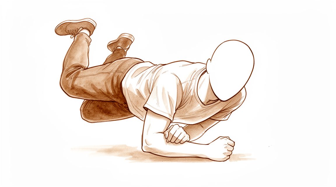
ஒரு ஓலெக்ரானோன் (முழங்கை முனை) முறிவின் அறுவை சிகிச்சைக்குப் பிறகு மீட்பு, ஒரு எளிய ஸ்லிங் மற்றும் செயலில் நீட்டிப்பின் படிப்படியான திரும்புவதன் மூலம் இயக்கத்தை மீட்டெடுக்கும் போது முழங்கைகள் பழுதுபார்க்கப்படுவதைப் பாதுகாக்கிறது.
மேட்டர் தனியார் மருத்துவமனை ராக்ஹாம்ப்டனில் டாக்டர் கீரன் ஹிர்பாரா நடத்திய திறந்த குறைப்பு மற்றும் உள் சரிசெய்தல் (ORIF) உடன் ஓலெக்ரானோன் (உங்கள் முழங்கையின் எலும்பு புள்ளி) முறிவின் அறுவை சிகிச்சைக்குப் பிறகு இந்த நெறிமுறை உங்கள் மீட்புக்கு வழிகாட்டுகிறது. இது உங்கள் வீட்டு உடற்பயிற்சி திட்டத்துடன் தொடங்குகிறது, அதைத் தொடர்ந்து உங்கள் உடற்பயிற்சி நிபுணர் அல்லது கை சிகிச்சையாளருக்காக எழுதப்பட்ட கட்டமைக்கப்பட்ட மருத்துவ நெறிமுறை. உங்கள் மறுவாழ்வு ஒருங்கிணைக்கப்படுவதற்காக இந்த பக்கத்தை அல்லது அதன் PDF ஐ உங்கள் முதல் சிகிச்சை வருகைக்கு கொண்டு வாருங்கள். உங்கள் மீட்பு எவ்வாறு முன்னேறுகிறது என்பதைப் பொறுத்து உங்கள் சிகிச்சையாளர் திட்டத்தை சரிசெய்யலாம்.
அறுவை சிகிச்சைக்குப் பிறகு உங்கள் காயத்தைப் பற்றி ஏதேனும் கவலைகள் இருந்தால், அறைகளைத் தொடர்பு கொள்ளுங்கள். காயத்தின் புகைப்படத்தை எடுத்து அதை மதிப்பாய்வு செய்ய மின்னஞ்சல் அனுப்புவது பெரும்பாலும் உதவியாக இருக்கும்.
எதிர்பார்ப்பது என்ன
ஒலெக்ரானோன் என்பது உங்கள் முழங்கையின் எலும்பு புள்ளியாகும். உங்கள் கையின் பின்புறத்தில் உள்ள பெரிய ட்ரைசெப்ஸ் தசை அதனுடன் இணைகிறது, மேலும் இது முழங்கையின் உள்ளே உள்ள கீல் மூட்டுவின் ஒரு பகுதியாகும். ஒலெக்ரானோன் உடைக்கும்போது, ட்ரைசெப்ஸின் இழுப்பு உடைந்த பகுதியை இழுக்க முனைகிறது, எனவே எலும்பு முறிவு அறுவை சிகிச்சையால் சரி செய்யப்படுகிறது, இது ஒரு கம்பி மற்றும் முள் "டென்ஷன் பேண்ட்" (ஒரு சுத்தமான, நேரான முறிவுக்கு) அல்லது ஒரு தட்டு மற்றும் திருகுகள் (மிகவும் சிதைந்த அல்லது கோண முறிவுக்கு). நீங்கள் முழங்கைகளை ஆரம்பத்தில் நகர்த்தத் தொடங்கும் அளவுக்கு எலும்பை ஒன்றாக உறுதியாகப் பிடிக்க இந்த உறுதிப்படுத்தல் கட்டப்பட்டுள்ளது.
இந்த புனர்வாழ்வின் முழு நோக்கம் ஒரு சமநிலைப்படுத்தும் செயலாகும். நீண்ட நேரம் சும்மா வைத்திருக்கும் முழங்கை மிக விரைவாக கடினமாகிவிடுகிறது, எனவே அதை ஆரம்பத்தில் நகர்த்த விரும்புகிறோம். ஆனால் முறிவைப் பிரிக்கும் அதே ட்ரைசெப்ஸ் தசைதான் உங்கள் முழங்கையை நிமிர்ந்து நிற்க வைக்கும் தசை, எனவே முதல் ஆறு வாரங்களுக்கு, முழங்கையை வளைத்து முன் காலை சுழற்றுவதில் சுதந்திரமாக வேலை செய்வதன் மூலம் சரிசெய்தலைப் பாதுகாக்கிறோம். எலும்புக்கு நெசவு நேரம் கிடைத்தவுடன், நிமிர்ந்து நிற்பது படிப்படியாக மீண்டும் அறிமுகப்படுத்தப்படுகிறது: முதலில் உங்கள் கையின் எடையைப் பயன்படுத்தி, பின்னர் எதிர்ப்புக்கு எதிராக மட்டுமே.
காயம், வீக்கம் மற்றும் வடுக்கள் மேலாண்மை, நடைமுறையில் பார்க்க காயம் பராமரிப்பு வழிகாட்டல்.
ஒலெக்ரானன் முறிவு நன்கு சரிசெய்யப்பட்ட பிறகும், பெரும்பாலான மக்கள் கடைசியாக ஒரு சிறிய நிரந்தர இழப்புடன் விடப்படுகிறார்கள் என்பதை ஆரம்பத்தில் இருந்தே அறிந்து கொள்வது மதிப்பு 1015 டிகிரி இது சாதாரணமானது, அன்றாட வாழ்க்கையில் அரிதாகவே கவனிக்கப்படுகிறது, மற்றும் ஏதேனும் தவறு நடந்ததற்கான அறிகுறியாக இல்லை.
முன்னெச்சரிக்கைகள் மற்றும் வரம்புகள்
செய்
- உங்கள் அணிய எளிய கயிறு உங்கள் உடற்பயிற்சிக்காக அதை கழற்றுவதன் மூலம் ஆறுதலையும் ஆதரவையும் பெறுவீர்கள். உங்களுக்கு கடினமான அடுக்கு அல்லது பிரேஸ் தேவையில்லை.
- வேலை வளைத்தல் முழங்கை மற்றும் சுழலும் முதுகெலும்பு (கையை மேலே / கையை கீழே) ஆரம்பத்தில் இருந்து, வசதியான வரம்புகளுக்குள்.
- உங்கள் கை, மணிக்கட்டு மற்றும் தோள்பட்டைகளை சுதந்திரமாக நகர்த்தி, உங்கள் பிடியை வலுவாக வைத்திருக்க ஒரு பந்தை அழுத்தவும்.
வேண்டாம்
- செய் தீவிரமாக நேராக இல்லை உங்கள் முழங்கை முதல் 6 வாரங்களுக்கு அதன் சொந்த தசை சக்தியின் கீழ்; அது ஈர்ப்பு சக்தியால் மட்டுமே நிமிர்ந்து நிற்கட்டும். செயலில் நிமிர்ந்து நிற்பது முழங்கை தசைகளை இழுக்கிறது மற்றும் எலும்பு முறிவை திசைதிருப்பலாம்.
- செய் இல்லை செய்வது எதிர்ப்பு அல்லது எடை கொண்ட நேராக்குதல் சுமார் வரை 3 மாதங்கள், உங்கள் சிகிச்சையாளர் அதை அனுமதிக்கும் போது.
- செய் இல்லை ஆரம்ப வாரங்களில், அறுவை சிகிச்சைக்கு உட்படுத்தப்பட்ட கையை உயர்த்தவும், தள்ளவும் அல்லது இழுக்கவும், அதன் வழியாக எடைகளை சுமக்கவும் வேண்டாம்.
உங்கள் முழங்கை மற்றும் முதுகெலும்புகளின் இயக்கத்தை மீட்டெடுப்பதற்கான உங்கள் கையேட்டில் உள்ள பயிற்சிகள் இவை. உங்கள் பயிற்சிகளுக்கு உங்கள் ஸ்லிங்கை எடுத்துக் கொள்ளுங்கள். டாக்டர் ஹிர்பாரா மற்றும் உங்கள் சிகிச்சையாளரின் வழிகாட்டுதலின்படி அவற்றைத் தொடங்குங்கள்.
உங்கள் பயிற்சிகள்
உங்கள் மருத்துவ நெறிமுறை
இந்த பக்கத்தின் மீதமுள்ள பகுதி திறந்த குறைப்பு மற்றும் உள் உறுதிப்படுத்தல் மூலம் சிகிச்சையளிக்கப்படும் ஒரு ஒலெக்ரானன் உடைந்த மருத்துவ நெறிமுறை ஆகும். இந்த பிரிவு உங்கள் உடலியல் சிகிச்சையாளர் அல்லது கை சிகிச்சையாளருக்கு வழங்கப்பட வேண்டும், மேலும் கீழே உள்ள ஒவ்வொரு கட்டமும் என்ன நடக்கிறது என்பதற்கான எளிய ஆங்கில விளக்கத்துடன் தொடங்குகிறது. இந்த நெறிமுறையை பொதுவான முழங்கை எலும்பு முறிவிலிருந்து வேறுபடுத்தும் மிக முக்கியமான விதி என்னவென்றால் த்ரிசெப்ஸ் பாதுகாப்பு: செயலில் நீட்டிப்பு ~ 6 வாரங்களுக்கு ஒத்திவைக்கப்படுகிறது மற்றும் நீட்டிப்பு ~ 3 மாதங்களுக்கு எதிர்க்கப்படுகிறது, ஏனெனில் ட்ரைசெப்ஸ் ஓலெக்ரானோனில் செருகப்பட்டு பத்திரத்தை ஏற்றுகிறது.
சிகிச்சைக்கு முன்னர், நோயாளியின் எக்ஸ்-ரே, அறுவை சிகிச்சை அறிக்கை மற்றும் கடந்தகால மருத்துவ வரலாறு ஆகியவற்றை சரிபார்த்து, சிகிச்சையளிக்கும் அறுவை சிகிச்சை நிபுணருடன் இணைப்பு கட்டுமானம் (டென்ஷன்-பேண்ட் வயரிங் Vs பிளேட்), அதன் நிலைத்தன்மை மற்றும் சாத்தியமான கணிப்பு ஆகியவற்றைப் பற்றி தொடர்பு கொள்ளுங்கள்.
வாரங்கள் 01: இயங்காத நிலை
முதல் வாரத்தில் மென்மையான திசுக்களை ஓய்வெடுப்பதில் கவனம் செலுத்தப்படுகிறது, வீக்கத்தை சரிசெய்து, மற்ற எல்லாவற்றையும் நகர்த்துகிறது. எளிய கயிறு சுமார் 90 டிகிரி, உடற்பயிற்சிக்காக எடுக்கப்பட்டது. இறுக்கமான பின்புற ஸ்பிளின்ட் அல்லது பிரேஸ் இல்லை.
உங்கள் பிசியோதெரபிஸ்ட்டுக்கு:
- இயலாமை: எளிய ஸ்லிங், ~ 90 ° க்கு முழங்கை, பயிற்சிகளுக்கு அகற்றப்பட்டது (கே. எச் ரத்துஃ பின்புற அடுக்கு அல்லது பிரேஸ் இல்லை).
- குறிக்கோள்கள்: மென்மையான திசு ஓய்வு; வீக்கம் கட்டுப்பாடு (உயர்வு, மென்மையான அழுத்தம், குளிர்).
- பயிற்சிகள்: செயலில் உள்ள கை, மணிக்கட்டு மற்றும் தோள்பட்டை இயக்க வரம்பு; மென்மையான செயலில் உள்ள முழங்கை மற்றும் முதுகெலும்பு இயக்க வரம்பு கட்டுமானம் நிலையானதாக இருந்தால் நாள் 2 3 முதல் தொடங்கலாம்.
- முன்னெச்சரிக்கைகள்: சுமை தாங்காத மேல் கால்; தூக்குதல், தள்ளுதல் அல்லது இழுத்தல் இல்லை; முழங்கை நீட்டிப்பு இல்லை.
- முன்னேற்றத்திற்கான அளவுகோல்கள்: காயம் மற்றும் வீக்கம் குறையும்.
வாரங்கள் 16: பாதுகாக்கப்பட்ட இயக்கம் (நீட்டிப்பு தடைசெய்யப்பட்டது)
இது திரிசெப்ஸ்-பாதுகாப்பு கட்டமாகும். வளைத்தல் சீராக முன்னேறப்படுகிறது, அதே நேரத்தில் நேராக்குதல் செயலற்றதாகவோ அல்லது ஈர்ப்பு சக்தியால் மட்டுமே செய்யப்படுகிறது, முழங்கையின் சொந்த நேராக்குதல் சக்தியின் கீழ் ஒருபோதும் இல்லை.
உங்கள் பிசியோதெரபிஸ்ட்டுக்கு:
- ROM உச்சவரம்புஃ மேம்பட்ட வளைவு தோராயமாக வாரத்திற்கு 10° ஒரு பதற்றம் இல்லாத வரம்பிற்குள்; இலக்கு 6 வாரங்களில் முழுமையான செயலற்ற நீட்டிப்பு, 120° வரை அழுத்தம் இல்லாத வளைவு, மற்றும் முழு முதுகெலும்பு சுழற்சி. செயலற்ற நீட்டிப்பு மட்டுமே: செயலில் நீட்டிப்பு இல்லை.
- பயிற்சிகள்: சுறுசுறுப்பான மற்றும் சுறுசுறுப்பான உதவியுடன் வளைவு மற்றும் புரோனேஷன்/சூபினேஷன்; செயலற்ற இயக்க வரம்பு ~ வாரம் 4 வரை தொடங்கலாம். கட்டி, பெரிஸ்காப்புலர் மற்றும் முன்கை ஐசோமெட்ரிக் மட்டும், முழங்கை நீட்டிப்பு அல்ல.
- ஆர்த்தோசிஸ்: கயிற்றில் கயிறு 4 வது வாரம் (வீட்டிற்கு வெளியே மற்றும் இரவில் அதன்பிறகு ஆறுதலுக்காக அணியப்படுகிறது).
- முன்னேற்றத்திற்கான அளவுகோல்கள்: முழு செயலற்ற நீட்டிப்பு, ~ 120 ° வரை வளைவு, முழு சுழற்சி; வலி ≤3/10.
வாரங்கள் 612: செயலில் நீட்டிப்பு அறிமுகப்படுத்தப்பட்டது
எலும்பு நெசவு செய்ய ஆறு வாரங்கள் கிடைத்தவுடன், சுறுசுறுப்பான நேராக்குதல் மீண்டும் அறிமுகப்படுத்தப்படுகிறது, ஈர்ப்பு சக்திக்கு எதிராக மட்டுமே மற்றும் கூடுதல் எடை இல்லாமல். வளைவு ஒரு முழு வளைவை நோக்கி முன்னேறப்படுகிறது, மேலும் கை வழியாக எடை தாங்கி மெதுவாக முன்னேறுகிறது.
உங்கள் பிசியோதெரபிஸ்ட்டுக்கு:
- குறிக்கோள்கள்: இயக்கம் வளைவு முடிக்க; மென்மையான செயலில் நீட்டிப்பு அறிமுகப்படுத்த.
- பயிற்சிகள்: சுறுசுறுப்பான முழங்கை நீட்டிப்பு ஈர்ப்பு சக்திக்கு எதிராக மட்டுமே 6 வாரத்தில் இருந்து; முன்கூட்டியே முழு வளைவு (முழு பதற்றம் இல்லாத வளைவை ~ 9 வாரத்தில் இலக்காகக் கொண்டது) எடை தாங்கி மேசை → சுவர் → நான்கு மடங்காக முன்னேற்றம் பொறுத்துக்கொள்ளப்படுகிறது. வளைவு / சுழற்சிக்கு முற்போக்கான எதிர்ப்பு உடற்பயிற்சி தொடங்கலாம், ஆனால் இன்னும் NO நீட்டிப்பு எதிர்ப்பு: இது 3 மாதங்கள் வரை நடைபெறுகிறது.
- முன்னேற்றத்திற்கான அளவுகோல்கள்: முழு செயலில் வில்; மோசமான நீட்டிப்பு தாமதம் இல்லை; ரேடியோகிராஃபிக் யூனியன்.
36 மாதங்கள்: நீட்டிப்பு எதிர்ப்பு மற்றும் திரும்ப
எலும்பு முறிவு ஒருங்கிணைந்த பிறகுதான் எதிர்ப்பு நேராகச் சரிசெய்தல் (டிரிசெப்ஸ் மற்றும் பழுதுபார்ப்பை நேரடியாகச் சுமக்கும் படி) தொடங்குகிறது.
உங்கள் பிசியோதெரபிஸ்ட்டுக்கு:
- எதிர்ப்பு முழங்கை நீட்டிப்பு தொடங்குகிறது ~3 மாதங்கள் (சிறிய எடைக்கு முன்னேறும் இசைக்குழுக்கள், ~0.52 கிலோ / 15 பவுண்டுகள், 3x/வாரம்).
- விளையாட்டு மற்றும் வேலை சார்ந்த பயிற்சிகள் ~4.5 மாதங்களிலிருந்து.
- தூக்குதல் மற்றும் கன உழைப்பு முழு வலிமையும் ஒற்றுமையும் அடைந்தவுடன் திரும்புதல், பொதுவாக 6 மாதங்களுக்குள்.
- ஹார்ட்வேர் அகற்றுதல் என்பது ஓலெக்ரானோன் (குறிப்பாக டென்ஷன்-பேண்ட் கம்பிகள், உடலில் அடிக்கடி அகற்றப்படும் உள்வைப்பு) இணைப்பிற்குப் பிறகு பொதுவாக செய்யப்படுகிறது, முக்கிய அல்லது எரிச்சலூட்டும் வன்பொருளின் நிவாரணத்திற்காக, வழக்கமான நடவடிக்கையாக அல்ல.
வேலை மற்றும் செயற்பாட்டிற்கு திரும்புதல்
சுய-பராமரிப்புக்காக கையை இலகுவாகப் பயன்படுத்துவது மேலே உள்ள முன்னெச்சரிக்கை நடவடிக்கைகளுக்குள் ஆரம்பத்திலிருந்தே பரவாயில்லை, ஆனால் முழங்கைகளை சுமக்கும் எதையும் அதன் முறைக்கு காத்திருக்கிறது. வளைத்தல் மற்றும் முன்கை சுழற்சி பொதுவாக ஆறு வாரங்களுக்குள் வசதியாக உணர்கிறது; செயலில் நேராக்குதல் ஆறு வாரங்களில் இருந்து திரும்புகிறது மற்றும் அடுத்த வாரங்களில் சீராக மேம்படுகிறது.
கனமான கோரிக்கைகள்ஃ சுமார் மூன்று மாதங்கள் வரை எதிர்ப்பு அல்லது எடையுள்ள நேராக்குதல் இல்லை, சுமார் நான்கரை மாதங்களிலிருந்து விளையாட்டு மற்றும் வேலை-குறிப்பிட்ட பயிற்சி, மற்றும் உங்கள் வலிமை மற்றும் எலும்பு குணமடைந்தவுடன் பொதுவாக சுமார் ஆறு மாதங்களில் தூக்குதல் மற்றும் கனமான கை வேலைக்கு திரும்புதல். மேசை அடிப்படையிலான மற்றும் ஒளி கடமைகள் வழக்கமாக மிக விரைவாக மீண்டும் தொடங்கலாம்; உங்கள் மதிப்பாய்வில் டாக்டர் ஹிர்பாராவுடன் நேரத்தைப் பற்றி விவாதிக்கவும், ஏனெனில் இது உங்கள் வேலை மற்றும் எந்தக் கைக்கு அறுவை சிகிச்சை செய்யப்பட்டது என்பதைப் பொறுத்தது. உங்கள் மதிப்பாய்வில் உறுதிப்படுத்தப்பட்டபடி, நீங்கள் ஸ்லிங்கிலிருந்து வெளியேறி, இரு கைகளாலும் பாதுகாப்பாக வாகனத்தை கட்டுப்படுத்த முடியும்.
கடைசி 1015 டிகிரி நேராக ஒரு சிறிய நிரந்தர இழப்பு எதிர்பார்க்க வேண்டும் என்பதை நினைவில் கொள்ளுங்கள்; இது சாதாரணமானது மற்றும் பொதுவாக தினசரி செயல்பாட்டில் எந்த விளைவையும் ஏற்படுத்தாது.
உங்கள் நெறிமுறை பிறகு
இந்த நெறிமுறை நடைமுறையின் பொதுவான மீட்பு ஆலோசனையுடன் இணைந்து செயல்படுகிறதுஃ அறுவை சிகிச்சைக்குப் பிந்தைய வலியை நிர்வகித்தல் மற்றும் காயம் பராமரிப்பு. காயம் பற்றி, பார்க்கவும் முதுகுவலி முறிவுமேலே உள்ள படிப்படியான திட்டம் ஒலெக்ரானோன் ORIF க்குப் பிறகு வெளியிடப்பட்ட மறுவாழ்வு நெறிமுறைகளுடன் ஒத்துப்போகிறது, மேலும் உங்கள் முழங்கை எவ்வாறு முன்னேறுகிறது என்பதைப் பொறுத்து உங்கள் இயற்பியல் சிகிச்சையாளர் அல்லது கை சிகிச்சையாளர் உங்கள் தொடர்ச்சியான மீட்புக்கு தனித்தனியாக வழிகாட்டுகிறார்.
Evidence & references
Olecranon Fracture — Open Reduction Internal Fixation (ORIF) — Post-operative Rehabilitation
Topic scope: Post-operative rehabilitation after ORIF of a displaced olecranon fracture — tension-band wiring (TBW) for simple transverse patterns, or plate-and-screw fixation for comminuted / oblique / Monteggia-type / proximal-ulna patterns. The olecranon fracture is an intra-articular fracture of the extensor mechanism: the fixation construct must resist the triceps pull that distracts the fragment, and the rehabilitation is built around protecting that construct while preventing the stiffness to which the elbow is highly prone.
Defining principle (the inverse of an arthrolysis): unlike a stiff-elbow release, here there is a fixation construct to protect, and the deforming force is the triceps. So active and resisted elbow EXTENSION — and lifting / pushing — are restricted early, while flexion and forearm rotation are advanced relatively freely to prevent stiffness. Dr Hirpara's practice override: the operated elbow is rested in a simple sling, not a posterior splint or brace; active extension is withheld for 0–6 weeks (protecting the tension band / triceps insertion), resisted extension is delayed to ~3 months, flexion is advanced ~10°/week, and the patient is counselled to expect a permanent 10–15° loss of terminal extension.
Evidence-strength flag: MODERATE-to-STRONG. Two concordant institutional physiotherapy protocols (Brigham & Women's Hospital; The Christ Hospital / Rao) give explicit phase timelines, and the biomechanical rationale — anatomic reduction plus stable fixation to permit early motion of an intra-articular fracture — is universally agreed. Consensus is strong on early protected motion + delayed active / resisted extension and triceps loading. The evidence is weaker (equipoise-level) on TBW vs plate, and on operative vs non-operative management in the elderly.
Phased rehabilitation timeline
| Phase | Window | Sling / immobilisation | ROM / use | Strengthening | Criteria to progress |
|---|---|---|---|---|---|
| I — Immobilisation | Weeks 0–1 | Simple sling at ~90° (KH — no posterior splint/brace), off for exercises | Active hand/wrist/shoulder ROM; gentle active elbow + forearm ROM from day 2–3 if stable; NO active extension | — (oedema control) | Wound and swelling settling |
| II — Protected motion, extension restricted | Weeks 1–6 | Wean sling at week 4 (out-of-house + night thereafter) | Flexion advanced ~10°/week in a tension-free zone; passive extension only; PROM may begin ~wk 4; goal full passive extension + flexion to 120° + full rotation by 6 wk | Cuff / periscapular / forearm isometrics only | Full passive extension, flexion ~120°, full rotation; pain ≤3/10 |
| III — Active extension introduced | Weeks 6–12 | Discontinued | Active extension against gravity only from wk 6; advance flexion to full (full tension-free arc by ~wk 9); progress weight-bearing tabletop → wall → quadruped | PRE to flexion/rotation; still NO resisted extension | Full active arc; no worsening extension lag; radiographic union |
| IV — Resisted extension + return | 3–6 months | — | Return to lifting/loading staged | Resisted extension from ~3 months (bands → light weights 1–5 lb, 3×/wk); sport/job-specific ~4.5 mo | Full strength + union; lifting/heavy labour ~6 mo |
Evidence by theme
The triceps-pull rule (the rehab-defining constraint) — Consensus, strong biomechanical rationale
The triceps inserts on the olecranon; active and resisted extension distract the fracture and load the tension band. Active extension against gravity is therefore deferred to ~6 weeks and resisted extension to ~3 months. This is the single most important rule distinguishing olecranon ORIF from a generic elbow-fracture rehabilitation, and both published protocols enforce it (The Christ Hospital / Rao explicitly prohibit active extension for the first six weeks).
Early protected motion to prevent stiffness — Strong consensus
There is universal agreement that an intra-articular extensor-mechanism fracture needs early motion to prevent stiffness; how early depends on construct stability and surgeon confidence. Brigham permits active ROM from day 2–3; the more conservative Christ / Rao protocol protects the first weeks with no active extension. The tension throughout is "prevent stiffness" versus "protect the triceps repair," resolved by advancing flexion and rotation freely while restricting extension.
Construct choice — TBW vs plate — Moderate / equipoise
Both constructs aim to be stable enough for early motion. TBW is simpler and adequate for simple transverse fractures but carries the highest symptomatic-hardware / removal rate and can lose compression in comminuted or osteoporotic bone. Plate fixation better controls comminution, oblique and diaphyseal-extension patterns; Hume & Wiss's randomized study favoured plate over TBW for comminuted patterns, while Anderson et al. reported a mean ~13.5° flexion contracture after plating. Rehabilitation phases are broadly the same — a more stable construct simply allows the surgeon to liberalise motion sooner.
Operative vs non-operative in the elderly — Moderate (RCT, equipoise)
A prospective RCT of displaced olecranon fractures in elderly patients was stopped early for a high operative complication rate, with comparable patient-reported outcomes — shifting practice toward non-operative management and earlier free mobilisation in low-demand older patients. Geriatric locking plates reduce fixation failure in osteoporotic bone but still carry meaningful complication and implant-failure rates.
Expected residual deficit — Moderate (corpus, observational)
Even with stable fixation and early motion, isolated olecranon fractures lose ~10–15° of terminal extension on average. Patients should be counselled that this is the expected norm, not a complication.
Hardware removal — Moderate (observational)
Olecranon hardware — especially TBW K-wires / wires — is the most commonly removed implant in the body; prominent subcutaneous hardware drives a high secondary-removal rate (up to roughly half of TBW cases in some series). Removal is typically performed after union (often ≥4–6 months) and largely for symptom relief, not as a routine staged step. Plate fixation has fewer wire-irritation issues but a non-trivial removal rate.
Evidence strength flags (summary)
- STRONG / CONSENSUS: early protected motion to prevent stiffness; the triceps-pull rule deferring active extension to ~6 weeks and resisted extension to ~3 months (biomechanical rationale
- two concordant institutional protocols).
- MODERATE: TBW vs plate construct choice (Hume & Wiss; Anderson ~13.5° contracture after plating); operative vs non-operative in the elderly (RCT stopped early for operative complications); expected 10–15° terminal-extension loss; high TBW hardware-removal rate.
- CONSENSUS / EXPERT: the precise phase timings (drawn from surgeon patient-guidance protocols rather than a rehabilitation RCT); the simple-sling immobilisation (KH practice override).
CITATIONS
RAG corpus (180,000+ Orthopaedic articles)
- Chalidis BE, Sachinis NC, Samoladas EP, Dimitriou CG, Pournaras JD. Is tension band wiring technique the "gold standard" for the treatment of olecranon fractures? A long-term functional outcome study. J Orthop Surg Res. 2008;3:9. DOI: 10.1186/1749-799X-3-9
- Hume MC, Wiss DA. Olecranon fractures: a clinical and radiographic comparison of tension band wiring and plate fixation (randomized comparison; plate favoured for comminuted patterns). (Cited in retrieved corpus text.)
- Anderson et al. — mean ~13.5° flexion contracture after olecranon plate fixation (retrieved corpus text).
- Prospective RCT, operative vs non-operative management of displaced olecranon fractures in the elderly — stopped early for a high rate of complications in the operative group, comparable patient-reported outcomes (retrieved corpus text).
- Geriatric olecranon locking-plate case series — major/minor complication and implant-failure rates (J Shoulder Elbow Surg, retrieved corpus text).
- Retrieved corpus text: isolated olecranon fractures lose an average of ~10–15° of terminal extension despite stable fixation and early ROM; anatomic reduction and early range of motion to restore functional elbow motion and strength; high re-operation / hardware-removal rate after TBW.
Published rehabilitation protocols (URLs)
- The Christ Hospital / Rao — "Olecranon ORIF Physical Therapy Protocol." https://www.thechristhospital.com/landingpages/Documents/Rao%20PT%20Protocols/Operative/Elbow/Rao%20Olecranon%20ORIF%20r1.pdf
- Brigham & Women's Hospital, Department of Rehabilitation Services — "Elbow Fracture Post-Op (Radial Head / Olecranon ORIF) Hand Therapy Guideline" (2021). https://www.brighamandwomens.org/assets/BWH/patients-and-families/rehabilitation-services/pdfs/elbow-fracture-orif-hand-therapy-protocol.pdf
- Physiopedia — "Olecranon Fracture" (general background). https://www.physio-pedia.com/Olecranon_Fracture


