钙化性肌腱炎
Patients › Rehabilitation
Recovery after arthroscopic excision of a calcific deposit, from early movement to full activity.
本康复方案涵盖由 Kieran Hirpara 医生在罗克汉普顿 Mater 私立医院进行的关节镜下钙化沉积物切除术后的康复:即通过微创(钥匙孔)手术从肩袖肌腱中清除钙质沉积,有时同时进行肩峰下减压术,以为肌腱腾出更多空间。请在您首次就诊物理治疗时带上本页或其 PDF,以便您的康复保持协调一致。您的康复将由物理治疗师根据肩部的恢复情况,按下列各阶段为您逐步推进。
如果您在手术后对伤口有任何顾虑,请与诊室联系。拍下伤口照片并通过电子邮件发送以供查看,通常会有所帮助。
如果切除沉积物需要进行肩袖修补(沉积物有时会在肌腱上留下需要缝合闭合的缺损),您的恢复将改为遵循肩袖修补的规则,此时肩袖修补方案的优先级高于本页。若此情况适用于您,Hirpara 医生会在手术后告知您。
您可以期待什么
两种路径都是早期活动路径。除非肩袖需要修补,否则没有任何缝合部位需要数月的保护;治疗的目标是消除疼痛的来源,而康复的目标是平复治疗后的急性发作,保持肩部活动以免其僵硬,然后重建力量。
- 在关节镜下切除术后,会提供一个吊带,仅供舒适之用。它只需短暂佩戴,通常为数天,很少超过两周,并且应尽可能少戴。您无需戴着它睡觉。恢复到完全、不受限制的活动通常约需三个月。
佩戴吊带时请勿驾车。驾车通常从约两周后恢复,前提是您已脱离吊带、感觉舒适,并能安全地完成紧急刹车。
无论采用哪种治疗,肩部都可能需要时间才能完全平复。不适往往是分阶段而非一次性地改善,手术后可能需要数月(偶尔长达九个月)治疗前的症状才会完全消退。稳步改善,而非即刻的舒适,才是预期的过程。
这是通过若干个小切口进行的微创日间手术。钙化沉积物位于肩袖肌腱内并被清除,同时往往还会进行肩峰下减压术以给肌腱更多空间。敷料通常在约两天后即可去除,伤口会在术后约一周至十天的首次复诊时进行检查。
第一阶段——早期活动(第0–2周)
您从手术中醒来时,手臂会置于吊带内,但吊带仅供舒适之用:请尽量少戴,多数人在数天内即可脱离吊带。从一开始,您就可以在肩部高度以下不受限制地使用手臂。将手臂举过肩部高度是允许且安全的,尽管起初会感到不适;每天数次,用健侧手臂帮助将术侧手臂举过肩部高度作为轻柔的伸展,以免肩部僵硬。在最初几周内,术侧手臂请避免携带超过约两公斤的物品,否则会引起疼痛。请尽快开始您的练习,每项动作以10次为目标,每天三次。请在练习前服用止痛药,并用冰敷以缓解不适。佩戴吊带时请勿驾车。
致您的物理治疗师:
目标
- 平复术后疼痛与肿胀
- 早期恢复活动范围:首要任务是防止僵硬,钙化性肌腱炎患者尤易发生僵硬
- 在肩部高度以下正常使用手臂
处理措施
- 吊带仅供舒适之用;在舒适允许的情况下尽快脱离,通常在数天内
- 从第一天起在肩部高度以下不受限制地主动使用手臂
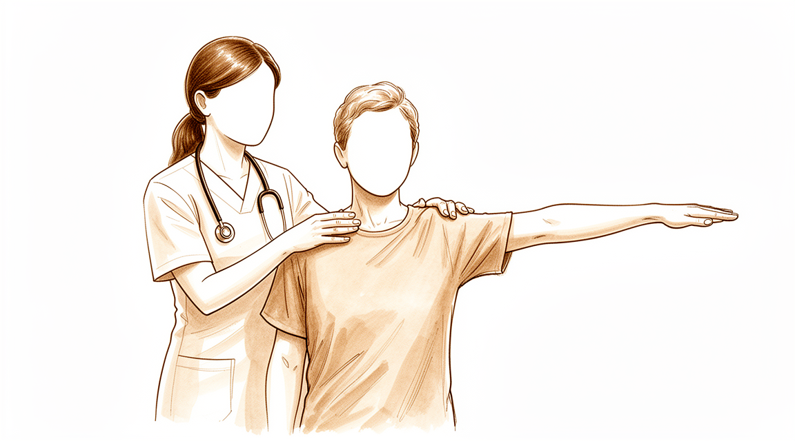
- 在可耐受的范围内主动上举至肩部高度以上
- 每天数次(使用另一只手臂)进行肩部高度以上的被动及主动辅助上举,以防止僵硬
- 家庭练习方案每项动作10次,每天三次
- 练习前给予镇痛;根据需要冷疗以缓解疼痛
注意事项
- 术侧手臂不得携带或提举超过约两公斤的物品
- 佩戴吊带时不得驾车
进阶标准
- 首次术后复诊时伤口检查满意
- 已脱离吊带并能在肩部高度以下舒适地使用手臂
第二阶段——恢复您的活动范围(第2–8周)
您将在约两到三周时于诊室接受复诊,届时会检查您的伤口和被动活动范围。本阶段的重点是活动范围:推进您的前向上举伸展,并加入向侧方的动作,由物理治疗给予进阶指导。典型目标是在六周时能主动将手臂抬至水平位,并在六周时使辅助(被动)活动范围(前向、侧向及旋转)恢复正常。驾车可从约两周后恢复,前提是您已脱离吊带、感觉舒适,并能安全地完成紧急刹车。
致您的物理治疗师:
目标
- 六周时主动前屈和外展达到水平位
- 六周时被动前屈、外展和外旋恢复正常
- 能独立完成日常活动
处理措施
- 推进被动及主动辅助前屈;引入并推进外展
- 在舒适允许的情况下进阶至各平面的主动活动范围
- 继续肩胛骨定位及姿势练习
- 继续在治疗前镇痛,并根据个人偏好在伸展前后热敷或冰敷
注意事项
- 在活动范围恢复期间保持轻量提举;进阶仍以症状为导向
- 伸展至明显不适是可以接受的;强行的、剧烈疼痛的伸展则不可取
进阶标准
- 被动活动范围达到或接近正常
- 主动上举达到水平位或更佳,且疼痛正在平复
第三阶段——力量训练与恢复完全活动(第8–16周)
您通常会在约八周时再次接受复诊。在活动范围恢复后,康复转向锻炼肩袖力量,通常在物理治疗师的监督下进行,并自由地在肩部高度以上使用手臂。目标是在约十二周时实现完全的主动前向上举和外展。仅切除沉积物后的恢复通常约需三个月,之后不再有任何限制;如果肩袖需要修补,恢复期会更长(通常约五个月),并改为遵循肩袖修补方案。如果部分酸痛持续到这一时间点之后也无需担忧:在这项手术后,术前症状可能需要长达九个月才能完全平复,而总体趋势则稳步朝着正确的方向发展。
致您的物理治疗师:
目标
- 约十二周时完全的主动前屈和外展
- 逐步恢复肩袖及肩胛骨的力量与耐力
- 约三个月时恢复完全、不受限制的活动
处理措施
- 从八周起进行渐进性肩袖力量训练:从等长练习进阶至弹力带及轻重量练习,低负荷、较高次数
- 推进在肩部高度以上主动使用手臂
- 在第十二至十六周期间,在可耐受的范围内推进针对健身、工作及运动的专项负荷
注意事项
- 力量训练不应以牺牲活动范围为代价;全程持续进行活动度练习
- 逐步增加重负荷及过头负荷;一旦出现疼痛发作,即意味着后退一步
进阶标准
- 完全的主动活动范围,力量正在恢复,症状持续平复
- 进展良好时从常规随访中出院,通常在约八至十六周之间
完成您的方案之后
上述各阶段改编自本手术已发表的患者指南和康复方案:伦敦肩部合作组织(The London Shoulder Partnership)的钙化沉积物切除术康复方案、ShoulderDoc(英国)关于钙化性肌腱炎手术的患者指南,以及 Kevin Ko 医生关于关节镜下切除术的患者指南。各周数范围为典型值而非固定值,您后续的康复将由物理治疗师与本诊所协作,根据您肩部的恢复情况为您个别指导。本页与本诊所的一般恢复建议配合使用:请参见术后疼痛管理和伤口护理。关于疾病本身以及这些治疗的原理,请参见钙化性肌腱炎。本方案背后的证据(自然病程、针刺抽吸术以及手术切除相关文献)总结于证据部分,可从本页顶部以 PDF 形式获取。
Evidence & references
Calcific Tendinitis of the Rotator Cuff — Staged Management & Post-operative Rehabilitation (Arthroscopic Excision)
Topic scope: (A) the natural history and stepped non-operative management of rotator-cuff calcific tendinitis (rest/analgesia → barbotage ± subacromial steroid → ESWT), and (B) post-operative rehabilitation after arthroscopic excision of the calcific deposit (± subacromial decompression; the cuff-repair pathway defers to the rotator-cuff-repair protocol).
Defining principle of the surgical rehab here: arthroscopic excision removes the source of pain and does not, by itself, create a construct that needs months of protection — provided the rotator cuff is left intact. So (like a debridement/decompression, and unlike a cuff repair) the rehab is an early-movement pathway: short sling for comfort only, unrestricted use below shoulder height from day one, assisted elevation to prevent stiffness, strengthening from ~8 weeks. The single branch point is whether removing the deposit left a tendon defect that needed repair — if so, the recovery converts to the slower, protected rotator-cuff-repair pathway.
A. NATURAL HISTORY & NON-OPERATIVE MANAGEMENT
Natural history (self-limiting in most)
Rotator-cuff calcific tendinitis is self-limiting in the majority: after a variable quiescent period the deposit enters a resorptive phase (peripheral vascularisation + phagocytosis), and spontaneous resorption occurs in roughly two-thirds of cases within 1–2 years [Uhthoff & Loehr; Chianca 2018 review]. This underpins a non-operative-first approach and explains why post-operative residual calcium that "dissolves spontaneously" does not harm the outcome.
Stepped non-operative interventions
- Analgesia / activity modification / physiotherapy — first line; many settle as the deposit resorbs. Consensus.
- Ultrasound-guided barbotage (needling + lavage), usually + subacromial corticosteroid — the best-supported interventional option. A systematic review of 908 patients and subsequent meta-analyses favour barbotage for medium-term pain/function; barbotage + subacromial steroid improves Constant–Murley score and reduces deposit size vs steroid alone. Notably, clinical improvement is NOT dependent on how much calcium is aspirated — perforating the deposit to trigger resorption is the active mechanism. Moderate–strong (SR/RCT).
- Extracorporeal shock-wave therapy (ESWT) — reduces deposit size and pain; broadly comparable to barbotage in several comparisons. Moderate (RCT).
B. POST-OPERATIVE REHABILITATION (arthroscopic excision ± subacromial decompression)
Surgery is reserved for deposits recalcitrant to adequate non-operative care. The operation locates and removes the deposit from within the cuff tendon, often with a subacromial decompression for room. Key surgical-outcome facts that shape the rehab:
- Preserving cuff integrity while removing as much deposit as possible gives good-to-excellent results in ~90% and avoids iatrogenic tendon defects [arthroscopic excision series].
- Complete vs near-complete removal gives equivalent outcomes — residual calcium resorbs spontaneously afterwards; the surgeon need not chase every fleck at the cost of the tendon.
- Arthroscopic decompression WITHOUT cuff repair is a validated strategy with good outcomes where the residual defect is not repaired [Bone & Joint 2023].
- Symptom settling is gradual — significant pain relief and ROM gains are the norm, but the pre-operative symptoms can take up to ~9 months to fade fully; recovery to unrestricted activity after excision alone is ~3 months.
Phased post-op timeline (no cuff repair)
| Phase | Window | Sling | ROM / use | Strengthening | Notes |
|---|---|---|---|---|---|
| I — Early movement | Week 0–2 | Comfort only, days (rarely > 2 wk), off ASAP | Unrestricted use below shoulder height from day 1; assisted elevation above shoulder height several × daily to prevent stiffness | — | Settle post-op flare; calcific patients are stiffness-prone → motion is the priority. ≤ ~2 kg, no driving while in sling |
| II — Regaining range | Week 2–8 | Off | Progress active elevation; restore full passive + active ROM | Begin gentle as pain allows | Most regain comfortable range through this window |
| III — Strengthening / return | Week 8–16 | Off | Full active elevation goal by ~12 wk | Cuff + scapular strengthening from ~8 wk, isometric → band/light weight; advance work/sport loading wk 12–16 | Full unrestricted activity ~3 months; discharge ~8–16 wk |
Branch point — if a rotator cuff repair was required: recovery converts to the protected rotator-cuff-repair pathway (sling ~6 wk, ROM restrictions, strengthening deferred), typically ~5 months total. The surgeon confirms post-operatively which pathway applies.
C. KEY CONTROVERSIES / EVIDENCE QUALITY
- Surgery is a last resort — given high spontaneous-resorption rates and effective barbotage/ESWT, excision is reserved for genuinely recalcitrant cases. Strong rationale.
- How much to remove / whether to repair the defect. Equivalent outcomes for complete vs partial removal, and viable decompression-without-repair, mean the surgeon balances deposit clearance against tendon integrity intra-operatively — which in turn decides the rehab pathway. Moderate.
- The post-op rehab protocol itself is consensus/expert, drawn from surgeon patient-guidance protocols rather than a rehab RCT — phase timings are typical, not trial-derived.
D. EVIDENCE STRENGTH FLAGS (summary)
- MODERATE–STRONG (SR / RCT): barbotage ± steroid for non-operative calcific tendinitis (908-patient SR; barbotage + steroid > steroid alone); ESWT efficacy.
- MODERATE (cohorts): arthroscopic excision outcomes (~90% good-excellent; equivalence of complete vs partial removal; decompression without repair, Bone & Joint 2023); spontaneous resorption ~2/3 within 1–2 years.
- WEAK / CONSENSUS: the post-operative rehabilitation protocol (surgeon patient-guidance documents; no defining rehab RCT).
CITATIONS
RAG corpus (180,000+ Orthopaedic articles) — adjacent rotator-cuff evidence
- Predictors of failure of non-operative treatment of chronic symptomatic rotator-cuff disease (2013 Neer Award). J Shoulder Elbow Surg. 2016. DOI: 10.1016/j.jse.2016.04.030
- Arthroscopic rotator cuff repair: scientific rationale, surgical technique, early clinical results. J Shoulder Elbow Surg. 2010. DOI: 10.1016/j.jse.2009.12.012
- Early versus delayed rehabilitation after arthroscopic rotator cuff repair. Knee Surg Sports Traumatol Arthrosc. 2024. DOI: 10.1002/ksa.12129
- Speed of recovery after arthroscopic rotator cuff repair. J Shoulder Elbow Surg. 2017. DOI: 10.1016/j.jse.2016.11.002
- (The corpus is thin on calcific-tendinitis-specific rehab; the evidence base below is the calcific literature + published surgeon protocols.)
Calcific tendinitis literature (URLs)
- Ultrasound-guided barbotage for calcific tendonitis of the shoulder: a systematic review including 908 patients (DARE). https://www.ncbi.nlm.nih.gov/books/NBK241935/
- Determining the efficacy of barbotage for pain relief in calcific tendinitis. JSES Int / ScienceDirect. 2024. https://pmc.ncbi.nlm.nih.gov/articles/PMC11401591/
- Needling and lavage in rotator-cuff calcific tendinitis: ultrasound-guided technique. PMC. 2024. https://pmc.ncbi.nlm.nih.gov/articles/PMC10805427/
- Calcific tendinitis of the rotator cuff: a review (natural history, phases, resorption). PMC. https://pmc.ncbi.nlm.nih.gov/articles/PMC3749672/
- Recovery pattern after arthroscopic treatment for calcific tendinitis of the shoulder. Orthop Traumatol Surg Res. 2020. https://www.sciencedirect.com/science/article/pii/S1877056820301043
- Arthroscopic decompression of calcific tendinitis without cuff repair. Bone Joint J. 2023. https://boneandjoint.org.uk/Article/10.1302/0301-620X.105B6.BJJ-2022-1137.R1
- Arthroscopic treatment of calcific tendonitis (preserve cuff, ~90% good-excellent; residuals resorb). PMC. https://pmc.ncbi.nlm.nih.gov/articles/PMC4044535/
Published rehab protocols (patient-guidance — basis for the phase structure)
- The London Shoulder Partnership — Calcific Deposit Excision Rehabilitation. http://thelondonshoulderpartnership.co.uk/shoulder/shoulder-rehabilitation/calcific-deposit-excision-rehabilitation/
- Ko K. Arthroscopic Excision of Calcific Tendonitis — What Can I Expect? (OPA Orthopedics). https://www.kevinkomd.com/pdf/calcific-tendonitis.pdf
- Funk L. Surgery for Calcific Tendinitis. ShoulderDoc. https://shoulderdoc.co.uk/pages/surgery-for-calcific-tendinitis


