肩关节镜
Patients › Rehabilitation
Recovery after simple or diagnostic shoulder arthroscopy with no repair — an early-mobilisation fast pathway.
本方案涵盖基兰·希尔帕拉(Kieran Hirpara)医生在罗克汉普顿 Mater 私人医院进行的单纯或诊断性肩关节镜手术后的康复:即关节镜下探查、冲洗或清理(清创)但未进行任何修复的微创手术。由于无需保护修复部位,这是恢复最快的肩关节手术之一:目标是早期活动并尽快恢复正常生活。请将此页面或其 PDF 文件带给您的首次物理治疗就诊,以确保康复过程协调一致。
本方案适用于未进行修复的关节镜手术:仅进行清创、冲洗或诊断性评估。 如果在您的关节镜手术中进行了修复、减压或稳定,请遵循该手术的康复方案:例如,如果您的肩袖进行了修复,请遵循肩袖修复方案。如果您不确定进行了何种操作,请在继续康复前查阅您的手术记录或致电医生办公室确认。
如果您对术后伤口有任何疑虑,请联系医生办公室。拍摄伤口照片并通过电子邮件发送以供审查通常很有帮助。
预期情况
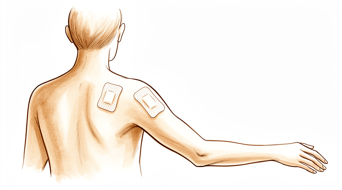
关节镜手术且未进行修复后,肩关节内部无需特别保护,因此没有严格的运动限制;早期即可开始活动,并根据舒适度而非日历时间逐步推进。提供肩带仅用于舒适目的:大多数人仅在术后头一两天使用,并在第一周内完全停用。佩戴肩带期间请勿驾驶。
简要过程如下:
- 第一阶段 — 早期活动与恢复 — 大约前两周
- 第二阶段 — 恢复全范围活动并开始力量训练 — 第2–6周
- 第三阶段 — 恢复全面活动 — 第6周起
大多数人可在数天至一周内恢复办公室工作,并在停用肩带、疼痛缓解且能自信控制车辆后的一至三周内恢复驾驶。较重的体力劳动和运动通常需要更长时间,一般在六至十二周之间,具体取决于肩部所承受的需求。肩关节在恢复期间常会持续数周的轻度酸痛;这属正常现象,随着活动度和力量的恢复会逐渐改善。
第一阶段 — 早期活动与恢复(第0–2周)
最初两周的重点是让肩部逐渐恢复稳定,同时保持其活动能力。仅在有助于舒适时(通常为第一天或第二天)使用吊带,并尽可能将其取下;您无需在睡觉时佩戴。从一开始就应自由活动手部、手腕和肘部,并在舒适允许的范围内,使用手臂进行进食、洗漱和穿衣等轻度日常活动。肩部轻柔活动应立即开始:进行钟摆运动及辅助运动,并随着肩部耐受情况逐步过渡到主动运动。冰敷和简单的止痛措施有助于使锻炼过程更加舒适。您的伤口敷料仅防溅而非防水;请尽量保持其干燥,敷料通常在术后十至十二天拆除。
致物理治疗师:
目标
- 控制疼痛和肿胀
- 早期活动范围,从辅助运动逐步过渡到主动运动,以耐受为度
- 实现轻度日常生活活动的独立
- 在最初几天内逐步停用吊带
管理措施
- 吊带仅用于舒适目的:鼓励在最初几天内逐步停用,最迟在一至两周内完全停用
- 钟摆运动;辅助主动活动范围训练(使用滑轮、手杖或魔杖),随着耐受情况逐步过渡到所有平面的主动活动范围训练
- 手部、手腕和肘部自由活动;在舒适范围内进行握力训练
- 肩胛骨稳定训练及姿势矫正训练
- 在舒适允许的范围内,进行轻柔的肩袖肌群和三角肌等长收缩训练
- 采用冷疗和镇痛措施以支持锻炼计划
注意事项
- 佩戴吊带期间禁止驾驶
- 活动范围根据舒适度逐步增加:若出现尖锐或持续性疼痛,应减轻强度,而非强行坚持
- 保持敷料干燥直至约10–12天拆除;若出现过度红肿或渗出,请报告
进阶标准
- 已停用吊带,且能舒适地进行轻度日常活动
- 疼痛已得到充分控制,能够进行主动活动范围训练
第二阶段——恢复完全活动度并启动力量训练(第2–6周)
随着肩部逐渐稳定,重点转向恢复完全的活动度并开始力量训练。主动活动度向各个方向的完全范围逐步推进,阻力训练也轻柔地开始,根据舒适度从等长收缩过渡到针对肩袖和肩胛骨肌肉的弹力带练习。如果尚未进行,大多数人会在第一或第二周恢复久坐办公工作;一旦摘下吊带、疼痛缓解且患者有信心在紧急情况下控制车辆,即可恢复驾驶(通常在术后一至三周内)。在物理治疗师的指导下,较轻的休闲活动在此期间恢复。
致物理治疗师:
目标
- 在各个平面达到完全或接近完全的主动活动度
- 开始对肩袖和肩胛稳定肌进行渐进式力量训练
- 恢复正常日常生活、工作和驾驶
管理
- 在各个平面逐步推进主动活动度至完全范围;典型的中间目标为前屈超过140–160°,外旋超过40–60°
- 从等长收缩过渡到弹力带肩袖训练(中立位附近的内旋和外旋),根据耐受情况逐步进阶
- 肩胛骨强化:耸肩、后缩、前伸和下沉练习逐步增加阻力
- 大约从第4周开始,进行轻负荷等张力量训练,使用低重量和高重复次数,以舒适度为准
- 针对任何残留的关节囊紧张进行手法治疗和拉伸,包括必要时进行后关节囊拉伸
注意事项
- 力量训练保持在舒适范围内,不应引发随后持续存在的疼痛
- 逐步增加外展位(90/90体位)下的负重旋转:仅在主动作旋转练习舒适时引入
- 在力量恢复期间,避免提举重物和用力的过头动作
进阶标准
- 主动活动度完全或接近完全,且疼痛轻微
- 弹力带和轻负荷力量训练耐受良好,无症状加重
第三阶段 — 恢复全面活动(第6周起)
最终阶段是逐步恢复较重的举重、体力劳动、健身房训练和体育运动。常规力量训练通常可在约6周后重新开始,从轻负荷开始并稳步增加,而 overhead(过顶)或对抗性运动通常在6至12周之间恢复,具体取决于运动类型以及肩部的恢复情况。康复完成的标志是肩部舒适、活动范围完全恢复且力量自信;大多数人约在3个月时能恢复所有想做的活动,此后任何残留的酸痛感会进一步缓解。
致物理治疗师:
目标
- 完全无痛的活动范围
- 恢复工作、运动所需的力量、耐力和信心
- 逐步恢复重体力劳动、健身房训练和体育运动
管理
- 从第6周左右开始进展至常规抗阻训练,随着控制能力的提高,从器械训练过渡到自由重量训练
- 根据耐受情况开展离心训练和闭链训练
- 针对特定运动的体能训练,包括在相关情况下分阶段的投掷或过顶训练计划
- 将肩袖强化训练限制在每周约3次,以避免过度负荷导致的肌腱病变
注意事项
- 进展应以症状为导向:如果疼痛随负荷增加或持续存在,则需减少负荷
- 恢复对抗性或过顶运动需等待完全无痛的活动范围和足够的力量
进展标准
- 完全无痛的活动范围,且力量与健侧相当,足以胜任目标活动
- 能完成特定运动或工作相关的任务且不诱发症状
术后方案
上述阶段改编自以下来源发表的简单及诊断性肩关节镜术后康复方案:Jorge Chahla 博士(鲁什大学医学中心)、Benedict Nwachukwu 博士(特殊外科医院)、Blake Obrock 博士(阿马里洛骨科运动医学)以及英国皇家国立骨科医院发布的诊断性肩关节镜患者指南。周数范围为典型区间,而非固定值;您的康复计划由您的物理治疗师根据肩部恢复情况,与诊所合作进行个体化指导。本页面与诊所的一般术后恢复建议配合使用:请参阅 管理术后疼痛 和 伤口护理。关于手术本身,请参阅 肩关节镜。本方案背后的证据(早期活动的理论依据、安慰剂对照手术试验以及所借鉴的已发表康复方案)已在证据部分进行总结,可从本页顶部下载 PDF 版本。
Evidence & references
Shoulder Arthroscopy (Diagnostic / Debridement / Washout) — Post-operative Rehabilitation
Topic scope: Post-operative rehabilitation after a generic keyhole shoulder arthroscopy in which nothing was repaired — diagnostic assessment, washout (lavage), debridement of degenerate tissue, removal of loose bodies, and isolated subacromial decompression or distal clavicle excision. Specific repair or reconstruction procedures have their own protocols that take priority — rotator-cuff repair, labral/instability stabilisation (anterior-Bankart, posterior-stabilisation, Latarjet), capsular release, biceps tenodesis and AC-joint stabilisation each convert to a slower, construct-protecting pathway. This page is the default keyhole pathway used only when the operation note confirms no repair was performed.
Defining principle of the rehab here: when nothing is repaired there is no construct to protect, so the rehab is an early-motion pathway — a sling for comfort only (days, not weeks), unrestricted use below shoulder height from day one, motion progressed on comfort rather than the calendar, and strengthening as soon as range and pain allow. The single branch point is whether anything was actually repaired or stabilised; if it was, recovery converts to that procedure's protected protocol. Unlike a cuff repair or a labral repair, there is no healing tissue that early movement can disrupt, so the usual risks of early motion (re-tear, construct failure) do not apply — the main thing early motion prevents here is post-operative stiffness.
The operation and why the rehab is fast
A keyhole (arthroscopic) shoulder operation in this scope involves looking inside the joint and subacromial space through small portals and doing one or more of: confirming a diagnosis, washing out the joint, trimming (debriding) frayed labrum, degenerate cuff or inflamed bursa, removing loose bodies, or shaving bone in a subacromial decompression or distal clavicle excision. None of these creates a repair that must heal under protection. That is the central fact that separates this pathway from cuff repair, stabilisation and the other audited protocols: the tissue is either removed or simply inspected, so the post-operative soreness — not a healing construct — is what paces recovery.
Because of this, recovery is among the quickest of any shoulder operation. Most people are back to desk-based work within days to a week, out of the sling within the first week, driving within one to three weeks once the sling is off and they can control the car confidently, and back to heavier manual work and sport somewhere between six and twelve weeks depending on the demands placed on the shoulder.
Evidence by theme
1. Early motion is the goal — there is no construct to protect
The case for early movement here is largely a mechanistic one rather than one settled by a dedicated trial: with no repair to disrupt, the only thing prolonged immobilisation achieves is avoidable stiffness, discomfort and delayed return to activity. The closest high-quality evidence comes by analogy from the cuff-repair literature, where — even with a real construct to protect — randomised trials and meta-analyses show early controlled motion does not increase re-tear and tends to reduce stiffness (number-needed-to-harm for re-tear in the order of several hundred). If early motion is safe when a repair is present, it is plainly safe when there is nothing to protect. Mechanistic + analogous moderate evidence; no debridement-specific RCT.
2. The procedures themselves: a candid note on efficacy
Two landmark placebo-controlled surgical trials bear directly on the commonest reason a no-repair arthroscopy is done — subacromial pain:
- FIMPACT (BMJ 2018) — a double-blind trial of 210 patients randomised to arthroscopic subacromial decompression, diagnostic arthroscopy (placebo surgery), or exercise therapy. At 24 months decompression gave no benefit over diagnostic arthroscopy; both surgical arms improved, but no more than each other. Strong (placebo-controlled RCT).
- CSAW (Lancet 2018) — a three-arm placebo-controlled UK trial reaching the same conclusion: decompression was no better than investigational (diagnostic) arthroscopy, and the small edge of either over no-treatment was not clinically important. Strong (placebo-controlled RCT).
The honest reading is that for subacromial pain the surgical element adds little over diagnostic arthroscopy or structured exercise — which reinforces why, when this operation is done, the rehabilitation (early motion, restoring strength and confidence) carries much of the recovery. A longer-term single RCT (Magnussen-class, 10-year follow-up, in the corpus) did favour decompression over therapy alone, so practice remains individualised — but the placebo-controlled data are the higher tier.
3. Debridement of degenerate tissue — limited, old evidence
Arthroscopic debridement of irreparable degenerative cuff lesions (Burkhart, J Bone Joint Surg 1995, in the corpus) can relieve pain and restore functional "force-couple" mechanics in selected patients, but the evidence base is small, old and uncontrolled. Debridement and washout are best understood as symptom-directed measures, not structural repairs — which again places the weight of recovery on rehabilitation rather than on a healing construct. Weak (historical case series).
4. The phased protocol is consensus, drawn from published surgeon protocols
The phase structure below is expert/consensus, compiled from published patient-guidance protocols for general/diagnostic shoulder arthroscopy and debridement (Chahla – Rush; Nwachukwu – HSS; Obrock; Royal National Orthopaedic Hospital). There is no rehabilitation RCT defining the optimal regimen for a no-repair arthroscopy; the week ranges are typical, not trial-derived. Weak/consensus.
Phased post-op timeline (no repair performed)
| Phase | Window | Sling | ROM / use | Strengthening | Notes |
|---|---|---|---|---|---|
| I — Early movement & settling | Week 0–2 | Comfort only, days (rarely > 1–2 wk), off ASAP; no sleeping in it | Free hand/wrist/elbow + light ADLs from day 1; pendulums and assisted ROM progressing to active ROM as comfort allows | Scapular setting; gentle cuff/deltoid isometrics as comfort allows | Settle the post-op flare. No driving while in the sling. Dressings off ~10–12 days |
| II — Restore movement, start strength | Week 2–6 | Off | Progress active ROM in all planes toward full (interim targets ~140–160° flexion, 40–60° ER) | Isometric → elastic-band cuff + scapular work; light isotonic from ~wk 4 | Desk work + driving once sling off, pain settled, confident to control the car (typically wk 1–3) |
| III — Return to full activity | Week 6 onward | Off | Maintain full, pain-free ROM | Conventional resistance training from ~wk 6; eccentric/closed-chain; sport-specific conditioning. Cap heavy cuff loading at ~3×/week | Heavier manual work & sport return ~6–12 wk by demand; most back to everything by ~3 months |
Branch point — if anything was repaired or stabilised: recovery converts to that procedure's protected protocol (e.g. rotator-cuff repair — sling ~6 weeks, restricted ROM, deferred strengthening, ~5 months total; or the relevant stabilisation/capsular-release pathway). The operation note and the rooms confirm which pathway applies.
Key controversies / evidence quality
- Does the surgery help at all (for subacromial pain)? Two placebo-controlled RCTs (FIMPACT, CSAW) found decompression no better than diagnostic arthroscopy, and arthroscopy little better than exercise. This is the strongest evidence in the topic — and it argues that, where a no-repair arthroscopy is performed, good rehabilitation is doing much of the work. Strong.
- Debridement evidence is thin and dated. The supportive data (e.g. Burkhart 1995) are small, uncontrolled case series; debridement is symptom-directed, not curative. Weak.
- The rehab protocol itself is consensus, not trial-derived. No RCT defines the optimal regimen after a no-repair arthroscopy; phase timings are typical surgeon-protocol values, and recovery is individualised by the treating physiotherapist. Weak/consensus.
- Safety of early motion is inferred, not directly tested here. It rests on a sound mechanism (nothing to protect) reinforced by analogy to the cuff-repair early-motion trials, rather than a debridement-specific RCT. Mechanistic + analogous moderate.
The evidence base for this generic pathway is genuinely limited. The high-quality data (placebo-controlled trials) speak to whether the operation helps, not to how best to rehabilitate it; the rehabilitation guidance is consensus-level. This is stated plainly because it is the honest position.
Evidence-strength flags (summary)
- STRONG (placebo-controlled RCT): subacromial decompression gives no benefit over diagnostic arthroscopy — FIMPACT (BMJ 2018), CSAW (Lancet 2018).
- MODERATE (analogous RCT/MA): safety of early controlled motion (extrapolated from cuff-repair early-motion trials — early motion does not raise re-tear and reduces stiffness even when a construct is present).
- WEAK (historical case series): arthroscopic debridement of irreparable degenerative cuff lesions (Burkhart 1995).
- WEAK / CONSENSUS: the post-operative rehabilitation protocol itself (published surgeon patient-guidance documents; no defining rehab RCT).
- SAFETY NOTE (rare complication): glenohumeral chondrolysis has been linked to post-arthroscopic intra-articular continuous bupivacaine infusion and to thermal capsulorrhaphy — a reason such adjuncts are avoided, not a reflection on standard debridement.
CITATIONS
RAG corpus (180,000+ Orthopaedic articles)
- Burkhart SS. Débridement of degenerative, irreparable lesions of the rotator cuff. J Bone Joint Surg Am. 1995. DOI: 10.2106/00004623-199506000-00006
- Magnussen R, et al. Subacromial decompression yields a better clinical outcome than therapy alone: a prospective randomized study with minimum 10-year follow-up. Am J Sports Med. 2018. DOI: 10.1177/0363546518755759
- Bailie DS, Ellenbecker TS. Severe chondrolysis after shoulder arthroscopy associated with continuous bupivacaine infusion. Arthroscopy. 2009. DOI: 10.1016/j.arthro.2009.08.024
- (The corpus is thin on no-repair / diagnostic-arthroscopy rehabilitation specifically; the higher-tier evidence below comes from the placebo-controlled surgical trials and published surgeon protocols.)
Literature (URLs)
- FIMPACT — Paavola M, et al. Subacromial decompression versus diagnostic arthroscopy for shoulder impingement: randomised, placebo surgery controlled clinical trial. BMJ. 2018. https://pmc.ncbi.nlm.nih.gov/articles/PMC6052435/
- CSAW — Beard DJ, et al. Arthroscopic subacromial decompression for subacromial shoulder pain: a multicentre, pragmatic, placebo-controlled, three-group, randomised surgical trial. Lancet. 2018. https://www.thelancet.com/journals/lancet/article/PIIS0140-6736(17)32457-1/fulltext
Published rehab protocols (patient-guidance — basis for the phase structure)
- Chahla J. Shoulder Arthroscopy Debridement — Rehab Protocol. Rush University Medical Center. https://www.jorgechahlamd.com/wp-content/uploads/2021/08/Shoulder-Arthroscopy-Debridement.pdf
- Nwachukwu BU. Post-Operative Shoulder Arthroscopy Debridement Rehab Protocol. Hospital for Special Surgery. https://manhattansportsdoc.com/post-operative-shoulder-arthroscopy-debridement-rehab-protocol/
- Obrock B. Post-operative Rehabilitation Protocol — General Shoulder Arthroscopy (Debridement, Subacromial Decompression, and/or Distal Clavicle Resection). https://www.drblakeobrock.com/pdf/obrock-shoulder-arthroscopy-general.pdf
- Royal National Orthopaedic Hospital. A Patient's Guide to Diagnostic Shoulder Arthroscopy. https://www.rnoh.nhs.uk/patients-and-visitors/patient-information-guides/diagnostic-shoulder-arthroscopy-patients-guide


