Flexor Sheath Ganglion Excision Info Evidence
Last reviewed
A quick, motion-led recovery plan after excision of a flexor tendon sheath ganglion (volar retinacular cyst) at the base of a finger, protecting a small palm wound while you start gentle finger movement within days, then add scar care and grip as the wound settles.
This protocol guides your recovery after a small operation to remove a flexor sheath ganglion — a firm cyst at the base of a finger on the palm side of the hand — with Dr Kieran Hirpara at Mater Private Hospital Rockhampton. It begins with your home exercise program, followed by the structured clinical protocol written for your hand therapist — bring this page or its PDF to your first therapy visit so your rehabilitation stays coordinated. Your hand therapist may adjust the plan depending on how your recovery progresses.
If you have any concerns about your wound after surgery, get in touch with the rooms. It is often helpful to take a photo of the wound and email it for review.
What to expect
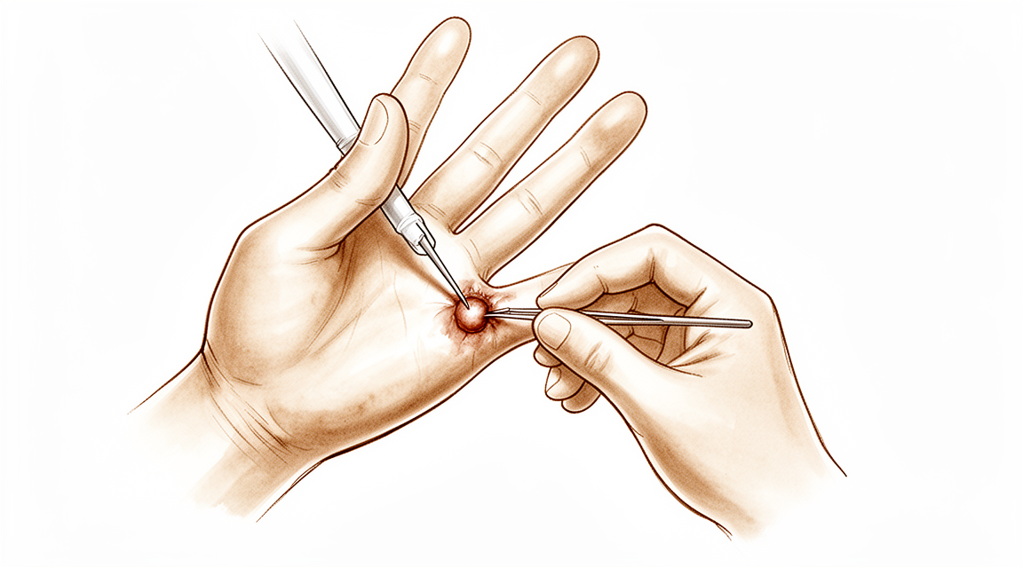
A flexor sheath ganglion (also called a volar retinacular cyst) is a small, firm — often tender — lump, usually only a few millimetres across, that grows from the sheath the flexor tendons run through, right at the base of a finger on the palm side (commonly over the firm band called the A1 pulley, at the crease where the finger meets the palm). It is fixed to the sheath and does not move when you bend the finger. It is a completely benign (non-cancerous) lump, and one of the more common ganglions in the hand and wrist.
The operation is a small day-case excision. Through a short zig-zag cut in the palm, Dr Hirpara removes the cyst together with a small cuff of the tendon sheath it grew from. The two small nerves and blood vessels that run along each side of the finger are carefully protected. The sheath itself is not repaired — leaving it open is deliberate and does not weaken the finger — and the skin is closed with sutures.
Because nothing inside the finger has to be protected while it heals, this is a quick recovery — weeks, not months. The plan is simple: protect the small palm wound, control swelling, and start gentle finger movement within a few days so the finger does not stiffen and the tendons do not stick down to the healing scar. Once the wound is healed, scar massage and desensitisation settle the area, and grip is built back up. A bit of tingling or tenderness around the wound is common early on as the small skin nerves recover, and it usually settles over the following weeks.
Precautions and limitations
- Keep the dressing clean and dry until the wound is healed and the sutures are out (usually around day 10–14). There is no plaster and usually no splint — just a soft dressing.
- Do start gentle finger movement within the first few days — bending, straightening and tendon glides — to prevent stiffness and tendon sticking.
- Do keep the hand elevated and use it for light everyday tasks within comfort.
- Do NOT do heavy gripping, lifting or forceful pinching until the wound has settled (around two to three weeks).
- Do NOT massage the scar or soak the hand until the wound is fully healed.
- Do NOT drive while the dressing stops you gripping the wheel safely — usually for about the first week.
For wound, swelling and scar management, see the practice's wound care guidance.
Your exercises
Kieran Hirpara 4.0
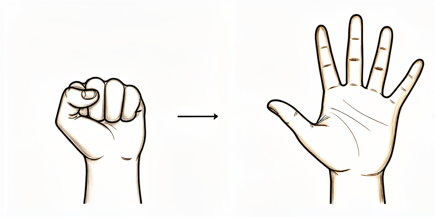
Finger movement (gentle fist and straighten)
Within the first few days, gently make a soft fist and then straighten your fingers out fully, moving slowly and only as far as is comfortable. Early movement keeps the finger from stiffening and stops the tendons sticking to the healing wound. Do not force it — let the dressing and a little discomfort be your guide.
10 times, 3–4 times a day, within comfort
Kieran Hirpara 4.0
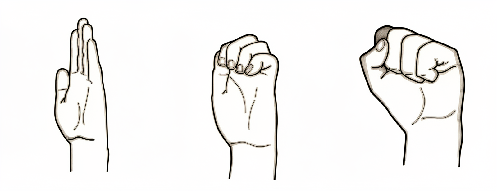
Tendon glides (hook, fist, straight)
Move your fingers through three shapes: a hook (bend the tips and middle joints but keep the knuckles straight, like a claw), a full fist, then a flat straight hand. These positions slide the flexor tendons through the surgical area so they keep gliding freely rather than sticking. Keep each one gentle and pain-free.
5 of each position, 3–4 times a day
Swelling control
Keep your hand raised up above the level of your heart as much as you can in the first week, and gently open and close the fingers to pump the swelling away. Less swelling means an easier, more comfortable finger and a freer recovery. A little swelling around the palm is normal early on.
Elevate often through the day; gentle finger pumps every hour or so while awake
Scar massage and desensitisation
Once the wound is fully healed and the sutures are out (usually after about two weeks), rub a little moisturiser into the scar with firm small circles for a few minutes, and stroke the area with different textures (a soft cloth, then a towel). This softens the scar and settles the tenderness and tingling that are common at first as the small skin nerves recover. Do NOT massage an open or unhealed wound.
A few minutes, 2–3 times a day, once fully healed
Kieran Hirpara 4.0
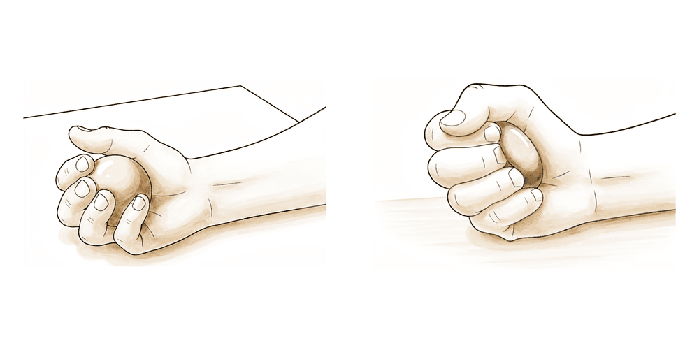
Grip strengthening
A LATER exercise — begin from around two to three weeks, once the wound has settled and your hand therapist is happy. Gently squeeze a soft ball or therapy putty, hold for a few seconds, then relax, building the effort up gradually. This rebuilds grip ready for full use. Stop if it is sharply painful over the wound.
10–15 squeezes, 2–3 times a day (from ~2–3 weeks)
These are the exercises from your handout. Start them only as guided by Dr Hirpara and your hand therapist, staying within whatever limits you have been given. The early exercises — gentle fist-and-straighten, tendon glides and swelling control — keep the finger moving and the tendons gliding from within the first few days, which is the single most important thing for a smooth recovery. Scar massage and desensitisation begin once the wound is fully healed, and grip strengthening belongs to a slightly later phase (from around two to three weeks). Stop anything that causes sharp pain over the wound.
Your clinical protocol
The rest of this page is the staged clinical protocol for rehabilitation after excision of a flexor sheath ganglion (volar retinacular cyst). This section is to be provided to your hand therapist, and each phase opens with a plain-English explanation of what is happening. This is an excision, not a repair — the tendon sheath is left open and there is no construct to protect — so the programme is an early-motion pathway built around wound protection, oedema control, tendon gliding to prevent adhesion, and scar/desensitisation work, not protected immobilisation.
Prior to treatment, check the operation report and past medical history, and liaise with the treating surgeon regarding the digit involved, the extent of sheath excision and the integrity of the digital neurovascular bundles. Dr Hirpara's excision is via a Bruner (zig-zag) palmar incision over the A1/proximal sheath, removing the cyst with a cuff of sheath; the sheath is not repaired and there is no immobilisation beyond a soft dressing. Transient digital-nerve paraesthesia is common and self-limiting.
Phase I — wound protection and early motion (week 0 to ~1)
The first week protects the small palm wound and gets the finger moving early so it does not stiffen or develop tendon adhesions. The hand is managed in a bulky soft dressing with no splint, kept elevated, with gentle active finger motion started within a few days.
For your hand therapist:
Education and precautions - Soft bulky dressing only, no splint; keep clean and dry until sutures out (~day 10–14) - Protect the wound from heavy load; light unloaded hand use within comfort - Counsel that transient digital-nerve paraesthesia / hypersensitivity around the wound is common and self-limiting
Management - Wound: surgical dressing as directed; monitor for infection - Oedema: elevation above heart level, gentle finger pumping, ice as needed - Exercises: gentle active finger AROM (gentle composite fist and full extension) and tendon glides (hook / fist / straight) started within a few days; active motion of the uninvolved digits, thumb and wrist; light functional use
Criteria to progress - Wound settling, no infection; comfortable early active arc; ready for full active/gentle passive motion as the wound permits
Phase II — full motion, oedema and scar work (week ~1 to 3)
From around one week, motion is progressed to full active and gentle passive range — full fist and full extension — and, once the wound is fully healed and sutures are out, scar massage and desensitisation begin. Oedema control continues.
For your hand therapist:
Assessments - Active and passive finger ROM (aim full fist and full extension); wound/scar status; swelling; digital-nerve sensitivity
Education and precautions - Progress to full active and gentle passive finger motion as comfort allows - Begin scar massage and desensitisation only once the wound is fully healed - Avoid heavy gripping and forceful pinching until the wound has settled
Management - Exercises: full composite fist and full extension; continued tendon glides; gentle passive stretch into any residual tightness - Scar: scar massage + textured desensitisation once healed; oedema management as needed
Criteria to progress - Full, pain-free active motion; wound healed; settling scar; ready to load
Phase III — strengthening and return (week ~3 to 6)
Once the wound is healed and motion is full (around three weeks), grip and pinch strengthening begins and is built up gradually towards full unrestricted use. Most patients return to full activity by around four to six weeks, with a routine follow-up at about two months.
For your hand therapist:
Assessments - Grip and pinch versus the other side; residual scar tenderness or sensitivity; functional / task-specific demands
Education and precautions - Begin grip and pinch strengthening from around 2–3 weeks once the wound has settled; build load gradually - Progress to full unrestricted use as comfort and strength allow
Management - Exercises: putty / soft-ball grip squeezes, pinch strengthening, progressive functional loading; continue any residual scar work and desensitisation - Discharge once motion is full and grip is comfortable and near-symmetrical; routine surgical follow-up at ~2 months - Refer back to the treating doctor if recovery plateaus, the scar stays markedly hypersensitive, or there is concern about recurrence
Criteria for full return - Full pain-free motion; comfortable grip and pinch; settled scar; able to meet work and activity demands
Getting back to work and activity
Light everyday hand use — eating, writing, light self-care — is encouraged from the start, within comfort, as long as it does not involve heavy gripping or forced pinching through the wound. Most people manage daily tasks within a few days. Driving usually resumes from about one week, once you can grip and control the wheel comfortably and are no longer limited by the dressing — confirmed with Dr Hirpara at your review.
Gripping and strengthening start from around two to three weeks, once the wound has settled, and are built up gradually. Full, unrestricted activity is usually reached by around four to six weeks. Office work can often resume within a few days to a week; heavier manual work follows the same staged progression as your grip returns. A routine follow-up is usually arranged at about two months.
After your protocol
This protocol works alongside the practice's general recovery advice — see managing post-operative pain, wound care and scar management. Because this ganglion sits at the base of the finger over the A1 pulley, the recovery has much in common with other small palm-side finger-base procedures such as trigger finger release. The phased plan above reflects published guidance after ganglion excision, and your ongoing recovery is guided individually by Dr Hirpara and your hand therapist according to how your finger progresses.
Evidence & references
Flexor Sheath Ganglion Excision — Lesion, Procedure Outcomes & Post-operative Rehabilitation (Volar Retinacular Cyst)
Topic scope: post-operative rehabilitation after surgical excision of a flexor tendon sheath ganglion (volar retinacular cyst / "seed" ganglion) at the base of a finger — a small, firm, often tender cyst arising from the flexor sheath, commonly over the A1 pulley at the metacarpophalangeal crease of the middle or ring finger. This is an excision, not a reconstruction: the cyst is removed with a small cuff of sheath, the sheath is not repaired, and there is no construct to protect — so the rehab is a brief early-motion pathway built around wound protection, oedema control, tendon gliding and scar/desensitisation work rather than months of protected healing.
Defining principle of the rehab here: a flexor sheath ganglion is a benign cyst tethered to the tendon sheath; excising it (with a cuff of sheath) removes the lesion without creating anything that needs to heal under protection. The sheath is meant to be left open — partial sheath excision does not weaken the digit or cause bowstringing at this level — so immediate, unrestricted light use and early gentle finger motion are the default. The therapy programme exists to keep the flexor tendons gliding through the healing palm wound so they do not adhere, to settle the transient digital-nerve hypersensitivity that commonly follows dissection between the neurovascular bundles, and to mature the scar — not to immobilise. The single branch point is wound healing: scar massage and grip loading wait until the wound is healed and sutures are out.
A. PROCEDURE OUTCOMES (excision of flexor sheath ganglion)
Excision of a flexor sheath ganglion is a small, reliable day-case hand operation: the great majority of patients are rendered symptom-free with a low recurrence rate, and the principal trade-off is the common but self-limiting transient digital-nerve paraesthesia from dissecting the cyst out from between the digital neurovascular bundles.
- Flexor sheath (volar retinacular) ganglions are a well-defined, common entity. They account for roughly 5–16% of ganglions of the hand and wrist, presenting as a small (typically 3–8 mm), firm, often tender nodule fixed to the flexor sheath at the digit base — classically over the A1/A2 pulley region — that does not move with the tendon [JAAOS 2022; JAAOS 1999; Hand Clin 2004]. Well-established (lesion nature).
- Surgical excision gives reliable symptom relief with low recurrence. Level-IV case series of flexor sheath / volar retinacular ganglion excision report durable resolution and low recurrence after complete excision of the cyst with a cuff of sheath; recurrence is the main long-term concern and is uncommon when the lesion and its sheath origin are fully removed [Hand 2007; J Hand Surg series; PMC surgical series]. Moderate (level-IV case series).
- Transient digital-nerve paraesthesia is the principal complication. Because the cyst sits immediately deep to, and is dissected free from, the digital nerves, temporary numbness or tingling in the finger is the commonest reported post-operative event; it is typically self-limiting and settles over weeks. True nerve injury is rare with careful protection of both bundles [Hand 2007; volar retinacular series; Medscape]. Moderate (case series + expert review).
- Partial excision of the sheath is biomechanically tolerated. Removing the cyst with a small cuff of the flexor sheath at the A1 level does not produce clinically significant bowstringing or weakness, which is the anatomical basis for not repairing the sheath and for an early-motion rehab without protected immobilisation [Hand Clin 2004 (palmar digital ganglia / A1–A2 origin)]. Mechanistic.
B. REHABILITATION / THERAPY EVIDENCE
There are no randomised rehab trials specific to flexor sheath ganglion excision; the post-operative programme is low-level / expert-consensus, but it is strikingly consistent across hand-therapy and surgical sources: dressing only (no splint), early gentle finger motion within days, tendon gliding to prevent adhesion, scar massage and desensitisation once healed, and return to full use by ~4–6 weeks.
- Dressing-only, no routine splinting. Aftercare guidance for ganglion (including flexor sheath) excision describes a soft dressing with no immobilisation, with the patient encouraged to move the finger early — there is no construct to protect, so splinting is not required and would risk stiffness [MSA aftercare; Medscape]. Weak / consensus.
- Early active finger motion and tendon gliding prevent stiffness and adhesion. Starting gentle active fist/extension and tendon glides within the first few days keeps the flexor tendons gliding through the palm wound so they do not adhere to the healing scar — the same adhesion-prevention rationale that underpins early-motion hand rehab generally. The benefit is mechanistic / consensus rather than trial-proven for this lesion [hand-therapy aftercare sources]. Weak (mechanism sound).
- Scar massage and desensitisation, started once healed, settle the wound and digital-nerve hypersensitivity. Palm scars at the digit base are prone to tenderness, and the transient digital-nerve paraesthesia from the dissection responds to graded desensitisation; both begin only after the wound is fully healed and sutures are out (~day 10–14) [MSA aftercare; Medscape]. Weak / consensus.
- Grip loading and full return are early. Because nothing is repaired, gripping and strengthening begin once the wound has settled (~2–3 weeks) and full unrestricted use is typically reached by ~4–6 weeks, with routine follow-up around two months — consistent across aftercare sources [MSA aftercare; PMC series; Medscape]. Weak / consensus.
Recovery trajectory (expected, evidence-anchored)
| Phase | Window | Restraint | Hand use / therapy focus | Strength / load | Notes |
|---|---|---|---|---|---|
| I — Wound protection & early motion | Week 0–1 | Soft dressing, no splint | Elevate above heart; gentle active fist + full extension and tendon glides within a few days; protect wound; light functional use | Light unloaded use only | Transient digital-nerve tingling is expected, not a complication |
| II — Full motion, oedema & scar work | Week ~1–3 | Heavy-grip avoidance | Progress to full active + gentle passive motion (full fist, full extension); scar massage + desensitisation once wound healed; oedema control | No forceful gripping/pinching until wound settled | Sutures out ~day 10–14; scar work only after full healing |
| III — Strengthening & return | Week ~3–6 | Restrictions lifted | Progress grip/pinch loading; task-specific use | Grip/pinch strengthening from ~2–3 wk; full unrestricted use by ~4–6 wk | Routine follow-up ~2 months; driving ~1 wk once gripping the wheel comfortably (surgeon discretion) |
(Phase windows mirror the precautions and return milestones in the patient protocol; they are typical guides, not trial-derived deadlines.)
C. KEY CONTROVERSIES / EVIDENCE QUALITY
- Lesion nature is well-established. The flexor sheath / volar retinacular ganglion is a recognised, characterised entity (firm, sheath-tethered, A1-pulley region, 5–16% of hand/wrist ganglions); its diagnosis and origin are not in dispute. Strong (descriptive).
- Excision outcomes are good but evidenced at level IV. Low recurrence and reliable symptom relief come from case series, not controlled trials — adequate for a small benign lesion, but the evidence tier is modest. Moderate (level-IV).
- Transient digital-nerve paraesthesia vs true nerve injury. Temporary tingling is common and self-limiting; framing it for patients up front avoids alarm, while careful intra-operative protection of both neurovascular bundles keeps true injury rare. Moderate.
- The rehab protocol is consensus, not trial-derived. No RCTs govern post-excision therapy; the dressing-only, early-motion, scar-care, ~4–6-week-return pathway is consistent across sources but rests on expert consensus and the general principles of early-motion hand rehab. Weak / consensus.
- Recurrence is the main long-term failure mode and is uncommon after complete excision of the cyst with its sheath origin; a residual or recurrent lump warrants reassessment rather than prolonged therapy. Moderate.
D. EVIDENCE STRENGTH FLAGS (summary)
- WELL-ESTABLISHED (descriptive): the nature, location and prevalence (5–16% of hand/wrist ganglions) of flexor sheath / volar retinacular ganglions.
- MODERATE (level-IV case series): reliable symptom relief and low recurrence after excision; transient digital-nerve paraesthesia as the principal, self-limiting complication; recurrence as the main long-term failure mode.
- WEAK / CONSENSUS: the dressing-only, early-motion, scar-care rehab programme and its phase timings (consistent across aftercare sources, mechanistically rationalised, but not trial-derived); ~4–6-week full return and ~1-week driving (surgeon discretion).
CITATIONS
RAG corpus (180,000+ Orthopaedic articles)
- Ganglions of the hand and wrist. J Am Acad Orthop Surg. 2022. DOI: 10.5435/jaaos-d-22-00105
- Ganglions of the hand and wrist. J Am Acad Orthop Surg. 1999. DOI: 10.5435/00124635-199907000-00003
- Surgical excision of flexor sheath ganglions of the hand: results and outcomes. Hand (N Y). 2007. DOI: 10.1007/s11552-007-9028-4
- Volar retinacular ganglions of the hand: a clinical series. J Hand Surg Am. 2011. DOI: 10.1016/j.jhsa.2011.05.013
- Palmar digital ganglia and the A1–A2 sheath origin. Hand Clin. 2004. DOI: 10.1016/j.hcl.2004.03.015
Flexor sheath ganglion / rehabilitation literature (URLs)
- Surgical excision of flexor sheath ganglions — case series (full text). PMC. https://pmc.ncbi.nlm.nih.gov/articles/PMC2527143/
- Volar retinacular ganglions of the hand. Journal of Hand Surgery (American). https://www.jhandsurg.org/article/S0363-5023(11)00627-7/abstract
- Ganglion cyst excision — post-operative aftercare (dressing-only, early motion, scar care, ~4–6 week return). Mississippi Sports & Arthritis (MSA) Hand Center. https://msapc.com/hand-center/aftercare/ganglion-cyst-excision/
- Ganglion treatment (surgical excision, recurrence and transient digital-nerve paraesthesia). Medscape. https://emedicine.medscape.com/article/1243525-treatment


