Hội chứng ống quay
Patients › Elbow
Radial tunnel syndrome — causes forearm pain, weakness straightening fingers, and is distinct from tennis elbow.
Những gì bạn đang cảm thấy
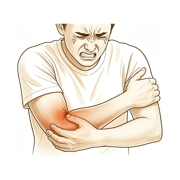
Bạn có thể đang trải qua cơn đau ở phần ngoài của cánh tay trên và cẳng tay. Sự khó chịu này xuất phát từ việc chèn ép một dây thần kinh gọi là dây thần kinh gian cốt sau. Hãy tưởng tượng dây thần kinh này như một sợi cáp chạy dọc phía sau cánh tay của bạn. Khi nó bị siết chặt trong đường hầm quay, nó sẽ gửi các tín hiệu cảm giác như một cơn đau âm ỉ sâu hoặc nhạy cảm.
Cơn đau thường trở nên nghiêm trọng hơn khi bạn sử dụng cánh tay. Bạn có thể nhận thấy nó bùng phát sau các hoạt động liên quan đến việc xoay cẳng tay hoặc nắm chặt đồ vật. Ví dụ, xoay tay nắm cửa, sử dụng tua vít hoặc nâng một túi đồ tạp hóa nặng có thể kích hoạt cảm giác này. Sự khó chịu cũng có thể rõ rệt hơn khi bạn vừa thức dậy vào buổi sáng.
Các hoạt động hàng ngày có thể trở nên khó khăn. Với tay ra sau lưng để cài áo ngực hoặc nhét áo vào quần có thể gây ra cơn đau nhói. Bạn có thể thấy khó ngủ ở bên cánh tay bị ảnh hưởng vì áp lực làm trầm trọng thêm dây thần kinh. Trong khi yếu cơ ít phổ biến hơn, một số người nhận thấy bàn tay của họ cảm thấy yếu hơn hoặc kém phối hợp hơn theo thời gian.
Điều quan trọng cần biết là tình trạng này hiếm gặp. Không có một xét nghiệm đơn lẻ nào khẳng định chắc chắn rằng bạn bị hội chứng đường hầm quay. Bác sĩ thường chẩn đoán dựa trên các triệu chứng và khám thực thể của bạn. Một số bệnh nhân thấy những thay đổi trên MRI, chẳng hạn như sưng trong các cơ được kiểm soát bởi dây thần kinh này. Tuy nhiên, những phát hiện này không phải lúc nào cũng xuất hiện.
Điều trị thường bắt đầu bằng các lựa chọn không phẫu thuật. Nghỉ ngơi, điều chỉnh hoạt động và vật lý trị liệu là những bước đầu tiên mà bác sĩ phẫu thuật của bạn có thể khuyên dùng. Nếu những biện pháp này không giúp ích sau một khoảng thời gian, giải phóng chèn ép bằng phẫu thuật có thể được xem xét. Thủ thuật này liên quan đến việc nới lỏng các vùng chặt chẽ xung quanh dây thần kinh để giảm áp lực.
Trải nghiệm của bạn có thể khác nhau. Một số người tìm thấy sự giảm nhẹ với chăm sóc bảo tồn, trong khi những người khác cần phẫu thuật. Mục tiêu là giảm đau và khôi phục chức năng để bạn có thể trở lại các hoạt động bình thường của mình. Hãy ghi lại những gì làm cho cơn đau của bạn tốt hơn hoặc tồi tệ hơn. Thông tin này giúp bác sĩ phẫu thuật của bạn điều chỉnh một kế hoạch phù hợp với bạn.
Những gì thực sự đang xảy ra
Hội chứng ống quay là một bệnh lý chèn ép dây thần kinh quay. Điều này có nghĩa là dây thần kinh quay bị ép hoặc chèn ép khi đi qua cẳng tay của bạn. Dây thần kinh quay là một bó mô lớn truyền tín hiệu từ não đến các cơ và da cánh tay của bạn. Khi bó mô này bị chèn ép, nó không thể truyền tải thông điệp một cách chính xác.
Dây thần kinh quay đi qua một ống hẹp, cấu tạo bởi mô xơ trong cẳng tay của bạn. Hãy tưởng tượng ống này giống như một ống tay áo chật hoặc một ống nước hẹp. Trong một số trường hợp, các cấu trúc xung quanh ống này đè lên dây thần kinh. Áp lực này gây kích thích dây thần kinh và dẫn đến đau, yếu hoặc tê ở cánh tay và bàn tay của bạn. Nguyên nhân chính xác của sự chèn ép này có thể khác nhau ở mỗi người.
Phần lớn thông tin về tình trạng này đến từ các nghiên cứu nhỏ hoặc báo cáo ca bệnh cá thể. Vì đây là một tình trạng hiếm gặp, không có nhiều bằng chứng lâm sàng chất lượng cao để hướng dẫn mọi quyết định. Đó là lý do tại sao bác sĩ phẫu thuật của bạn có thể dựa vào kinh nghiệm lâm sàng và các triệu chứng cụ thể của bạn để xác định hướng điều trị tốt nhất.
Quản lý không phẫu thuật là phương pháp điều trị đầu tay cho hội chứng ống quay. Điều này thường bao gồm nghỉ ngơi, điều chỉnh hoạt động và có thể bao gồm vật lý trị liệu để giảm áp lực lên dây thần kinh. Nhiều người tìm thấy sự giảm nhẹ triệu chứng với các biện pháp bảo tồn này.
Nếu các phương pháp điều trị không phẫu thuật không mang lại hiệu quả, giải phóng chèn ép bằng phẫu thuật là một lựa chọn khả thi cho các trường hợp kháng trị. Thủ thuật này liên quan đến việc nới lỏng các cấu trúc chặt chẽ xung quanh dây thần kinh để tạo thêm không gian cho nó. Nó thường được xem xét khi các triệu chứng vẫn tiếp diễn mặc dù đã thực hiện các phương pháp điều trị khác. Vẫn còn nhiều tranh cãi xoay quanh việc chẩn đoán và kết quả điều trị của hội chứng ống quay, đó là lý do tại sao việc hiểu rõ tình trạng cụ thể của bạn là rất quan trọng.
Những gì chúng tôi có thể làm về vấn đề này
Bác sĩ phẫu thuật của bạn có khả năng sẽ bắt đầu bằng việc quản lý không phẫu thuật như phương pháp điều trị đầu tay cho hội chứng đường quay. Cách tiếp cận này tập trung vào nghỉ ngơi và tránh gập khuỷu tay để giảm áp lực lên dây thần kinh. Hầu hết các trường hợp chèn ép dây thần kinh tại khuỷu tay sẽ cải thiện với việc chăm sóc bảo tồn này. Bạn nên dành đủ thời gian cho cách tiếp cận không phẫu thuật kéo dài này để phát huy tác dụng, vì nó được chỉ định trong hầu hết các trường hợp.
Nếu nghỉ ngơi đơn thuần không mang lại sự giảm đau, bác sĩ phẫu thuật của bạn có thể khuyến nghị các bài tập hoặc liệu pháp cụ thể. Mặc dù bằng chứng nhấn mạnh việc nghỉ ngơi và tránh vận động, vật lý trị liệu thường nhằm mục đích khôi phục chức năng bình thường mà không làm trầm trọng thêm tình trạng chèn ép. Mục tiêu là để dây thần kinh bị kích thích ổn định lại. Nhiều bệnh nhân nhận thấy rằng những thay đổi đơn giản trong các hoạt động hàng ngày và vận động nhẹ nhàng là đủ để kiểm soát các triệu chứng một cách hiệu quả.
Quản lý bằng thuốc có thể giúp kiểm soát cơn đau trong khi bạn hồi phục. Bác sĩ phẫu thuật của bạn có thể đề xuất các loại thuốc chống viêm để giảm sưng xung quanh dây thần kinh. Trong một số trường hợp, tiêm có thể được xem xét để cung cấp sự giảm đau có mục tiêu. Những phương pháp điều trị này nhằm mục đích làm dịu tình trạng viêm và giảm bớt khó chịu. Hiệu quả của các can thiệp này khác nhau, nhưng chúng thường được sử dụng để duy trì trong khoảng thời gian chờ đợi cho đến khi dây thần kinh tự lành.
Phẫu thuật chỉ được xem xét nếu hội chứng đường quay không đáp ứng với quản lý không phẫu thuật. Đây vẫn là một lựa chọn khả thi cho các trường hợp không cải thiện với chăm sóc bảo tồn. Bác sĩ phẫu thuật của bạn sẽ đánh giá xem giải phóng chèn ép bằng phẫu thuật có cần thiết hay không. Thủ thuật này liên quan đến việc giải phóng áp lực lên dây thần kinh để khôi phục chức năng bình thường. Nó thường được dành riêng cho các trường hợp bệnh lý chèn ép dây thần kinh quay cao kháng lại các phương pháp điều trị khác.
Nếu cần phẫu thuật, bác sĩ phẫu thuật của bạn sẽ thảo luận về cách tiếp cận tốt nhất cho tình trạng cụ thể của bạn. Thủ thuật nhằm mục đích giải phóng chèn ép dây thần kinh bằng cách tách đường hầm xơ dọc theo toàn bộ chiều dài của nó. Điều này giúp giảm bớt sự chèn ép gây ra cơn đau của bạn. Việc hồi phục sau phẫu thuật khác nhau, nhưng hầu hết bệnh nhân trải qua sự cải thiện đáng kể về các triệu chứng của họ. Bác sĩ phẫu thuật của bạn sẽ hướng dẫn bạn qua quá trình chăm sóc sau phẫu thuật để đảm bảo quá trình hồi phục suôn sẻ.
Cần lưu ý rằng hội chứng đường quay là một hội chứng đau do chèn ép dây thần kinh gian cốt sau ở phần gần của cẳng tay. Chẩn đoán dựa chủ yếu vào đánh giá lâm sàng, vì các xét nghiệm hình ảnh có thể không luôn luôn cho thấy các dấu hiệu rõ ràng. MRI có thể hữu ích trong việc xác định các thay đổi cơ liên quan đến tình trạng này. Tuy nhiên, bác sĩ phẫu thuật của bạn sẽ chủ yếu dựa vào các triệu chứng và khám thực thể của bạn để đưa ra quyết định điều trị.
Hầu hết các trường hợp chèn ép dây thần kinh sẽ cải thiện với điều trị không phẫu thuật hoặc phẫu thuật. Bác sĩ phẫu thuật của bạn sẽ điều chỉnh kế hoạch theo nhu cầu của bạn, bắt đầu với các lựa chọn ít xâm lấn nhất. Giao tiếp cởi mở với nhóm chăm sóc sức khỏe của bạn là chìa khóa để quản lý quá trình hồi phục của bạn. Bằng cách làm theo các bước được khuyến nghị, bạn có thể giải quyết nguyên nhân gốc rễ của cơn đau và trở lại các hoạt động bình thường của mình.
Những điều cần biết
Hội chứng đường quay là tình trạng chèn ép dây thần kinh quay ở cẳng tay. Tình trạng này hiếm gặp. Do tính chất hiếm gặp, hầu hết thông tin y khoa đều dựa trên các nghiên cứu quy mô nhỏ thay vì các thử nghiệm lâm sàng lớn. Vẫn còn tranh luận giữa các chuyên gia về cách chẩn đoán và hiệu quả của các phương pháp điều trị. Điều này có nghĩa là chưa có một tiêu chuẩn chẩn đoán duy nhất được chấp nhận rộng rãi.
Quản lý không phẫu thuật là phương pháp điều trị đầu tay cho hầu hết bệnh nhân. Nhiều bệnh nhân tìm thấy sự thuyên giảm mà không cần phẫu thuật. Nếu các triệu chứng của bạn không cải thiện với các biện pháp bảo tồn, thì giải phóng chèn ép bằng phẫu thuật là một lựa chọn khả thi. Điều này đặc biệt đúng nếu bạn bị chèn ép dây thần kinh quay ở vị trí cao và không đáp ứng với các phương pháp điều trị khác. Bác sĩ phẫu thuật của bạn có thể cần phải thăm dò cẩn thận toàn bộ chiều dài của đường hầm sợi bao quanh dây thần kinh để giảm áp lực.
Kết quả điều trị có thể khác nhau. Một số bệnh nhân hồi phục tốt, trong khi những người khác có thể tiếp tục có các triệu chứng. Do việc chẩn đoán phức tạp, kết quả không phải lúc nào cũng có thể dự đoán trước. Nếu cần phẫu thuật, điều quan trọng là phải chọn một bác sĩ phẫu thuật giàu kinh nghiệm. Một số kết quả không tốt đôi khi có thể được tránh bằng sự chú ý cẩn thận trong suốt quá trình phẫu thuật.
Nếu không được điều trị, các triệu chứng có thể kéo dài. Tuy nhiên, nhiều trường hợp sẽ tự thuyên giảm theo thời gian và với các biện pháp chăm sóc không phẫu thuật. Nếu bạn cần phẫu thuật, quá trình hồi phục là một tiến trình. Bạn nên mong đợi sự cải thiện dần dần trong vài tuần đến vài tháng. Đừng mong đợi sự thuyên giảm ngay lập tức. Mục tiêu là giảm đau và khôi phục chức năng.
Nói một cách trung thực là không phải trường hợp nào cũng khỏi hoàn toàn. Một số bệnh nhân có thể vẫn không hài lòng với kết quả điều trị. Nếu các triệu chứng quay trở lại hoặc kéo dài sau phẫu thuật lần đầu, phẫu thuật chỉnh sửa (revision surgery) có thể được xem xét. Tuy nhiên, kết quả từ phẫu thuật chỉnh sửa khó dự đoán hơn và thường kém hài lòng hơn so với cuộc phẫu thuật đầu tiên. Nguy cơ cần phẫu thuật lần hai nói chung là thấp đối với hầu hết bệnh nhân, nhưng có thể cao hơn ở những người dưới 50 tuổi hoặc những người có các tình trạng sức khỏe nhất định như bệnh thận mạn tính.
Tiên lượng của bạn phụ thuộc vào cách cơ thể bạn đáp ứng với điều trị. Hãy kiên nhẫn với quá trình này. Giao tiếp cởi mở với bác sĩ phẫu thuật của bạn về tiến độ hồi phục. Những kỳ vọng thực tế giúp bạn đối mặt với tình trạng này một cách tự tin.
Khi nào cần gặp bác sĩ
Hội chứng ống trụ ngoài là một tình trạng đau hiếm gặp do chèn ép dây thần kinh ở cẳng tay. Vì không có xét nghiệm chuẩn để xác nhận chẩn đoán này, việc đánh giá chuyên khoa là rất quan trọng. Hãy gặp bác sĩ đa khoa nếu bạn có cơn đau dai dẳng không cải thiện khi nghỉ ngơi. Hãy yêu cầu đánh giá bởi chuyên gia nếu bạn nhận thấy yếu cơ, mất ổn định, hoặc nếu các triệu chứng ảnh hưởng đến giấc ngủ hoặc công việc của bạn. Sự gia tăng đột ngột của các triệu chứng cũng cần được chú ý ngay lập tức. Mặc dù những thay đổi cơ có thể xuất hiện trên MRI, chỉ có bác sĩ lâm sàng mới có thể xác định hướng điều trị tốt nhất. Đánh giá sớm giúp tránh các thủ thuật không cần thiết và đảm bảo bạn nhận được sự chăm sóc phù hợp cho vấn đề thần kinh hiếm gặp này.
Evidence & references
Overview
- Radial tunnel syndrome is a compression neuropathy of the radial nerve [2].
- Most publications regarding uncommon upper extremity compression syndromes, including radial tunnel syndrome, are small retrospective series or case reports [2].
- Treatment decisions for uncommon upper extremity compression syndromes are not typically based on high levels of evidence [2].
- Nonsurgical management is the first-line treatment for radial tunnel syndrome [1].
- Surgical decompression is a viable option for refractory cases of radial tunnel syndrome [1].
- There is ongoing controversy regarding the diagnosis and outcomes of radial tunnel syndrome [1].
- High radial nerve entrapment neuropathy cases resistant to conservative treatment are advocated for surgical intervention [14].
- Surgical treatment for high radial nerve entrapment neuropathy requires dissecting the entire length of the fibrous tunnel [14].
Anatomy & Pathophysiology
- Bony encasement of the ulnar nerve can occur secondary to heterotopic ossification of the elbow [3].
- Chronic structural adaptations of the shoulder and elbow are correlated in professional baseball pitchers, but adaptations in shoulder strength or range of motion are not significantly related to chronic structural adaptations of the elbow [35].
- The humeral trochlea protrudes into the cubital tunnel during elbow flexion, causing dynamic morphologic changes in the ulnar nerve [38].
- Shoulder position increases ulnar nerve strain at the elbow in patients with cubital tunnel syndrome [45].
- Increased elbow flexion influences the intraneural blood flow of the ulnar nerve in patients with cubital tunnel syndrome [47].
- The mechanism of symptom provocation by the elbow flexion test in cubital tunnel syndrome cannot be explained simply by dynamic pressure in the cubital tunnel, suggesting other pathophysiological factors contribute [51].
- Ulnar nerve gliding is most severe during passive wrist movement in elbow flexion and forearm supination [53].
- A cadaveric study could not detect a definitive effect of elbow deformity (cubitus valgus/varus) on ulnar nerve strain or demonstrate the extent of acceptable clinical elbow deformity [57].
- The throwing elbow is a common source of nerve injuries due to the unique combination of anatomy, high forces, and sheer repetition associated with throwing sports [58].
- Dynamic ulnar nerve compression at the elbow can be caused by the anconeus epitrochlearis muscle, an uncommon disorder with pathophysiologic mechanisms that remain to be elucidated [64].
Classification
- Radial tunnel syndrome is defined as a pain syndrome caused by compression of the posterior interosseous nerve at the proximal forearm [11].
- Radial tunnel syndrome is considered an illness construct based on speculative pathophysiology with no verifiable pathophysiology or accepted reference standard for diagnosis [5].
- Radial tunnel syndrome and posterior interosseous nerve syndrome are viewed as a single condition presenting along a spectrum of nerve entrapment [26].
- Radial tunnel syndrome and posterior interosseous nerve syndrome are distinct entities within the review of history, anatomy, and clinical presentation [6].
- Radial tunnel syndrome is classified as an unusual compression neuropathy of the forearm [12].
- Radial tunnel syndrome is classified as an uncommon compression syndrome of the radial nerve [2].
- Radial neuropathies are rare compared to other entrapment neuropathies [4].
Clinical Presentation
- Radial tunnel syndrome is a pain syndrome caused by compression of the posterior interosseous nerve at the proximal forearm [11].
- Radial tunnel syndrome is an illness construct based on a speculative pathophysiology with no verifiable pathophysiology or accepted reference standard for diagnosis [5].
- Radial tunnel syndrome and posterior interosseous nerve syndrome are viewed as a single condition presenting along a spectrum of nerve entrapment [26].
- The deep branch of the radial nerve and the posterior interosseous nerve are distinct entities requiring consistent terminology distinction [26].
- Radial neuropathies are rare [4].
- Muscle denervation edema or atrophy along the distribution of the posterior interosseous nerve is the most common MR finding in radial tunnel syndrome [27].
Investigations
- Radial tunnel syndrome is an illness construct based on a speculative pathophysiology with no verifiable pathophysiology or accepted reference standard for diagnosis [5].
- Radial tunnel syndrome is a pain syndrome caused by compression of the posterior interosseous nerve at the proximal forearm [11].
- Muscle denervation edema or atrophy along the distribution of the posterior interosseous nerve is the most common MR finding in radial tunnel syndrome [27].
- MRI has emerged as the imaging modality of choice for the evaluation of elbow pain in the athlete due to its high spatial resolution, excellent soft tissue contrast, and multiplanar imaging capabilities [67].
- Clinical evaluation is paramount in the diagnosis of cubital tunnel syndrome because electrodiagnostic testing often is not sufficiently sensitive to detect changes associated with the syndrome [10].
- Ultrasound may be able to better identify patients with early stages of ulnar neuropathy with negative electrodiagnostic findings [69].
- MRI is an effective diagnostic modality, and clinicians should be aware of primary synovial chondromatosis as a causative factor of cubital tunnel syndrome [21].
- Only a small number of individuals with MRI evidence of an anconeus epitrochlearis muscle (AEM) had clinical evidence of ulnar neuropathy [46].
- Radial neuropathies are rare [4].
- Ulnar neuropathies are more frequent in men [4].
Treatment
Non-Operative Management
- Nonsurgical management is the first-line treatment for radial tunnel syndrome [1].
- Initial treatment of most compressive neuropathies at the elbow is nonoperative, consisting of rest and avoidance of elbow flexion [36].
- Most cases of ulnar nerve compression improve with nonsurgical treatment [43].
- A prolonged nonsurgical approach is warranted in most cases for median nerve or anterior interosseous nerve (AIN) compression, as surgical decompression is rarely indicated [44].
Operative Management: Radial Tunnel Syndrome
- Surgical decompression remains a viable option for radial tunnel syndrome cases that are refractory to nonsurgical management [1].
- Surgery is advocated for high radial nerve entrapment neuropathy cases that are resistant to conservative treatment [14].
- In cases of high radial nerve entrapment requiring surgery, it is important to dissect the entire length of the fibrous tunnel [14].
Operative Management: Cubital Tunnel Syndrome (General)
- Treatment decisions for cubital tunnel syndrome are not typically based on high levels of evidence, as most publications are small retrospective series or case reports [2].
- Surgery was effective in treating cubital tunnel syndrome, with more than 90% of patients cured or showing improvement in a multicenter study with a mean follow-up of 92 months [20].
- Most cases of ulnar nerve compression get better with surgical decompression [43].
- Reoperation after primary surgery of cubital tunnel syndrome gave satisfactory results for patients who fail conservative treatment [18].
Operative Techniques: In Situ Decompression
- In situ decompression of the ulnar nerve is a reliable treatment for cubital tunnel syndrome with a low failure rate [39].
- In situ decompression represents an efficient and safe method for cubital tunnel syndrome management [54].
- The endoscopic technique has proven effective in the treatment of cubital tunnel syndrome [42].
Operative Techniques: Transposition and Epicondylectomy
- Medial epicondylectomy is recommended for patients with cubital tunnel syndrome associated with abnormal nerve-conduction velocity [13].
- Both minimal medial epicondylectomy and anterior subcutaneous transposition can be used for the treatment of cubital tunnel syndrome with a high rate of satisfaction [37].
- Partial epicondylectomy represents an efficient and safe method for cubital tunnel syndrome management [54].
Operative Techniques: Comparative Outcomes
- Current evidence suggests that different surgical methods to treat ulnar neuropathy at the elbow do not differ in their clinical outcomes [15].
- There is similar effectiveness between endoscopic (ECTuR) and open (OCTuR) techniques for the treatment of idiopathic cubital tunnel syndrome, with similar outcomes, complication profiles, and reoperation rates [40].
- The patient-reported outcome of surgical treatment of cubital tunnel syndrome is good but is affected by preoperative symptom severity [9].
Operative Considerations and Diagnosis
- Clinical evaluation is paramount in the diagnosis of cubital tunnel syndrome because electrodiagnostic testing often is not sufficiently sensitive to detect changes associated with the syndrome [10].
- Elbow arthroscopy is not necessarily contraindicated in patients with a subluxating or transposed ulnar nerve [52].
Complications
- Radial tunnel syndrome is an illness construct based on a speculative pathophysiology with no verifiable pathophysiology or accepted reference standard for diagnosis [5].
- Most publications regarding uncommon compression syndromes of the radial, ulnar, and median nerves are small retrospective series or case reports [2].
- Treatment decisions for uncommon compression syndromes are not typically based on high levels of evidence [2].
- Radial neuropathies are rare [4].
- The short-term complication rate of cubital tunnel surgery is 3.2% [28].
- The short-term complication rate of cubital tunnel surgery is higher for patients with chronic kidney disease [28].
- Reoperation after primary surgery of cubital tunnel syndrome gave satisfactory results for patients who fail conservative treatment [18].
- Results of revision surgery for recurrent or persistent cubital tunnel syndrome are less predictable and satisfying than primary surgery [29].
- For patients with idiopathic cubital tunnel syndrome, the risk of revision surgery following in situ ulnar nerve decompression is low [66].
- The risk of revision surgery following in situ ulnar nerve decompression for idiopathic cubital tunnel syndrome is increased in patients younger than 50 years [66].
- Poor outcomes and unnecessary revision surgeries for cubital tunnel syndrome can be avoided with intraoperative attention to 7 structures distal to the medial epicondyle [17].
Recovery
- Surgical decompression is a viable option for refractory cases of radial tunnel syndrome despite ongoing controversy regarding diagnosis and outcomes [1].
- Most publications on uncommon compression syndromes of the radial nerve are small retrospective series or case reports, and treatment decisions are not typically based on high levels of evidence [2].
- Radial neuropathies are rare [4].
Key Evidence
- [L4] The article reviews the anatomy, diagnosis, and treatment of radial tunnel syndrome, noting that while nonsurgical management is first-line, surgical decompression remains a viable option for refractory cases despite ongoing controversy regarding diagnosis and outcomes. [1] (10.5435/jaaos-d-23-00314)
- [L4] This article reviews uncommon compression syndromes of the radial, ulnar, and median nerves, noting that most publications are small retrospective series or case reports and treatment decisions are not typically based on high levels of evidence. [2] (10.1016/j.hcl.2013.04.014)
- [L4] This treatment approach leads to superior range of motion, improved or resolved ulnar neuropathy, and good to excellent long-term functional outcomes. [3] (10.1016/j.jse.2023.12.003)
- [L3] Ulnar and radial neuropathies were less common, with ulnar neuropathies more frequent in men and radial neuropathies being rare. [4] (10.1177/1753193419886741)
- [L5] Radial tunnel syndrome is an illness construct based on a speculative pathophysiology with no verifiable pathophysiology or accepted reference standard for diagnosis. [5] (10.1016/j.jhsa.2010.03.020)
- [Paper] This article is a review of the history, anatomy, and clinical presentation of radial tunnel syndrome (RTS) and posterior interosseous nerve syndrome (PINS). [6] (10.1016/s0749-0712(21)00357-7)
- [L3] The patient-reported outcome of surgical treatment of cubital tunnel syndrome is good but is affected by preoperative symptom severity. [9] (10.1016/j.jhsa.2009.05.014)
- [L4] Clinical evaluation is paramount in the diagnosis of cubital tunnel syndrome because electrodiagnostic testing often is not sufficiently sensitive to detect changes associated with the syndrome. [10] (10.1016/j.hcl.2013.08.019)
- [L5] Radial tunnel syndrome is a pain syndrome caused by compression of the posterior interosseous nerve at the proximal forearm. [11] (10.1016/j.ocl.2012.07.022)
- [L5] This article is a review examining unusual compression neuropathies of the forearm, specifically focusing on the radial nerve, including posterior interosseous nerve syndrome, radial tunnel syndrome, and superficial radial nerve compression (Wartenberg's syndrome). [12] (10.1016/j.jhsa.2009.10.016)
- [L4] The procedure is recommended for patients with cubital tunnel syndrome associated with abnormal nerve-conduction velocity. [13] (10.2106/00004623-198062060-00016)
- [Case_report] The authors advocate for surgery in high radial nerve entrapment neuropathy cases resistant to conservative treatment, emphasizing the importance of dissecting the entire length of the fibrous tunnel. [14] (10.1016/j.jse.2025.02.060)
- [L4] Current evidence suggests that different surgical methods to treat ulnar neuropathy at the elbow do not differ in their clinical outcomes. [15] (10.1016/j.hcl.2013.04.013)
- [L5] Poor outcomes and unnecessary revision surgeries for cubital tunnel syndrome can be avoided with intraoperative attention to 7 structures distal to the medial epicondyle. [17] (10.1177/1558944718771390)
- [L4] Surgery was effective in treating cubital tunnel syndrome with more than 90% of patients cured or showing improvement. [20] (10.1016/j.otsr.2014.03.009)
- [Case_report] MRI is an effective diagnostic modality, and clinicians should be aware of primary synovial chondromatosis as a causative factor of cubital tunnel syndrome. [21] (10.1177/1758573216683396)
- [L5] The authors advocate for consistent use of the terminology distinguishing the deep branch of the radial nerve (DBRN) and the posterior interosseous nerve (PIN), and recommend viewing radial tunnel syndrome and posterior interosseous nerve syndrome as a single condition presenting along a spectrum of nerve entrapment. [26] (10.1177/17531934241254706)
- [L4] Muscle denervation edema or atrophy along the distribution of the posterior interosseous nerve is the most common MR finding in radial tunnel syndrome. [27] (10.1148/radiol.2401050028)
- [L4] The short-term complication rates of cubital tunnel surgery are low (3.2%), but higher for patients with chronic kidney disease. [28] (10.1016/j.jhsa.2017.01.020)
- [L4] Results of revision surgery for recurrent or persistent cubital tunnel syndrome are less predictable and satisfying than primary surgery. [29] (10.1016/j.jhsa.2011.11.024)
- [L3] However, no significant relationships between adaptations in shoulder strength or ROM were related to chronic structural adaptations of the elbow. [35] (10.1177/03635465251317509)
- [L5] Initial treatment of most compressive neuropathies at the elbow is nonoperative, consisting of rest and avoidance of elbow flexion. [36] (10.5435/00124635-199809000-00004)
- [L3] Both methods can be used for the treatment of cubital tunnel syndrome with a high rate of satisfaction. [37] (10.1016/j.jse.2005.10.007)
- [L5] The humeral trochlea protrudes into the cubital tunnel during elbow flexion, causing dynamic morphologic changes in the ulnar nerve. [38] (10.1016/j.jse.2022.05.026)
- [L4] In situ decompression of the ulnar nerve is a reliable treatment for cubital tunnel syndrome with a low failure rate. [39] (10.1177/1753193408101467)
- [L1] The current study demonstrates similar effectiveness between the endoscopic (ECTuR) and open (OCTuR) techniques for treatment of idiopathic cubital tunnel syndrome with similar outcomes, complication profiles, and reoperation rates. [40] (10.1177/1558944715616097)
- [L4] The technique has proven effective in the treatment of cubital tunnel syndrome. [42] (10.1177/1753193408094443)
- [L5] Surgical decompression of the median nerve or the AIN in the forearm is rarely indicated; a prolonged nonsurgical approach is warranted in most cases. [44] (10.5435/jaaos-d-16-00010)
- [L4] To the best of our knowledge, this is the first study showing that shoulder position changes the ulnar nerve strain around the elbow in living patients with CubTS. [45] (10.1016/j.jse.2015.01.014)
- [L4] Only a small number of individuals with MRI evidence of an AEM had clinical evidence of ulnar neuropathy. [46] (10.1016/j.jse.2018.03.021)
- [L3] Increased elbow flexion in patients with CuTS influences the intraneural blood flow of the ulnar nerve. [47] (10.1016/j.jhsa.2021.06.024)
- [L3] The mechanism of provocation of symptoms of cubital tunnel syndrome by the elbow flexion test could not be explained simply by dynamic pressure in the cubital tunnel, and other pathophysiological factors could also be contributing. [51] (10.1016/j.jhsa.2010.11.013)
- [L4] Elbow arthroscopy is not necessarily contraindicated in patients with a subluxating or transposed ulnar nerve. [52] (10.1016/j.arthro.2009.04.024)
- [L4] Ulnar nerve gliding was most severe during passive wrist movement in elbow flexion and forearm supination. [53] (10.5397/cise.2024.00934)
- [L3] In situ decompression and partial epicondylectomy both represent efficient and safe methods for cubital tunnel syndrome management. [54] (10.1016/j.jse.2009.10.014)
- [L5] The study could not detect a definitive effect of elbow deformity on ulnar nerve strain or demonstrate the extent of acceptable clinical elbow deformity. [57] (10.1186/s12891-022-05786-9)
- [L5] The throwing elbow is a common source of nerve injuries due to the unique combination of anatomy, high forces, and sheer repetition associated with throwing sports. [58] (10.1016/j.csm.2004.04.012)
- [L4] Dynamic ulnar nerve compression at the elbow due to the anconeus epitrochlearis muscle is an uncommon, little-known disorder with much remaining to be elucidated about its incidence and pathophysiologic mechanisms. [64] (10.1016/j.jhsg.2022.11.002)
- [L3] For patients with idiopathic cubital tunnel syndrome, the risk of revision surgery following in situ ulnar nerve decompression is low, but increased in patients younger than 50 years. [66] (10.1016/j.jhsa.2015.12.012)
- [L5] MRI has emerged as the imaging modality of choice for the evaluation of elbow pain in the athlete due to its high spatial resolution, excellent soft tissue contrast, and multiplanar imaging capabilities. [67] (10.1016/j.csm.2010.06.004)
- [L4] Ultrasound may be able to better identify patients with early stages of ulnar neuropathy with negative electrodiagnostic findings. [69] (10.1016/j.jhsa.2023.08.014)
References
[1] Radial Tunnel Syndrome: Review and Best Evidence. Journal of the American Academy of Orthopaedic Surgeons. 2023. DOI: 10.5435/jaaos-d-23-00314 [2] Uncommon Upper Extremity Compression Neuropathies. Hand Clinics. 2013. DOI: 10.1016/j.hcl.2013.04.014 [3] Bony encasement of the ulnar nerve secondary to heterotopic ossification of the elbow: an evaluation of long-term outcomes. Journal of Shoulder and Elbow Surgery. 2024. DOI: 10.1016/j.jse.2023.12.003 [4] Incidence and operations of median, ulnar and radial entrapment neuropathies in Finland: a nationwide register study. Journal of Hand Surgery (European Volume). 2019. DOI: 10.1177/1753193419886741 [5] Radial Tunnel Syndrome. The Journal of Hand Surgery. 2010. DOI: 10.1016/j.jhsa.2010.03.020 [6] RADIAL TUNNEL SYNDROME. Hand Clinics. 1996. DOI: 10.1016/s0749-0712(21)00357-7 [9] Patient-Rated Outcome of Ulnar Nerve Decompression: A Comparison of Endoscopic and Open In Situ Decompression. The Journal of Hand Surgery. 2009. DOI: 10.1016/j.jhsa.2009.05.014 [10] Minimal-Incision In Situ Ulnar Nerve Decompression at the Elbow. Hand Clinics. 2014. DOI: 10.1016/j.hcl.2013.08.019 [11] Radial Tunnel Syndrome. Orthopedic Clinics of North America. 2012. DOI: 10.1016/j.ocl.2012.07.022 [12] Unusual Compression Neuropathies of the Forearm, Part I: Radial Nerve. The Journal of Hand Surgery. 2009. DOI: 10.1016/j.jhsa.2009.10.016 [13] Cubital tunnel syndrome. Treatment by medial epicondylectomy.. The Journal of Bone & Joint Surgery. 1980. DOI: 10.2106/00004623-198062060-00016 [14] High radial nerve entrapment neuropathy: an anatomical cadaver study and case report. Journal of Shoulder and Elbow Surgery. 2025. DOI: 10.1016/j.jse.2025.02.060 [15] Ulnar Neuropathy at the Elbow. Hand Clinics. 2013. DOI: 10.1016/j.hcl.2013.04.013 [17] The 7 Structures Distal to the Elbow That Are Critical to Successful Anterior Transposition of the Ulnar Nerve. HAND. 2018. DOI: 10.1177/1558944718771390 [18] 10.1055-s-2001-19937. 2001. [20] Cubital tunnel syndrome: Comparative results of a multicenter study of 4 surgical techniques with a mean follow-up of 92 months. Orthopaedics & Traumatology: Surgery & Research. 2014. DOI: 10.1016/j.otsr.2014.03.009 [21] Ulnar nerve palsy caused by synovial protrusion in synovial chondromatosis of the elbow: a case report and literature review. Shoulder & Elbow. 2016. DOI: 10.1177/1758573216683396 [26] Nomenclature of the radial nerve: distinguishing between the deep branch of the radial nerve and the posterior interosseous nerve. Journal of Hand Surgery (European Volume). 2024. DOI: 10.1177/17531934241254706 [27] MR Imaging Features of Radial Tunnel Syndrome: Initial Experience. Radiology. 2006. DOI: 10.1148/radiol.2401050028 [28] Rates of Complications and Secondary Surgeries After In Situ Cubital Tunnel Release Compared With Ulnar Nerve Transposition: A Retrospective Review. The Journal of Hand Surgery. 2017. DOI: 10.1016/j.jhsa.2017.01.020 [29] Recurrent or Persistent Cubital Tunnel Syndrome. The Journal of Hand Surgery. 2012. DOI: 10.1016/j.jhsa.2011.11.024 [35] Chronic Structural Adaptations of the Shoulder and Elbow Are Correlated in Professional Baseball Pitchers. The American Journal of Sports Medicine. 2025. DOI: 10.1177/03635465251317509 [36] Compressive Ulnar Neuropathies at the Elbow: II. Treatment. Journal of the American Academy of Orthopaedic Surgeons. 1998. DOI: 10.5435/00124635-199809000-00004 [37] Comparative study between minimal medial epicondylectomy and anterior subcutaneous transposition of the ulnar nerve for cubital tunnel syndrome. Journal of Shoulder and Elbow Surgery. 2006. DOI: 10.1016/j.jse.2005.10.007 [38] Dynamic analysis of the ulnar nerve and cubital tunnel morphology using ultrasonography: a cadaveric study. Journal of Shoulder and Elbow Surgery. 2022. DOI: 10.1016/j.jse.2022.05.026 [39] Incidence of Re-Operation and Subjective Outcome Following in Situ Decompression of the Ulnar Nerve at the Cubital Tunnel. Journal of Hand Surgery (European Volume). 2009. DOI: 10.1177/1753193408101467 [40] Endoscopic Versus Open Cubital Tunnel Release. HAND. 2016. DOI: 10.1177/1558944715616097 [42] Endoscopic Anatomical Nerve Observation and Minimally Invasive Management of Cubital Tunnel Syndrome. Journal of Hand Surgery (European Volume). 2008. DOI: 10.1177/1753193408094443 [43] 10.1007-s12593-009-0020-9. n.d.. [44] Ulnar Tunnel Syndrome, Radial Tunnel Syndrome, Anterior Interosseous Nerve Syndrome, and Pronator Syndrome. Journal of the American Academy of Orthopaedic Surgeons. 2017. DOI: 10.5435/jaaos-d-16-00010 [45] Shoulder position increases ulnar nerve strain at the elbow of patients with cubital tunnel syndrome. Journal of Shoulder and Elbow Surgery. 2015. DOI: 10.1016/j.jse.2015.01.014 [46] Quantitative magnetic resonance imaging analysis of the cross-sectional areas of the anconeus epitrochlearis muscle, cubital tunnel, and ulnar nerve with the elbow in extension in patients with and without ulnar neuropathy. Journal of Shoulder and Elbow Surgery. 2018. DOI: 10.1016/j.jse.2018.03.021 [47] Dynamic Evaluation of Intraneural Microvascularity of the Ulnar Nerve Using Contrast-Enhanced Ultrasonography in Patients With Cubital Tunnel Syndrome. The Journal of Hand Surgery. 2022. DOI: 10.1016/j.jhsa.2021.06.024 [51] Association Between the Elbow Flexion Test and Extraneural Pressure Inside the Cubital Tunnel. The Journal of Hand Surgery. 2011. DOI: 10.1016/j.jhsa.2010.11.013 [52] Is Elbow Arthroscopy Safe in Patients with a Subluxating ulnar nerve or Previous Ulnar Nerve Transposition? (SS‐24). Arthroscopy. 2009. DOI: 10.1016/j.arthro.2009.04.024 [53] Biomechanical analysis of ulnar nerve gliding and elongation: implications for nonsurgical ulnar nerve release in cubital tunnel syndrome. Clinics in Shoulder and Elbow. 2025. DOI: 10.5397/cise.2024.00934 [54] Comparative study of surgical treatment of ulnar nerve compression at the elbow. Journal of Shoulder and Elbow Surgery. 2010. DOI: 10.1016/j.jse.2009.10.014 [57] A cadaveric study of ulnar nerve strain at the elbow associated with cubitus valgus/varus deformity. BMC Musculoskeletal Disorders. 2022. DOI: 10.1186/s12891-022-05786-9 [58] Nerve injuries in the throwing elbow. Clinics in Sports Medicine. 2004. DOI: 10.1016/j.csm.2004.04.012 [64] Dynamic Compression of the Ulnar Nerve Associated With the Anconeus Epitrochlearis Muscle: Do We Really Know Everything?. Journal of Hand Surgery Global Online. 2023. DOI: 10.1016/j.jhsg.2022.11.002 [66] Predicting Revision Following In Situ Ulnar Nerve Decompression for Patients With Idiopathic Cubital Tunnel Syndrome. The Journal of Hand Surgery. 2016. DOI: 10.1016/j.jhsa.2015.12.012 [67] Magnetic Resonance Imaging of the Elbow in Athletes. Clinics in Sports Medicine. 2010. DOI: 10.1016/j.csm.2010.06.004 [69] Diagnosis of Ulnar Neuropathy at the Elbow Using Ultrasound — A Comparison to Electrophysiologic Studies. The Journal of Hand Surgery. 2023. DOI: 10.1016/j.jhsa.2023.08.014


