Giải phóng ngón tay kích
Patients › Rehabilitation
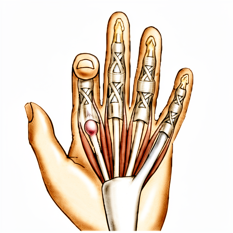
Post-operative exercises and precautions after trigger finger release, including tendon glides and joint blocking exercises.
Hướng dẫn này hỗ trợ quá trình phục hồi của bạn sau phẫu thuật cắt dây chằng gây ngón tay cái bập bênh cùng với Bác sĩ Kieran Hirpara tại Bệnh viện Tư nhân Mater Rockhampton. Hướng dẫn giải thích những điều bạn có thể mong đợi, các biện pháp phòng ngừa cần tuân thủ và chương trình bài tập sau phẫu thuật: hãy mang theo trang này hoặc file PDF của nó đến gặp nhà vật lý trị liệu hoặc chuyên gia trị liệu bàn tay của bạn để đảm bảo quá trình phục hồi chức năng được phối hợp nhịp nhàng.
Nếu bạn có bất kỳ lo ngại nào về vết mổ sau phẫu thuật, hãy liên hệ với phòng khám. Việc chụp ảnh vết mổ và gửi qua email để bác sĩ xem xét thường rất hữu ích.
Những điều cần biết
Việc chăm sóc vết thương được giải thích riêng: xem trang chăm sóc vết thương được liên kết ở cuối phác đồ này.
Các bài tập dưới đây rất quan trọng để ngăn ngừa gân bị dính khi vết thương của bạn lành lại. Đôi khi các khớp ở ngón tay có thể bị cứng sau thủ thuật này. Việc ngăn ngừa tình trạng cứng khớp sớm là rất quan trọng, vì vậy bạn được khuyến khích thực hiện các động tác kéo giãn mạnh và kéo dài cho ngón tay (sử dụng tay kia), đặc biệt là để duỗi thẳng. Hình thức kéo giãn thụ động này an toàn và sẽ không ảnh hưởng đến phẫu thuật: dây chằng vòng (pulley) đã được giải phóng, vì vậy không có gì bên trong mà động tác kéo giãn có thể làm xáo trộn.
Sau khi vết thương đã lành, hãy chườm nóng cho tay trong 15 phút trước khi thực hiện các bài tập này. Sau khi hoàn thành các bài tập, việc chườm đá có thể hữu ích để ngăn ngừa viêm.
Đôi khi bàn tay hoặc vết thương có thể trở nên nhạy cảm. Đây là điều bình thường và có thể được ngăn ngừa hoặc giảm thiểu bằng cách bắt đầu quá trình giảm nhạy cảm hàng ngày: nhẹ nhàng vỗ hoặc xoa lên vết thương (khi băng gạc vẫn còn), bắt đầu ngay sau phẫu thuật. Loại "phản hồi cảm giác" này cho phép da bình thường hóa khả năng tiếp xúc với chạm và kết cấu.
Trong 48 giờ đầu, hãy cố gắng giảm sưng: giữ bàn tay ở tư thế nâng cao, sử dụng đá lạnh, áp dụng ép nén nếu chuyên viên vật lý trị liệu đã cung cấp, và nhẹ nhàng "bơm" các ngón tay (mở và đóng) để di chuyển dịch sưng đi.
Bắt đầu sử dụng tay cho các công việc nhẹ (mặc quần áo, ăn uống và tương tự) ngay khi cơn đau cho phép, và tăng dần mức độ. Đừng làm quá sức: nếu cơn đau hoặc tình trạng sưng tăng rõ rệt sau một hoạt động, hãy giảm bớt cho đến khi bàn tay ổn định, sau đó tăng dần trở lại.
Một khi vết thương đã lành hoàn toàn, hãy bắt đầu mát-xa sẹo: thực hiện các vòng tròn mạnh lên đường rạch. Trang chăm sóc vết thương có thêm thông tin về quản lý sẹo.
Những gì bằng chứng cho thấy về quá trình hồi phục
Phẫu thuật mở giải phóng dây chằng A1 là một thủ thuật đã được thiết lập vững chắc, với hồ sơ thành công mạnh mẽ trong các tài liệu nghiên cứu đã công bố. Hiện tượng bắt và khóa khớp được phẫu thuật khắc phục: một khi dây chằng được cắt, gân sẽ trượt tự do trở lại và hiện tượng kẹt ngón thường không tái phát. Trong một loạt gần 1.600 ca giải phóng mở, ít hơn 1% bệnh nhân cần phẫu thuật lần hai để điều trị hiện tượng kẹt ngón dai dẳng hoặc tái phát, và không có trường hợp tổn thương thần kinh hay nhiễm trùng sâu [4]. Một nghiên cứu so sánh với thời gian theo dõi hơn ba năm cũng không tìm thấy sự tái phát sau phẫu thuật mở [5].
Cảm giác đau nhức ở lòng bàn tay giảm đáng kể trong khoảng một đến hai tuần đầu. Trong một nghiên cứu so sánh, thời gian trung bình để giảm đau đáng kể sau phẫu thuật mở là khoảng một tuần [5]. Một số cảm giác đau khi ấn mạnh, sưng nhẹ hoặc cứng ngón tay có thể kéo dài trong vài tuần sau đó. Điều này là bình thường và phản ánh quá trình sẹo đang trưởng thành, mất khoảng ba tháng [3]; chương trình giảm nhạy cảm, mát-xa sẹo và bài tập trong phác đồ này được thiết kế để quản lý chính xác tình trạng này. Trong loạt ca bệnh lớn nêu trên, khoảng một trên hai mươi ngón tay có vấn đề được ghi nhận sau phẫu thuật, phổ biến nhất là cứng tạm thời hoặc đau vùng sẹo, những vấn đề này sẽ giảm dần với điều trị; khả năng phục hồi tầm vận động có xu hướng chậm hơn ở những người mắc bệnh tiểu đường, do đó chương trình tập luyện càng quan trọng hơn đối với nhóm này [4].
Các phác đồ trị liệu tay được công bố bắt đầu các bài tập vận động ngón tay chủ động và bị động, cùng các bài tập trượt gân trong những ngày đầu tiên sau phẫu thuật, thêm các biện pháp quản lý sẹo và giảm nhạy cảm một khi vết thương đã lành, và tái đưa vào các bài tập tăng cường lực cầm nắm theo mức độ sau đó [2][3], cùng một cách tiếp cận từng giai đoạn như chương trình trên trang này. Việc bắt đầu các bài tập sớm là yếu tố giúp gân trượt tự do và các khớp duy trì độ linh hoạt trong khi vết thương đang lành.
Việc trở lại làm việc phụ thuộc vào yêu cầu công việc đối với bàn tay. Trong một nghiên cứu so sánh, một nửa số bệnh nhân đã trở lại làm việc trong khoảng hai tuần sau phẫu thuật mở [5]; những người làm các công việc nhẹ hoặc văn phòng thường có thể trở lại sớm hơn, trong khi những công việc nặng nhọc phải chờ đến khi các hạn chế về nâng và cầm nắm nêu dưới đây được dỡ bỏ.
Một thử nghiệm có đối chứng ngẫu nhiên đã so sánh ba tháng trị liệu có giám sát sau phẫu thuật mở với chương trình tập tại nhà tự hướng dẫn: chức năng tổng thể, tầm vận động và mức độ đau tương tự nhau giữa các nhóm sau sáu tháng, lực cầm nắm phục hồi tốt hơn với trị liệu có giám sát, và những bệnh nhân rõ ràng được hưởng lợi từ trị liệu chính thức là những người có hiện tượng kẹt ngón kéo dài hơn mười hai tháng trước phẫu thuật và những người làm việc nhà hoặc công việc nhẹ [1]. Về mặt thực tiễn, một chương trình tại nhà được thực hiện tốt (các bài tập trên trang này) giúp đa số bệnh nhân vượt qua giai đoạn hồi phục, trong khi trị liệu tay chính thức mang lại giá trị gia tăng khi ngón tay bị cứng trong thời gian dài trước phẫu thuật hoặc tiến triển chậm.
Các biện pháp phòng ngừa và hạn chế
Việc sử dụng tay ở mức độ chức năng nhẹ được khuyến khích cho các hoạt động sinh hoạt hàng ngày như chăm sóc bản thân, ăn uống, mặc quần áo, viết và đánh máy. Các giới hạn cần lưu ý:
- Tránh nâng vật, nắm chặt và chịu tải trọng trong tối đa 4 tuần sau phẫu thuật.
- Việc lái xe bị hạn chế trong tuần đầu tiên; có thể quay trở lại khi cơn đau cho phép, bạn có thể nắm chặt bàn tay hoàn toàn và có thể kiểm soát phương tiện một cách an toàn.
Dành cho nhà vật lý trị liệu của bạn:
Quản lý điều trị
- Chương trình bài tập tại nhà theo các thẻ hướng dẫn bên dưới: căng duỗi cổ tay (gập/mở); khóa khớp DIP (khớp liên đốt xa) và PIP (khớp liên đốt gần); các bài trượt gân (Dãy A và Dãy B)
- Các bài căng duỗi thụ động kéo dài và mạnh vào ngón tay, đặc biệt là ở tư thế duỗi, để phòng ngừa cứng khớp sớm
- Chườm nóng cho bàn tay 15 phút trước khi tập sau khi vết thương đã lành; chườm đá sau khi tập để phòng ngừa viêm
- Giảm nhạy cảm hàng ngày (chạm nhẹ / xoa nhẹ lên vết thương, băng gạc vẫn còn tại chỗ) bắt đầu ngay sau phẫu thuật
- Quản lý sưng nề trong 48 giờ đầu: kê cao, chườm đá, băng ép khi cần thiết, và các bài tập co duỗi ngón tay nhẹ nhàng
- Tăng dần mức độ sử dụng chức năng nhẹ cho sinh hoạt hàng ngày khi cơn đau cho phép, theo dõi các đợt đau/sưng nề tăng lên sau khi vận động
- Xoa sẹo (vỗ tròn mạnh lên đường rạch) sau khi vết thương đã lành hoàn toàn
Biện pháp phòng ngừa
- Chỉ sử dụng ở mức độ chức năng nhẹ cho các hoạt động sinh hoạt hàng ngày (chăm sóc bản thân, ăn uống, mặc quần áo, viết, đánh máy)
- Không nâng vật, nắm chặt hoặc chịu tải trọng trong tối đa 4 tuần sau phẫu thuật
- Việc lái xe bị hạn chế trong tuần đầu tiên; có thể quay trở lại khi cơn đau cho phép, nắm chặt bàn tay hoàn toàn được thực hiện và bệnh nhân có thể kiểm soát phương tiện một cách an toàn
Các mốc dự kiến (dựa trên tiêu chí, hướng dẫn bởi các phác đồ đã công bố [1][2][3])
- Cơn đau giảm xuống mức dễ chịu với thuốc giảm đau đơn giản trong vòng 1–2 tuần [5]
- Vết thương đã lành, bắt đầu xoa sẹo và giảm nhạy cảm liên tục, vào khoảng 2–3 tuần [2][3]
- Khớp ngón tay có thể gập và duỗi chủ động hoàn toàn (nắm chặt hoàn toàn và duỗi composite hoàn toàn) vào khoảng 3 tuần, được duy trì và phục hồi thông qua chương trình khóa khớp và trượt gân [2]
- Bắt đầu tăng dần sức mạnh của lực nắm và lực kẹp (ví dụ: dùng đất sét trị liệu) sau khi hết hạn chế nâng vật/nắm chặt ở tuần thứ 4, tiến tới sử dụng chức năng hoàn toàn
- Cân nhắc chuyển lên liệu pháp tay có giám sát nếu tình trạng kẹt gân (triggering) tồn tại hơn 12 tháng trước phẫu thuật, nếu vai trò của bệnh nhân đòi hỏi sử dụng tay nhẹ/tinh tế kéo dài, hoặc nếu phạm vi vận động hoặc phục hồi lực nắm chậm chạp [1]
Đây là các bài tập từ tài liệu hướng dẫn của bạn, bắt đầu sau phẫu thuật và tiếp tục tại nhà theo sự hướng dẫn của nhà vật lý trị liệu hoặc chuyên gia trị liệu tay.
Bài tập của bạn
Sau giao thức của bạn
Giao thức này được viết cùng với Sarah Farrell, BOccThy (Cử nhân Vật lý trị liệu nghề nghiệp), Chuyên trị liệu tay được chứng nhận, và bao gồm hướng dẫn quản lý sau phẫu thuật cập nhật (tháng 4 năm 2025) từ Ruby Doolan, Chuyên trị liệu tay được chứng nhận, Extend Rehabilitation. Giao thức này đi kèm với các lời khuyên phục hồi chung của phòng khám: xem quản lý đau sau phẫu thuật, chăm sóc vết thương và cơ bản trị liệu tay. Về ca phẫu thuật, xem phẫu thuật cắt dây chằng ngón tay cái.
Khung phục hồi và các cột mốc cũng được thông tin bởi các giao thức phục hồi sau phẫu thuật cắt dây chằng ngón tay cái đã được công bố, bao gồm cả của Trung tâm Tay Đại học Virginia và Twin Cities Orthopedics, và bởi các nghiên cứu kết quả đã được công bố về phẫu thuật cắt dây chằng ngón tay cái mở, bao gồm một thử nghiệm ngẫu nhiên có kiểm soát về phục hồi sau phẫu thuật (Saito et al., Journal of Clinical Medicine, 2023) và một loạt lớn các biến cố bất lợi (Bruijnzeel et al., Journal of Hand Surgery, 2012).
Tài liệu tham khảo
[1] Saito T, Nakamichi R, Nakahara R, Nishida K, Ozaki T. Hiệu quả của phục hồi chức năng sau giải phẫu mở cho ngón tay cái cò: một nghiên cứu ngẫu nhiên có đối chứng, tiến cứu. J Clin Med. 2023;12(22):7187. https://pmc.ncbi.nlm.nih.gov/articles/PMC10671987/ [2] Trung tâm Tay, Đại học Virginia. Hướng dẫn giải phóng ngón tay cái cò (phác đồ điều trị sau phẫu thuật). https://med.virginia.edu/orthopaedic-surgery/wp-content/uploads/sites/242/2015/11/Triggerfingerreleaseprotocol.pdf [3] Meletiou SD, Twin Cities Orthopedics. Quản lý sau phẫu thuật giải phóng ngón tay cái cò (giải phóng dây chằng vòng A1). https://tcomn.com/wp-content/uploads/2017/10/Trigger-Release-A1.pdf [4] Bruijnzeel H, Neuhaus V, Fostvedt S, Jupiter JB, Mudgal CS, Ring DC. Các biến cố bất lợi của giải phẫu mở dây chằng vòng A1 cho ngón tay cái cò vô căn. J Hand Surg Am. 2012;37(8):1650–1656. https://pubmed.ncbi.nlm.nih.gov/22763058/ [5] Chanthanapodi P, Aodsup S. Kết quả so sánh giữa phẫu thuật qua da và phẫu thuật mở cho ngón tay cái cò: phân tích điểm số xu hướng. Front Surg. 2025;12:1509292. https://pmc.ncbi.nlm.nih.gov/articles/PMC11922895/
Evidence & references
Trigger Finger Release (A1 Pulley Release) — Surgical Outcomes & Post-operative Rehabilitation
Topic scope: (A) the place of surgery in stenosing tenosynovitis (trigger finger/thumb) after failed conservative care (splinting, corticosteroid injection), and (B) post-operative rehabilitation after surgical division of the A1 pulley — open or percutaneous. This is an early-motion pathway: nothing is reconstructed, the catching is mechanically abolished the moment the pulley is divided, and the rehab exists to keep the now-free tendon gliding and the finger joints supple while the wound heals.
Defining principle of the rehab here: A1 pulley release removes the obstruction; it does not create a construct that needs protecting. Once the pulley is divided the flexor tendon glides freely and triggering does not usually recur. So — unlike a tendon repair, and like a carpal-tunnel decompression — the pathway is immediate active motion: full active finger flexion/extension and tendon glides from the first days, oedema and scar care, early light functional use, and a quick return. Most patients need no formal hand therapy at all; supervised therapy is reserved for the minority with pre-existing joint stiffness, long-standing triggering, or slow recovery. The single branch point is whether the finger was already stiff before surgery (long-standing fixed flexion / PIP contracture) — those patients need active therapy to recover motion the release alone cannot restore.
A. WHERE SURGERY SITS IN THE PATHWAY
Trigger finger is usually managed non-operatively first: activity modification, splinting, and corticosteroid injection, which resolves a substantial proportion of digits without surgery. Surgery (A1 pulley release) is reserved for digits that fail injection, recur, or present with a fixed deformity. The corpus contains the comparative evidence underpinning this stepped approach (percutaneous release vs steroid injection; one- vs two-injection regimens; corticosteroid solution choice) — Moderate (RCT). The rehab protocol on the patient page begins after that decision has been made, so this brief concentrates on the surgical and post-surgical evidence.
B. SURGICAL OUTCOMES & RESOLUTION RATES
Open release of the A1 pulley is one of the most reliable operations in hand surgery. The mechanical problem — a thickened tendon catching under a tight pulley — is solved by dividing the pulley, and the result is durable:
- In a series of 1,598 open releases, fewer than 1% required a second operation for persistent or recurrent triggering, with no nerve injuries and no deep infections [Bruijnzeel 2012]. About one digit in twenty had a documented post-operative problem, almost all minor and self-limiting (transient stiffness, scar tenderness). Strong (large cohort).
- Recovery of motion is slower in patients with diabetes, reinforcing the value of the exercise program in that group [Bruijnzeel 2012]. Moderate.
- A propensity-matched comparison with >3 years follow-up found no recurrences after open release, with median time to significant pain reduction of about one week and roughly half of patients back at work within ~2 weeks [Chanthanapodi 2025]. Moderate.
Take-home for rehab: because the operation itself abolishes the triggering, the rehabilitation is not "earning back" a surgical result — it is preventing the two things that can go wrong during healing: tendon adhesion and joint stiffness. Early glide and early extension are the levers.
C. OPEN vs PERCUTANEOUS RELEASE
Both techniques divide the same structure and converge to the same place.
- A Level I meta-analysis of 8 RCTs (548 patients) found no significant difference between open and percutaneous release in revision, complication, or pain rates — both are appropriate options [Casey 2024, J Hand Surg Am]. Strong (meta-analysis of RCTs).
- Larger RCT syntheses show percutaneous release confers faster early functional recovery — better short/mid-term Q-DASH, ~12 days earlier return to work, and shorter analgesic use — while long-term function, grip, motion and complication/revision rates are equivalent. Strong.
- Percutaneous (including ultrasound-guided/sonographically-controlled) technique is supported by multiple corpus series for efficacy and safety, with the main theoretical risks being incomplete release and digital nerve proximity, mitigated by surface landmarks and imaging [corpus percutaneous series]. Moderate.
Rehab implication: the post-operative program is essentially the same for both approaches — early active motion, glides, oedema and scar care. The patient page applies regardless of whether the release was open or percutaneous; percutaneous patients simply tend to be comfortable and back to activity a little sooner.
D. THE ROLE — AND LIMITS — OF POST-OPERATIVE HAND THERAPY
This is the central evidence point for the protocol, and it is one where "more therapy" is not automatically better.
- A prospective RCT compared 3 months of supervised rehabilitation after open
release against a self-directed home exercise program: at six months,
overall function, motion and pain were similar between groups. Supervised
therapy added further grip-strength recovery, and the patients who clearly
benefited from formal therapy were those whose **triggering had been present
12 months pre-operatively and those in housework/lighter-work roles [Saito 2023, J Clin Med]. Moderate (single RCT).
- Published surgeon and hand-therapy protocols (e.g. University of Virginia Hand Center; Twin Cities Orthopedics) start active and passive finger motion and tendon glides within the first days, add scar massage and desensitisation once the wound is healed, and reintroduce graded grip strengthening later — precisely the staged structure of the patient page. Consensus.
Bottom line: a well-performed home program carries most patients through. Formal hand therapy is reserved, not routine — escalate it for long-standing pre-operative triggering, pre-existing joint stiffness/contracture, manual or fine-use occupational demands, or slow motion/grip recovery.
E. COMPLICATIONS
Serious complications are uncommon (roughly <1–4% across series) and most "complications" are minor, self-limiting healing phenomena:
- Digital nerve injury — the most feared complication, particularly relevant to percutaneous technique (blind division near the radial digital nerve of the thumb and index) and to scar/retraction in open release. Rare in experienced hands; transient paraesthesia is more common than true division [corpus complication series]. Moderate.
- Incomplete release / persistent triggering — failure to fully divide the A1 pulley (or an A2/FDS slip contribution); a recognised cause of revision, more often discussed with percutaneous technique. Moderate.
- Recurrent triggering — uncommon after adequate open release (<1% reoperation in the 1,598-digit series) [Bruijnzeel 2012]. Strong.
- Infection — usually superficial; deep infection rare (none in the large open series) [Bruijnzeel 2012]. Strong.
- Bowstringing — a rare complication from excessive proximal pulley loss (A1 plus encroachment on A2); largely avoided by limiting division to A1 [bowstringing case literature]. Weak (case-level).
- Stiffness / flexion contracture / "flare" — the commonest self-limiting problem; transient PIP stiffness, scar tenderness and a post-operative inflammatory flare that settle with the motion, desensitisation and scar program. Recovery is slower in diabetes. Moderate. This is the category the rehabilitation program actively targets.
F. PHASED POST-OP TIMELINE (matches the patient protocol)
| Phase | Window | Protection | Motion / use | Therapy add-ons | Notes |
|---|---|---|---|---|---|
| I — Immediate active motion & oedema control | Day 0–2 | None beyond dressing | Active finger flexion/extension and finger "pumps" from day 1; tendon glides commenced | Elevation, ice, compression if provided; desensitisation (tap/rub over dressed wound) from day 1 | Nothing reconstructed -> motion is the priority; manage swelling actively |
| II — Glide & joint motion | Week 0–2 | None | Tendon glides (Series A/B), DIP & PIP blocking, composite extension; firm passive stretch into extension | Continue desensitisation | Goal: keep tendon gliding, prevent adhesion & stiffness; pain settles substantially (~1 wk) [Chanthanapodi 2025] |
| III — Scar maturation & function | Week 2–4 | Light functional use only | Full active fist + full composite extension by ~3 wk; build light daily-living use | Scar massage (firm circles) once wound healed; heat before / ice after exercises | No lifting/gripping/weight-bearing to ~4 wk; driving limited ~first week (full fist + safe control) |
| IV — Strengthening & return | Week 4+ | None | Graded grip/pinch (e.g. putty) once 4-wk precaution lifts -> full function | Supervised therapy if indicated (long-standing trigger, stiffness, slow recovery, occupational demand) [Saito 2023] | Manual workers return later than desk/light roles |
Timings are criteria-based and drawn from published surgeon/hand-therapy protocols; they are typical, not trial-mandated.
G. KEY CONTROVERSIES / EVIDENCE QUALITY
- Is routine post-op hand therapy necessary? The best available evidence (Saito 2023 RCT) says no for most — home exercise matches supervised therapy on function/pain/motion at six months, with supervised therapy adding grip strength and benefiting a defined subgroup (long-standing trigger, lighter-work roles). The protocol's "therapy reserved, not routine" stance is evidence-aligned. Moderate.
- Open vs percutaneous. Equivalent long-term outcomes and safety (Casey 2024 meta-analysis); percutaneous offers faster early recovery. The rehab is the same either way. The live debate is technique-side (nerve safety, completeness of release), not rehab-side. Strong on equivalence.
- The rehab protocol structure itself is consensus/expert, built from surgeon patient-guidance documents plus one rehabilitation RCT — there is no large trial dictating exact phase timings.
- Diabetes modifies recovery — slower motion recovery and a lower threshold to involve a hand therapist; not a different protocol, a different pace. Moderate.
H. EVIDENCE STRENGTH FLAGS (summary)
- STRONG (meta-analysis / RCTs / large cohort): open vs percutaneous equivalence in revision/complication/pain (Casey 2024, 8 RCTs); percutaneous faster early functional recovery (RCT syntheses); durability of open release (<1% reoperation, no nerve injury/deep infection in 1,598 digits, Bruijnzeel 2012).
- MODERATE (single RCT / cohorts): home exercise ~ supervised therapy at 6 months with grip-strength edge for supervised therapy (Saito 2023); percutaneous efficacy/safety series; slower recovery in diabetes; injection-vs- surgery comparative data.
- WEAK / CONSENSUS: the post-operative rehabilitation protocol structure and exact phase timings (surgeon/hand-therapy patient-guidance documents); bowstringing risk (case-level).
CITATIONS
RAG corpus (180,000+ Orthopaedic articles)
- Open Versus Percutaneous Fixation of Trigger Finger: Meta-Analysis of Clinical Outcomes. J Hand Surg Am. 2024. DOI: 10.1016/j.jhsa.2024.03.010
- Complications of Open Trigger Finger Release. J Hand Surg Am. 2010. DOI: 10.1016/j.jhsa.2009.12.040
- Differential Pulley Release in Trigger Finger: A Prospective, Randomized Clinical Trial. Hand (N Y). 2021. DOI: 10.1177/1558944721994231
- Percutaneous A1 pulley release vs steroid injection for trigger digit. J Hand Surg Eur. 2010. DOI: 10.1177/1753193410381824
- Comparative Study of A1 Pulley Release and Ulnar Superficialis Slip Resection in Trigger Finger. J Hand Surg Am. 2022. DOI: 10.1016/j.jhsa.2022.04.021
- Risk Factors for Requiring Ulnar Superficialis Slip Resection During Trigger Finger Release. J Hand Surg Am. 2024. DOI: 10.1016/j.jhsa.2024.08.013
- Impact of Flexor Tendon Traction Tenolysis on Clinical Outcomes in Open A1 Pulley Release. J Hand Surg Glob Online. 2024. DOI: 10.1016/j.jhsg.2024.09.010
- Ultrasound-Assisted Percutaneous Trigger Finger Release: Is It Safe? Hand (N Y). 2008. DOI: 10.1007/s11552-008-9137-8
- Evaluation of Percutaneous First Annular Pulley Release: Efficacy and Complications. J Hand Surg Am. 2016. DOI: 10.1016/j.jhsa.2016.04.009
- Sonographically controlled minimally-invasive A1 pulley release using a new guide. BMC Musculoskelet Disord. 2023. DOI: 10.1186/s12891-023-06982-x
- Percutaneous A1 pulley with corticosteroid injection for trigger finger release. J Orthop Surg Res. 2025. DOI: 10.1186/s13018-025-05776-2
- A Cost and Efficiency Analysis of the WALANT Technique for the Management of Trigger Finger. Plast Reconstr Surg Glob Open. 2019. DOI: 10.1097/gox.0000000000002509
- Management of Pediatric Trigger Thumb and Trigger Finger. J Am Acad Orthop Surg. 2012. DOI: 10.5435/jaaos-20-04-206
- What's New in Hand Surgery. J Bone Joint Surg Am. 2024. DOI: 10.2106/jbjs.23.01343
Trigger finger surgical & rehabilitation literature (URLs)
- Saito T, et al. The Effectiveness of Rehabilitation after Open Surgical Release for Trigger Finger: A Prospective, Randomized, Controlled Study. J Clin Med. 2023;12(22):7187. https://pmc.ncbi.nlm.nih.gov/articles/PMC10671987/
- Bruijnzeel H, et al. Adverse Events of Open A1 Pulley Release for Idiopathic Trigger Finger. J Hand Surg Am. 2012;37(8):1650-1656. https://pubmed.ncbi.nlm.nih.gov/22763058/
- Casey JC, et al. Open Versus Percutaneous Fixation of Trigger Finger: Meta-Analysis of Clinical Outcomes. J Hand Surg Am. 2024;49(6):570-575. https://pubmed.ncbi.nlm.nih.gov/38727666/
- Chanthanapodi P, Aodsup S. Comparative results of percutaneous and open surgery for trigger fingers: a propensity score analysis. Front Surg. 2025;12:1509292. https://pmc.ncbi.nlm.nih.gov/articles/PMC11922895/
- Complications of Percutaneous Release of the Trigger Finger. PMC. https://pmc.ncbi.nlm.nih.gov/articles/PMC6485534/
- Trigger Finger. StatPearls, NCBI Bookshelf. https://www.ncbi.nlm.nih.gov/books/NBK459310/
- Bowstringing as a complication of trigger finger release. J Hand Surg Am. 1988. https://www.jhandsurg.org/article/S0363-5023(88)80097-2/abstract
- Trigger Finger (patient information). British Society for Surgery of the Hand (BSSH). https://www.bssh.ac.uk/patients/conditions/15/trigger_finger
Published rehab protocols (patient-guidance — basis for the phase structure)
- University of Virginia Hand Center. Trigger Finger Release Guidelines (post-operative therapy protocol). https://med.virginia.edu/orthopaedic-surgery/wp-content/uploads/sites/242/2015/11/Triggerfingerreleaseprotocol.pdf
- Meletiou SD, Twin Cities Orthopedics. Post-operative Management of Trigger Release (A1 pulley release). https://tcomn.com/wp-content/uploads/2017/10/Trigger-Release-A1.pdf
- EmergeOrtho. Trigger Finger Release - Post-operative Instructions. https://emergeortho.com/wp-content/uploads/2022/06/Trigger-Finger-Release.pdf


