肱二头肌腱固定术
Patients › Rehabilitation
Rehabilitation after isolated biceps tenodesis, protecting the tenodesis fixation through staged return of biceps loading.
本康复方案适用于基兰·希尔帕拉(Kieran Hirpara)医生在罗克汉普顿 Mater 私人医院进行的肱二头肌腱固定术后的康复,无论手术是通过关节镜(微创)进行,还是通过在腋窝前方的小切口(开放下肌腱下技术)进行。请在首次物理治疗就诊时携带此页面或其 PDF 文件,以确保康复过程协调一致。您的物理治疗师将根据您肩部和手臂的恢复情况,按以下阶段个体化推进您的康复进程。
本方案仅适用于单纯性肱二头肌腱固定术。如果您的手术同时进行了肩袖修复,请遵循肩袖修复方案;修复后的肌腱会要求更缓慢的康复进度。
如果您对术后伤口有任何疑虑,请联系诊所。拍摄伤口照片并通过电子邮件发送以供审查通常很有帮助。
预期情况
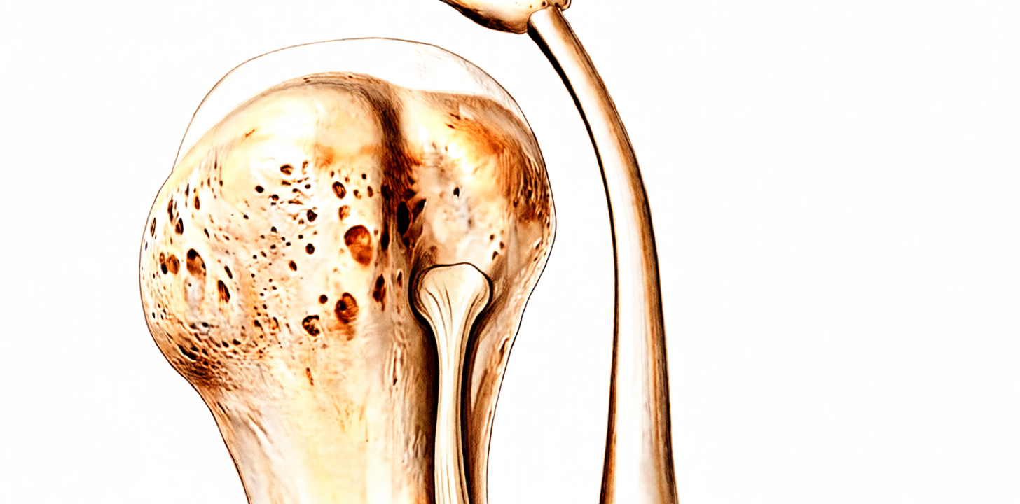
在肱二头肌腱固定术中,肱二头肌长头腱从其肩关节内的原始附着点分离,并使用锚钉或螺钉将其固定在上臂骨(肱骨)上。新的附着点需要时间才能牢固地愈合于骨骼,因此康复的早期阶段主要围绕保护该部位展开。
肱二头肌具有两个功能:屈肘和使手掌向上旋转(旋后)。因此(这与肩部手术通常的情况不同),早期的限制主要集中在肘部:在最初几周,肘部仅进行被动活动(由另一只手或物理治疗师协助弯曲),以避免愈合中的肌腱承受牵拉力。出于相同原因,术后早期禁止使用患侧手臂进行提举和搬运。此外,早期还会限制一些会使肌腱产生张力的肩关节位置:例如外旋超过约 40 度,以及将手臂置于身体后方。
您需要佩戴悬吊带约三至四周,包括睡眠时。从第三周左右开始,根据舒适度逐渐减少佩戴时间。佩戴悬吊带期间严禁驾驶。
康复进程概览:
- 第一阶段 — 保护固定术(大约前四周)
- 第二阶段 — 主动活动(第 4–6 周)
- 第三阶段 — 力量训练(第 6–12 周,从第 10 周开始进行肱二头肌抗阻训练)
- 第四阶段 — 恢复全面活动(第 12 周起)
上述周数范围仅为典型参考,并非固定不变。物理治疗师将根据您手臂的愈合和活动情况推进康复进程,而非依据日历时间。物理治疗通常在术后第一或第二周内开始,您的首次预约详情见出院资料包,除非您选择自行安排。
第一阶段——保护肌腱固定术(第0–4周)
最初几周的重点是让肌腱在骨头上愈合,同时保持周围组织的活动。从开始起,您的手、腕和手指就保持活动。您的肘部每天进行被动活动:由另一只手协助进行屈曲和手掌向上/向下的旋转,以使肱二头肌本身保持放松。肩部在以下限制范围内进行轻柔活动,包括钟摆运动和辅助运动。使用冰敷缓解疼痛,在锻炼和物理治疗课程前服用止痛药,并持续佩戴悬吊带,包括在卧床时。切勿使用患侧手臂提举或携带任何物品,且在需要佩戴悬吊带期间请勿驾驶。在感觉舒适的前提下,前臂有支撑的轻体力活动(如写字或打字)通常是可以的。
致您的物理治疗师:
目标
- 在肌腱固定术愈合至骨头的过程中保护固定
- 控制疼痛、肿胀及炎症反应
- 实现肘关节和前臂的全范围被动活动;在以下限制内实现舒适的肩部被动活动范围
- 维持肩胛骨功能和姿势
管理方案
- 佩戴悬吊带约3–4周,包括夜间,从第3周左右开始逐渐减少佩戴
- 肘关节被动活动范围:屈曲/伸展及前臂旋后/旋前
- 腕关节和手部的主动活动范围;握球练习
- 在限制范围内进行肩部被动及轻柔的主动辅助活动范围:钟摆运动,屈曲和外展初期至约90度,随舒适度进展;外旋至40度;内旋至约45度
- 肩胛骨定位和回缩(手臂有支撑),进展至肩胛骨等长收缩;颈椎活动范围及姿势训练
- 冷疗用于缓解疼痛和肿胀;在锻炼和课程前给予镇痛
- 仰卧时在肘下放置毛巾卷或小枕头,以避免肩关节过伸
注意事项
- 禁止主动肘关节屈曲及抗阻前臂旋后;肱二头肌保持无负荷状态
- 禁止主动肩部活动范围;外旋不得超过40度;禁止肩关节过伸或水平外展超过中立位
- 禁止使用患侧手臂提举或携带物品
- 禁止在肱二头肌近端/肌腱固定术部位进行摩擦按摩
- 在需要佩戴悬吊带期间禁止驾驶
进展标准
- 伤口愈合且疼痛控制良好
- 肘关节屈曲/伸展及前臂旋转的全范围被动活动
- 在处方限制内实现舒适的肩部被动活动范围
第二阶段——主动活动期(第4–6周)
去除吊带后,手臂开始依靠自身力量进行活动。肩关节从辅助活动逐步过渡到各个方向的主动活动,肘关节现在可以进行主动屈曲和旋转,但仍不施加负荷。肱二头肌处于活动状态,但尚未承担工作负荷:尽量减少抬举动作(使用该手臂提举的物品重量不得超过一杯茶),避免用该手臂进行推、拉和搬运。此阶段通常可以舒适地进行轻度的伏案工作。一旦去除吊带,当您可以舒适且安全地控制车辆时,即可恢复驾驶。
致您的物理治疗师:
目标
- 逐步恢复肩关节和肘关节的完全主动活动范围
- 运动中肩胛骨力学正常
- 开始进行次最大强度的肩关节等长收缩训练
- 手臂在腰部高度的轻度功能性使用
管理措施
- 肩关节主动辅助活动逐步过渡到各个平面的主动活动范围(例如躺椅式进展、墙壁和栏杆滑动、仰卧屈曲至站立外展)
- 肘关节主动屈曲/伸展及前臂旋后/旋前,无阻力
- 次最大强度肩关节等长收缩:内旋、外旋、外展、内收
- 继续进行肩胛骨稳定训练和姿势矫正
- 伤口成熟后进行疤痕按摩;腱固定术部位不进行交叉摩擦按摩
- 根据需要进行后关节囊拉伸(交叉身体拉伸、睡眠者拉伸)
- 步行或固定自行车进行体能训练;患肢不负重
注意事项
- 禁止进行肱二头肌抗阻训练;禁止对肘屈肌或旋后肌施加负荷
- 禁止使用手术侧手臂提举物品;暂禁止跑步
- 避免通过激进的拉伸或手法治疗过度牵拉愈合中的肌腱
进阶标准
- 肩关节、肘关节和前臂的完全无痛主动活动范围
- 运动和轻度功能活动中肩胛骨力学正常
- 疼痛控制良好
第三阶段——强化训练(第6–12周)
强化训练从轻柔开始,由肩胛骨向外周扩展:首先强化肩袖和肩胛骨肌肉,使用弹力带和小重量进行低负荷、高重复次数的训练。抗阻肱二头肌训练(弯举和抗阻旋后)需等到第10周才开始,初期负荷较轻并缓慢增加进度。已发表的方案中引入该训练的时间在第6周至第10周之间;本方案遵循该范围中更为保守的一端,以便在负荷施加前为肌腱固定提供最长的愈合时间。在此阶段,日常活动应基本恢复正常,且从第8周左右开始,在物理治疗师的指导下,通常可以恢复跑步、骑自行车和打高尔夫球。
致物理治疗师:
目标
- 使力量、耐力和神经肌肉控制恢复正常
- 恢复从腰部水平经胸部水平至过头水平的功能
管理
- 全程保持肩关节和肘关节的全范围活动度
- 等张肩袖强化训练:内旋和外旋,使用轻阻力,从中立位逐渐进展至外展90度
- 肩胛骨训练计划:俯卧系列贯穿本阶段;抗阻前伸(serratus punch)、低拉、俯卧撑加(progression)(墙壁、台面、跪姿、地面)
- 从第10周开始进行抗阻肱二头肌弯举、旋后和旋前训练;先使用短力臂,低负荷,高重复次数,谨慎推进
- 节律性稳定训练和对角线(D1/D2)模式;闭链稳定训练
- 从第8周左右开始恢复跑步、骑自行车和打高尔夫球,注意正确的动作力学
注意事项
- 本阶段早期避免长力臂的抗阻肘屈曲和旋后
- 在力量恢复之前,禁止提重物或搬运重物
- 本阶段禁止游泳或投掷
- 强化训练需等到活动度接近完全且无痛时才开始,并全程保持无痛
进阶标准
- 全范围、无痛的主动活动度,且肩肱节律正常
- 肩胛平面外展90度时肩袖力量为5/5,且肩胸关节力量为5/5
- 强化训练耐受良好,无症状加重
第四阶段——恢复全面活动(第12周起)
最终阶段是逐步恢复较重的体力工作、过头活动及体育运动。大多数人可在三至四个月内获准进行健身训练、手工劳动及娱乐性体育运动,并逐步增加负荷;对于需要过头动作及对抗性运动的运动员(如投掷、游泳、隔网对抗类运动),其回归过程通常更为漫长,往往需要四至五个月或更长时间。在此之后,肱二头肌的力量和信心仍会持续数月,因此本阶段的进展应基于手臂的功能表现,而非单纯依据时间。
致您的物理治疗师:
目标
- 在不出现代偿性运动模式的前提下,恢复完全的力量和爆发力
- 在高速度及专项运动动作中保持稳定和控制
- 恢复正常体育运动和工作活动
管理措施
- 多关节及复合力量训练,稳步增加负荷
- 增强式训练:从双臂低于肩高度的动作开始,逐步过渡至单臂及过头动作
- 根据具体情况,制定投掷、游泳或隔网对抗类运动的间歇性回归运动计划
- 改善核心及髋部力量与控制能力,防止肩部出现代偿
注意事项
- 缓慢推进对肩前侧产生应力的练习(例如卧推、直立划船)
- 回归运动需经许可,且患侧力量应达到健侧的约90%,并能无痛地控制高速度及专项运动动作
术后方案
上述阶段改编自已发表的孤立性肱二头肌腱固定术康复方案:马萨诸塞州总医院布里格姆运动医学科、弗吉尼亚大学骨科系、俄亥俄州立大学韦克斯纳医疗中心以及威斯康星大学健康系统(UW Health)。周数范围为典型区间而非固定值,您的持续康复由物理治疗师根据您手臂的恢复情况,与诊所合作进行个体化指导。本页面与诊所的一般康复建议配合使用;请参阅术后疼痛管理和伤口护理。关于手术本身,请参阅肱二头肌腱固定术。本方案背后的证据(固定与愈合、腱固定术与腱切断术的决策、并发症与失败率,以及康复节奏的选择依据)在证据部分进行了总结,可从本页面顶部下载PDF版本。
Evidence & references
Biceps Tenodesis — Post-operative Rehabilitation Evidence Summary
Topic scope: the evidence behind post-operative rehabilitation after isolated biceps tenodesis (re-anchoring the long head of biceps to the humerus) — fixation methods and their healing implications, when to protect versus load the construct (active elbow flexion and resisted supination), shoulder ROM restrictions, return-to-activity timing, and the failure/complication profile (Popeye deformity, fixation failure). The combined cuff-repair pathway defers to the rotator-cuff-repair protocol and is out of scope here.
Defining principle of the surgical rehab here: a tenodesis is a healing construct that must be protected — the tendon has been detached from its native anchor and fixed into bone, and that bone–tendon interface needs weeks to heal. Because the biceps bends the elbow and turns the palm up (supination), the early restrictions are unusually centred on the elbow, not the shoulder: active elbow flexion and resisted supination are the loads that pull directly on the fresh tenodesis, so they are deferred while the construct heals. The shoulder itself is generally less restricted — only positions that tension the tendon (external rotation past ~40°, extension/horizontal abduction behind the body) are limited early. This is the inverse of a rotator-cuff or labral repair, where the shoulder is the protected structure.
A. THE PROCEDURE & WHY THE REHAB IS SHAPED THIS WAY
In a biceps tenodesis the long head of biceps is released from its origin on the superior glenoid/labrum and re-fixed into the proximal humerus, most commonly with an interference screw, a suture anchor, or a cortical/unicortical button (or combinations). The location is either suprapectoral (higher, usually all-arthroscopic, in or above the bicipital groove) or subpectoral (lower, usually open, below the pectoralis major tendon). Both achieve the same goal — remove the painful intra-articular biceps as a pain generator while preserving the muscle's length-tension relationship to avoid a Popeye deformity and cramping.
The rehab is built around the time for the tendon to incorporate into the bone tunnel/socket. The biceps' two actions — elbow flexion and forearm supination — are precisely the movements that load the tenodesis, so resisted/active use of these is staged in last. Shoulder motion is restored earlier because most shoulder positions do not pull hard on the construct.
B. EVIDENCE BY THEME
1. Tenodesis vs tenotomy — the decision upstream of rehab
Tenotomy (simply releasing the tendon) and tenodesis give broadly equivalent pain relief and function, but tenodesis trades a slightly more demanding recovery for a lower rate of Popeye deformity and cramping/fatigue, particularly relevant in younger, leaner and more active patients. A prospective double-blinded RCT and multiple reviews support this trade-off [Castricini RCT; Frost/Hackney review; Slenker review]. Moderate–strong (RCT + SR). This is why a tenodesis — and therefore a protected construct needing staged rehab — is chosen in the first place.
2. Fixation strength and what it permits
Interference screws and suture anchors both provide clinically adequate fixation; biomechanical load-to-failure figures vary between studies and clinical outcomes do not differ meaningfully by fixation type [biomechanical cadaver series; clinical comparisons]. The practical point for rehab: the construct is strong at time zero but the biological bond to bone is what is healing over the first 6–12 weeks — which is why loaded elbow flexion is deferred regardless of the hardware used. Moderate (biomechanical + clinical cohorts).
3. Does the rehab pace actually need to be slow? — the key controversy
The traditional protocol protects the elbow for ~6 weeks before active flexion and reserves resisted biceps work for ~10–12 weeks. However, Mazzocca et al. challenged this: in 105 open subpectoral tenodeses (dual-fixation button + interference screw) rehabilitated with immediate, unrestricted motion and no postoperative restrictions, the failure rate was only 2.2% (2 of 98) at minimum 2-year follow-up, with excellent ASES/DASH scores. Both failures occurred early (5 and 9 weeks). The conclusion: with a robust dual-fixation construct, early mobilisation is reasonable and may improve outcomes [Mazzocca, JSES 2018, DOI 10.1016/j.jse.2018.02.061]. A subsequent comparison of early versus delayed active ROM reached similar reassurance [PMID 34458384]. Moderate (single-arm cohort + comparative). This protocol nonetheless follows the more protective published pace — it is the safer default across mixed fixation methods and does not assume a dual-fixation construct.
4. Complications and failure rates — what rehab is protecting against
A review of 1,526 shoulders found a low overall complication rate: persistent anterior shoulder pain ~11–13%, Popeye deformity ~4.6–4.7%, with no meaningful difference by fixation type or location; reported fixation failure/re-rupture is ~0.8% [Nho/Virk review, DOI 10.1016/j.jse.2018.09.005]. The small early-failure window (the Mazzocca failures at 5 and 9 weeks) is exactly the period the protective phases cover. Moderate (large pooled review).
5. Suprapectoral vs subpectoral — does it change rehab?
A systematic review and meta-analysis of arthroscopic suprapectoral versus open subpectoral tenodesis found comparable clinical outcomes and complication profiles [DOI 10.1177/2325967120945322]. The rehabilitation pathway is therefore the same for both techniques — this protocol covers arthroscopic and open subpectoral alike. Moderate (SR-MA).
C. PHASED POST-OP TIMELINE (isolated tenodesis)
Consistent with the synthesis protocol. The hallmark is protect the elbow (no active flexion / no resisted supination) while keeping the hand, wrist, scapula and most of the shoulder moving.
| Phase | Window | Sling | ROM | Strengthening | Notes |
|---|---|---|---|---|---|
| I — Protecting the tenodesis | Week 0–4 | Yes, incl. at night; wean from ~wk 3 | Passive elbow flexion/extension + forearm rotation; active hand/wrist; gentle shoulder PROM/AAROM — flexion/scaption to ~90°, ER to 40°, IR to ~45°; pendulums | None (biceps stays unloaded) | No active elbow flexion, no resisted supination; no shoulder extension / horizontal abduction past neutral; no lifting/carrying. No driving while in the sling. |
| II — Active movement | Week 4–6 | Off | Progress shoulder AAROM → AROM all planes; active (unloaded) elbow flexion/extension + supination/pronation begins | Submaximal shoulder isometrics | Biceps moving but not working — keep lifting minimal (≤ a cup of tea). Driving once out of sling and able to control the car safely. |
| III — Strengthening | Week 6–12 | Off | Maintain full ROM | Cuff + scapular strengthening from wk 6; resisted biceps curls / resisted supination from ~week 10 | Resisted biceps deliberately last; published protocols introduce it wk 6–10, this protocol uses the protective end. Running/cycling/golf from ~wk 8. |
| IV — Return to full activity | Week 12+ | Off | Full | Progressive load, power, sport-specific | Gym/manual work/recreational sport ~3–4 months; overhead/throwing/contact staged over ~4–5 months+. |
Branch point — combined cuff repair: if a rotator cuff repair was performed at the same time, the rotator-cuff-repair protocol takes priority (sling ~6 weeks, ROM restrictions, slower strengthening, ~5 months total). The surgeon confirms post-operatively which pathway applies.
D. KEY CONTROVERSIES / EVIDENCE QUALITY
- How fast is safe? The strongest single piece of rehab-specific evidence (Mazzocca 2018) suggests immediate unrestricted motion is safe with a robust dual-fixation construct (2.2% failure). But this is a single-arm cohort with one specific construct; it does not license fast rehab across all fixation methods. The protective default remains the prudent generalisation. Moderate.
- Tenodesis vs tenotomy is well studied (RCT + SRs) and favours tenodesis for cosmesis/cramping — but this is the indication decision, not a rehab-timing trial. Moderate–strong.
- The rehab protocol timings themselves are consensus/expert, drawn from published academic physiotherapy guidelines (MGH Brigham, UVA, Ohio State, UW Health) rather than head-to-head rehab RCTs. Week ranges are typical, not trial-derived. Weak/consensus.
- Resisted-biceps start week varies (6–10) across published protocols; the choice of week 10 here is a deliberate protective bias, not a trial-supported threshold. Weak/consensus.
E. EVIDENCE-STRENGTH FLAGS (summary)
- MODERATE–STRONG (RCT / SR-MA): tenodesis vs tenotomy equivalence with lower Popeye/cramping after tenodesis; suprapectoral vs subpectoral outcome equivalence (SR-MA).
- MODERATE (cohorts / large pooled review): immediate-unrestricted-motion safety with dual fixation (Mazzocca, 2.2% failure); low overall complication profile (1,526-shoulder review — Popeye ~4.6%, fixation failure ~0.8%); fixation-type clinical equivalence.
- WEAK / CONSENSUS: the post-operative rehabilitation protocol itself (academic PT guidelines, no defining rehab RCT); the specific week-10 resisted-biceps threshold.
CITATIONS
RAG corpus (180,000+ Orthopaedic articles)
- Mazzocca AD, et al. Immediate physical therapy without postoperative restrictions following open subpectoral biceps tenodesis: low failure rates and improved outcomes at a minimum 2-year follow-up. J Shoulder Elbow Surg. 2018. DOI: 10.1016/j.jse.2018.02.061
- Complications of biceps tenodesis based on location, fixation, and indication: a review of 1526 shoulders. J Shoulder Elbow Surg. 2019. DOI: 10.1016/j.jse.2018.09.005
- Biceps tenotomy versus tenodesis: a review of clinical outcomes and biomechanical results. J Shoulder Elbow Surg. 2011. DOI: 10.1016/j.jse.2010.08.019
- Biceps Tenodesis Versus Tenotomy in the Treatment of Lesions of the Long Head of the Biceps Tendon in Patients Undergoing Arthroscopic Shoulder Surgery: A Prospective Double-Blinded Randomized Controlled Trial. Am J Sports Med. 2020. DOI: 10.1177/0363546520912212
- Outcomes and Complications After Primary Arthroscopic Suprapectoral Versus Open Subpectoral Biceps Tenodesis for SLAP Tears or Biceps Abnormalities: A Systematic Review and Meta-analysis. Orthop J Sports Med. 2020. DOI: 10.1177/2325967120945322
- Arthroscopic Proximal Biceps Tenodesis at the Articular Margin: Evaluation of Outcomes, Complications, and Revision Rate. Arthroscopy. 2014. DOI: 10.1016/j.arthro.2014.08.024
- Clinical and Biomechanical Evaluation of an All-Arthroscopic Suprapectoral Biceps Tenodesis. Orthop J Sports Med. 2014. DOI: 10.1177/2325967114553558
- All-Arthroscopic Suprapectoral Versus Open Subpectoral Tenodesis of the Long Head of the Biceps Brachii Without the Use of Interference Screws. Arthroscopy. 2016. DOI: 10.1016/j.arthro.2016.07.007
Literature (URLs)
- Early Versus Delayed Active Range of Motion After Open Subpectoral Biceps Tenodesis. PubMed. https://pubmed.ncbi.nlm.nih.gov/34458384/
- Complications of biceps tenodesis based on location, fixation, and indication: a review of 1526 shoulders. PubMed. https://pubmed.ncbi.nlm.nih.gov/30573431/
- Mazzocca — immediate PT without restrictions (minimum 2-year follow-up). PubMed. https://pubmed.ncbi.nlm.nih.gov/29804912/
- Interference Screw vs. Suture Anchor Fixation for Open Subpectoral Biceps Tenodesis: Does it Matter? PMC. https://www.ncbi.nlm.nih.gov/pmc/articles/PMC2553411/
Published rehab protocols (patient-guidance — basis for the phase structure)
- Massachusetts General Brigham Sports Medicine. Rehabilitation Guidelines for Biceps Tenodesis. https://www.massgeneral.org/assets/MGH/pdf/orthopaedics/sports-medicine/physical-therapy/rehabilitation-protocol-for-biceps-tenodesis.pdf
- UVA Department of Orthopaedic Surgery, University of Virginia. Isolated Biceps Tenodesis Post-operative Rehabilitation Protocol. https://med.virginia.edu/orthopaedic-surgery/wp-content/uploads/sites/242/2021/06/Isolated-Biceps-Tenodesis.pdf
- The Ohio State University Wexner Medical Center. Biceps Tenodesis Clinical Practice Guideline. https://medicine.osu.edu/-/media/files/medicine/departments/sports-medicine/medical-professionals/shoulder-and-elbow/bicep-tenodesis-2020.pdf
- UW Health Sports Medicine, University of Wisconsin. Rehabilitation Guidelines for Biceps Tenodesis. https://bynder.uwhealth.org/m/8a7c2438102f495f/original/Rehab-Guideline-Biceps-Tenodesis.pdf


