锤状指
Patients › Rehabilitation
针对锤状指的夹板主导康复计划,在约六至八周内不间断地将指尖关节完全保持伸直状态,以使伸肌腱得以愈合,同时其他指间关节保持活动。
本方案旨在指导您在基兰·希尔帕拉(Kieran Hirpara)医生于罗克汉普顿 Mater 私立医院(Mater Private Hospital Rockhampton)的诊疗下,从锤状指(因伸肌腱损伤导致手指最远端关节下垂)中康复。大多数锤状指无需手术,而是通过佩戴夹板使指尖保持伸直状态以促进愈合。本方案首先从您的家庭康复计划开始,随后是专为您的手部治疗师制定的结构化临床方案。请在首次治疗访视时携带此页面或其 PDF 版本,以确保您的康复过程协调一致。您的治疗师可能会根据您的康复进展调整该计划。
如果您对手指、夹板下的皮肤或康复进展有任何疑虑,请与诊室联系。拍摄照片并通过电子邮件发送以供审查通常很有帮助。
预期情况
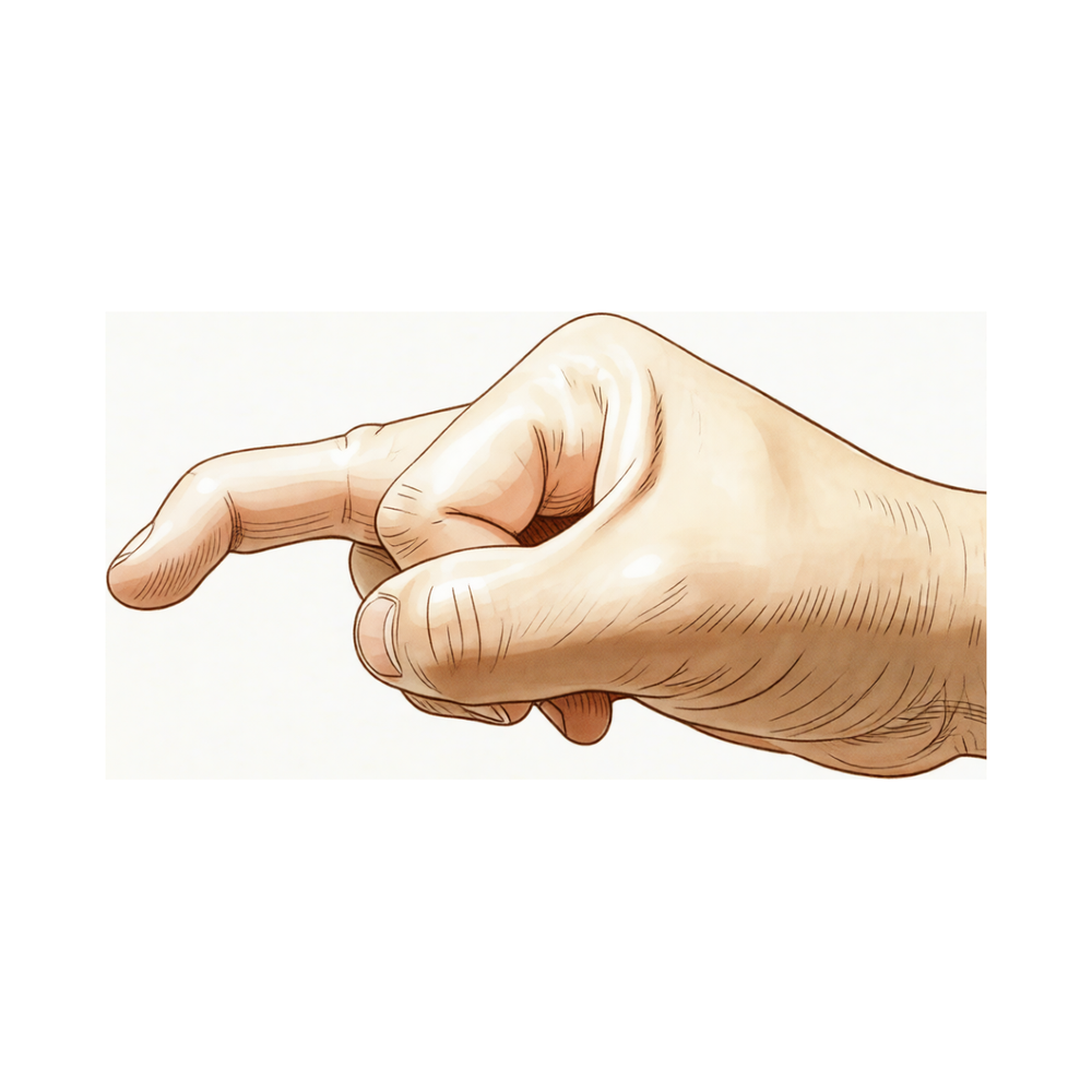
锤状指(Mallet finger)是指终末伸肌腱(即伸直手指最远端关节、靠近指甲的指间关节[DIP]的纤细肌腱)从骨头上撕脱所致。它通常发生在笔直的指尖被强行弯曲时,例如球类撞击或碰撞导致指尖“戳伤”。有时肌腱会撕下一小块骨片(称为骨性锤状指);有时肌腱则自行撕裂(称为肌腱性锤状指)。无论哪种情况,结果都是一样的:指尖下垂,且您无法自行伸直它,尽管手指的其他部分功能正常。
好消息是,这种损伤仅通过夹板固定即可非常可靠地愈合;大多数人无需手术。整个治疗基于一个简单原则:
- 在肌腱愈合期间,指尖关节必须保持完全伸直,且不得中断。 夹板使最后一个关节保持伸直(或略微过伸),以便撕裂的断端能够愈合。对于肌腱性锤状指,需全天佩戴(白天和晚上)约八周;对于骨性锤状指,则约需六周。
- 在此期间,绝不允许指尖弯曲。 如果指尖即使短暂下垂(例如在更换夹板或清洗时),愈合过程就会中断,时间需从零开始重新计算。因此,您保持指尖伸直的程度是决定手指恢复效果的最关键因素。
- 手指的其他关节保持自由活动。 近端指间关节(PIP)和掌指关节(MCP)不包含在夹板内,应从一开始就自由活动;活动这些关节不会干扰指尖的愈合。
在全天佩戴期结束后,逐渐减少夹板使用时间(首先仅用于夜间和高风险活动,然后完全停用),同时您开始缓慢地重新弯曲指尖。遗留约五至十度的轻微永久性下垂是正常的;这是预期内的情况,通常不影响手指功能,且大多数人对此结果非常满意。
注意事项与限制
- 在夹板固定期间,绝对不要让指尖弯曲,即使在清洗或更换夹板时,哪怕一秒钟也不行。如果指尖下垂,愈合过程将重置,固定期需重新开始计算。
- 全天佩戴夹板(白天和夜间),持续时间为治疗师设定的整个周期:肌腱型锤状指约8周,骨性锤状指约6周。
- 仅在清洁和干燥皮肤时取下夹板,且前提是你能全程保持指尖完全伸直(将其平放在桌面上,或用另一只手将其保持伸直状态)。
- 从开始起,保持中间关节和掌指关节活动自如;仅最后一个关节(远端指间关节)保持静止。
- 每日检查皮肤。如果关节上方的皮肤出现苍白、发白或疼痛,请告知手部治疗师;这可能意味着夹板将指尖过度向后牵拉,需要调整。
- 在手部治疗师开始减用夹板阶段之前,不要开始弯曲指尖。
关于伤口、肿胀和皮肤管理,请参阅本诊所的伤口护理指南。
您的锻炼
这些是您讲义中的锻炼方法。其中最重要的“锻炼”就是正确佩戴您的夹板,并确保指尖时刻保持伸直;其他所有内容都是围绕这一点展开的。在早期阶段,您的任务是保持夹板佩戴、保持皮肤健康,并确保其他手指关节活动自如。轻柔的指尖屈曲和限制伸直锻炼属于后期的夹板逐步停用阶段,只有在您的手部治疗师明确指示开始后才应进行。如果任何动作导致指尖下垂,请立即停止并恢复全天佩戴夹板。
您的临床方案
本页其余部分为夹板引导的锤状指康复分期临床方案。本节内容将提供给手部治疗师,每个阶段均以通俗易懂的语言解释当前发生的情况。愈合取决于远端指间关节(DIP)伸直的连续性:仅当在夹板固定期间远端指间关节从未发生过屈曲时,终腱(或撕脱的骨性碎片)才能愈合,而近端指间关节(PIP)和掌指关节(MCP)保持自由活动,因为它们的运动不会干扰终腱的愈合。患者依从性是决定预后的主要因素。
治疗前,需确认锤状指是肌腱型还是骨性,并复查影像学检查。使用远端指间关节伸直矫形器:Stack 型、热塑性材料型或掌侧/背侧铝泡沫型;夹板类型对预后无实质性影响,因此应根据贴合度、皮肤耐受性和患者依从性进行选择。将远端指间关节保持在完全伸直或轻微过伸位,但避免过度过伸(以防背侧皮肤苍白/溃疡风险)。对于骨性锤状指,优选远端指间关节保持伸直/中立位而非过伸位,以避免远节指骨掌侧半脱位。近端指间关节始终保持自由活动。
第一阶段——不间断的全职伸直位夹板固定(第0至6/8周)
指尖关节(DIP)在白天和夜间持续保持伸直位,以使肌腱或骨性碎片愈合。仅在皮肤护理时可取下夹板,且在整个过程中DIP必须保持伸直;任何一次DIP屈曲都会使愈合计时重新开始。近端指间关节(PIP)和掌指关节(MCP)自由活动。
致您的手治疗师:
教育与注意事项 - 适配DIP伸直矫形器(Stack/热塑性/铝泡沫),DIP处于完全伸直或轻微过伸位;避免过度过伸(皮肤苍白/溃疡);骨性锤状指→保持伸直/中立位,而非过伸位(半脱位风险) - 持续佩戴:肌腱性约8周,骨性约6周;在此窗口期内,DIP绝不可屈曲 - 教导平面更换夹板技术,确保DIP从不允许下垂;如果患者无法维持伸直位,则由治疗师进行更换 - PIP和MCP保持自由,并从第1天开始主动活动
管理 - 皮肤:每日检查DIP背侧和甲皱处的皮肤;如果出现苍白/受压,调整矫形器;保持清洁干燥 - 水肿:抬高患肢;轻柔的近端关节活动 - 锻炼:PIP和MCP完全主动活动范围(ROM);禁止DIP活动 - 骨性锤状指:在夹板固定期间保持影像学监测(对位/半脱位),因为夹板固定在伸肌滞后方面与克氏针固定相当,但必须监测碎片位置
进展标准 - 全职佩戴期已完成(肌腱性约8周/骨性约6周),且无超出可接受范围的DIP伸肌滞后,皮肤健康
第二阶段——逐步停用夹板并开始受控的远侧指间关节(DIP)活动(第6/8周,随后+2至6周)
全天候佩戴期结束后,若不存在或仅存在可接受的伸肌滞后,则夹板佩戴时间减少至夜间及高风险活动时佩戴,同时开始轻柔的受控DIP屈曲。夜间夹板佩戴可根据当前证据(一项I级研究认为其非必需)作为可选措施,并基于临床实际情况灵活使用。若出现明显的滞后复发,患者需重新回到全天候伸肌夹板佩戴。
供您手治疗师参考:
评估 - DIP主动伸肌滞后(度数)及主动屈曲;皮肤状况;患者指尖脱离夹板时的信心
教育与注意事项 - 逐步过渡至夜间及高风险活动时夹板佩戴,持续约2至6周;根据当前证据,夜间佩戴为可选 - 若夹板佩戴后伸肌滞后(>20°)复发,则恢复全天候伸肌夹板佩戴约4至6周
管理 - 练习:开始轻柔、分级DIP主动屈曲(先小范围)和DIP主动伸肌阻滞练习(稳定近侧指间关节PIP,伸展DIP);随着滞后改善逐步增加屈曲范围 - 一旦DIP能主动维持伸直位且无或仅有可接受(≤10–20°)的滞后,则减少日间夹板佩戴时间 - 继续进行PIP/MCP的全范围活动;根据需要处理瘢痕/皮肤护理 - 慢性或延迟就诊的锤状指仍对伸肌夹板治疗有反应;延迟开始治疗并非禁忌证
进阶标准 - DIP能主动维持伸直位且滞后在可接受范围内;恢复受控、无痛的DIP屈曲;皮肤完整
第三阶段——强化与回归(约第8至12周)
随着肌腱愈合且主动活动度恢复,手指完全脱离夹板,开始分级强化训练并逐步恢复活动。预期会出现轻微且持久的伸肌滞后(平均约8°),这与极佳的功能结果相容。
供您的手部治疗师参考:
评估 - 远侧指间关节(DIP)主动伸肌滞后及屈曲弧;握力;负荷及运动准备情况
教育与注意事项 - 日常使用无需夹板;回归运动期间进行接触性运动时需佩戴保护性夹板 - 告知患者残留约5–10°的伸肌滞后属正常现象,且不影响满意度
管理 - 练习:分级握力与捏力强化训练;全手指关节活动度(ROM);任务及运动特异性进阶训练 - 约第8至12周根据标准恢复运动或重体力工作(接触性运动需佩戴保护性夹板) - 当力量与功能充足且滞后稳定时出院;若滞后明显持续或复发,则转诊
恢复工作与活动
从开始即可对夹板固定的手进行轻度使用:夹板保持佩戴,指尖保持伸直,可在该限制范围内用于日常任务。小指尖夹板通常不会妨碍驾驶,只要您能够握住方向盘并安全操控车辆,但请在复查时咨询Hirpara医生。随着夹板逐渐停用,握力与力量训练约在6至8周时开始恢复。重返运动和较重体力劳动通常约在8至12周,依据的是恢复受控活动度而非仅凭日历时间,且在重返接触性运动期间需佩戴保护性夹板。预计指尖会有约5至10度的轻微永久性下垂;这属于正常现象,不影响手部功能,大多数人几乎不会注意到。
协议之后
本协议与诊所的一般康复建议并行;请参阅术后疼痛管理、伤口护理和瘢痕管理。上述分阶段计划反映了关于锤状指夹板固定的已发表指南,您的持续康复将由Hirpara医生和您的手部治疗师根据您指尖的进展情况个体化指导。
关于您的锤状指接受手术治疗时的说明
大多数锤状指患者无需手术。仅当为骨性锤状指,且骨折累及关节面较大(超过约三分之一),或末节指间关节出现半脱位(掌侧半脱位)时,才会考虑手术。若进行固定,通常采用伸肌腱阻滞(Ishiguro)克氏针固定,有时会在指尖关节临时穿入一根克氏针以维持伸直位。该克氏针通常保留约四至六周,在术后五至六周左右拔除,随后开始主动活动指尖,并可能继续佩戴夜间夹板约四周。现有证据表明,在最终下垂畸形程度方面,夹板固定与克氏针固定疗效相当(非劣效),因此手术仅保留用于上述特定情况,而非常规使用。
Evidence & references
Mallet Finger — Injury Outcomes & Splint-Led Rehabilitation (Terminal Extensor Tendon, DIP)
Topic scope: non-operative (and, where indicated, post-fixation) management of a mallet finger — disruption of the terminal extensor tendon at the distal interphalangeal (DIP) joint, either purely tendinous or with an avulsion bony fragment (bony mallet). This is a healing injury, not a reconstruction: the entire treatment is uninterrupted DIP extension splinting that holds the tendon (or fragment) in apposition while it unites, with the PIP and MCP left free.
Defining principle of the rehab here: the terminal extensor tendon heals only if the DIP is held in continuous extension and is never allowed to flex during the splinting period. Any single lapse into DIP flexion separates the healing ends and restarts the healing clock, which is why patient compliance is the dominant outcome driver. The PIP is deliberately kept mobile because proximal-joint motion does not disturb terminal-tendon healing. Splint type (Stack, thermoplastic, volar/dorsal alumifoam) does not materially change the outcome — fit, skin tolerance and compliance matter more than the device. The single branch point is the bony mallet with a large articular fragment or DIP volar subluxation, where surgical fixation is considered; even there, splinting is non-inferior to pinning for the final extensor lag, so operation is reserved rather than routine.
A. INJURY OUTCOMES (tendinous vs bony mallet; splinting vs fixation)
Mallet finger is one of the most reliably treated closed tendon injuries in the hand: the great majority heal well with splinting alone, and the principal debate is over the bony mallet — when, if ever, to fix it.
- Continuous extension splinting is the standard of care and works well for both tendinous and bony mallets, including chronic and delayed presentations, which still respond to splinting weeks after injury [Valdes systematic review LoE 1a; Salazar Botero review; Medscape; StatPearls]. Strong (SR + reviews).
- Splint type makes no meaningful outcome difference. A randomised comparison of splint designs found no superiority of one orthosis over another; the determinant is uninterrupted DIP extension and compliance, not the device [Pike RCT]. Strong (RCT).
- Splinting is non-inferior to extension-block pinning for the final extensor lag. A randomised trial comparing conservative extension splinting with operative extension-block K-wiring for bony mallet found no advantage to pinning in the residual lag, supporting non-operative management as the default even for many bony mallets [Thillemann RCT]. Strong (RCT).
- Surgery is reserved for the large bony fragment or subluxating DIP. Operative fixation is
considered when the fracture involves a large part of the articular surface (often cited as
~30%) or there is volar subluxation of the distal phalanx; common techniques are extension-block (Ishiguro) K-wiring with or without a trans-articular DIP pin. Single-K-wire constructs perform less well in non-compliant settings [Aksan; Salazar Botero; Medscape]. Moderate.
- Stack splints can subluxate a bony mallet. Volar-based Stack-type orthoses holding the DIP in hyperextension can displace a bony-mallet fragment / promote subluxation, which is why a straight/neutral DIP is preferred for bony mallets rather than hyperextension [Kaplan]. Moderate (mechanistic/clinical).
- The underlying mechanism is a terminal tendon avulsion at the distal phalanx. Anatomical and injury studies characterise the lesion as avulsion of the terminal extensor at its distal-phalanx insertion, and a very small amount of tendon lengthening translates into a large extensor lag — roughly 1 mm of lengthening ≈ 25° of lag — which is the biomechanical reason apposition must be maintained so strictly [Tuttle; Yeh; PMC current concepts]. Mechanistic.
B. REHABILITATION / THERAPY EVIDENCE
The central rehab questions are (1) how long and how strictly to splint, (2) whether the PIP should be included, and (3) whether night-time and post-splinting splinting are needed. The evidence supports uninterrupted full-time DIP extension splinting (~6–8 weeks) with the PIP free, followed by a weaning phase, and downgrades routine night-splinting to optional.
- Uninterrupted DIP extension is the active ingredient; the PIP must stay free. Splinting holds the DIP in full extension (or slight hyperextension) continuously; the PIP and MCP are mobilised from the outset because proximal-joint motion does not load the terminal tendon. Full-time wear is about 8 weeks for tendinous and 6 weeks for bony mallets [Valdes SR 1a; Salazar Botero; StatPearls; Physiopedia]. Strong (SR + guideline-level reviews).
- Compliance is the dominant outcome driver. Because any DIP flexion restarts healing, outcome tracks adherence to continuous extension more than any device choice; patient education and a safe flat-surface splint-change technique are central [Valdes SR; Cook BAHT survey of therapist practice]. Strong (mechanism + practice consensus).
- Avoid excessive hyperextension. Holding the DIP in marked hyperextension risks dorsal-skin blanching and pressure ulceration over the joint; slight hyperextension or neutral is sufficient, and bony mallets should be held straight/neutral to avoid fragment subluxation [Azad dorsal splinting outcomes; Kaplan]. Moderate.
- Night-time splinting after the full-time phase is non-essential (optional). A Level-I study found that continued night-splinting after the primary full-time period was not essential to the result, so the ~2–6 week post-splinting night/risky-activity phase is framed as optional and pragmatic rather than mandatory [Valdes SR 1a evidence base]. Moderate (Level I within SR).
- Recurrent lag responds to re-splinting. If an extensor lag (>~20°) recurs after the splinting period, a further ~4–6 weeks of full-time extension splinting is appropriate; chronic/delayed mallets likewise still respond [Salazar Botero; Medscape; StatPearls]. Moderate.
- A small residual extensor lag is the expected, satisfactory result. Most patients are left with a slight permanent lag (mean ~8°, typically 5–10°) that does not impair function or satisfaction; this should be counselled as normal rather than as failure [Salazar Botero; PMC current concepts; Physiopedia]. Moderate–strong (natural history).
Recovery trajectory (expected, evidence-anchored)
| Phase | Window | Restraint | Hand use / therapy focus | Strength / load | Notes |
|---|---|---|---|---|---|
| I — Continuous DIP extension splinting | Week 0–6/8 (bony ~6, tendinous ~8) | DIP held continuously extended; never flex the DIP | Full-time extension orthosis (Stack/thermoplastic/alumifoam); flat-surface splint changes only; PIP + MCP moved freely from day 1; daily dorsal-skin checks | No DIP loading; light splinted hand use | Any DIP flexion resets the clock; bony mallet held straight/neutral + radiographic surveillance |
| II — Weaning & controlled DIP motion | +2–6 weeks after full-time phase | Night / high-risk-activity splinting (night wear optional) | Begin gentle graded active DIP flexion + blocked active DIP extension; reduce day wear once lag ≤10–20° | Light functional load | Lag >20° recurring → re-splint full-time ~4–6 wk; chronic mallets still respond |
| III — Strengthening & return | From ~week 8–12 | None (protective splint for contact sport) | Splint-free use; graded grip/pinch strengthening; full ROM; sport-/work-specific progression | Grip/strength built up; driving once able to grip the wheel safely | Expect ~5–10° permanent lag (mean ~8°) — normal, satisfaction preserved |
(Phase windows mirror the precautions in the patient protocol; they are typical guides, not trial-derived deadlines.)
C. KEY CONTROVERSIES / EVIDENCE QUALITY
- Splint type. Stack vs thermoplastic vs volar/dorsal alumifoam — randomised data show no meaningful outcome difference; the determinant is uninterrupted extension and compliance, not the device [Pike RCT]. Strong evidence of equivalence.
- Splinting vs operative fixation for bony mallet. Randomised data show extension splinting is non-inferior to extension-block pinning for the residual lag; surgery is reserved for the large articular fragment (>~30%) or volar DIP subluxation, not used routinely [Thillemann RCT; Aksan; Salazar Botero]. Strong (RCT) for non-inferiority; moderate for the fixation indications.
- Hyperextension vs neutral. Slight hyperextension aids tendinous apposition but excessive hyperextension risks dorsal-skin ischaemia/ulcer, and in bony mallets can subluxate the fragment — hence straight/neutral for bony mallets [Azad; Kaplan]. Moderate.
- Is night-splinting necessary? A Level-I study found continued night-splinting after the full-time phase non-essential; the post-splinting phase is therefore optional/pragmatic rather than mandatory [Valdes SR 1a]. Moderate.
- Residual lag as expected outcome, not failure. A small permanent lag (mean ~8°) is the norm and is compatible with full function and satisfaction; mislabelling it as failure drives unnecessary intervention [Salazar Botero; PMC current concepts]. Strong natural-history data.
D. EVIDENCE STRENGTH FLAGS (summary)
- STRONG (RCT / SR): uninterrupted DIP extension splinting as standard of care (6–8 wk full-time, tendinous ~8 / bony ~6); splint-type equivalence; PIP-free mobilisation; compliance as the key outcome driver; expected ~5–10° residual lag; splinting non-inferior to pinning for bony mallet (with radiographic surveillance during splinting).
- MODERATE: exact length of the weaning/night-splinting phase (night wear non-essential per a Level-I study); strengthening and return-to-sport/work timing (~8–12 weeks, criterion-based); hyperextension-vs-neutral splint positioning and the bony-mallet subluxation caveat; surgical indications (>~30% articular fragment / volar subluxation) and fixation technique.
- WEAK / CONFIRM: driving — a fingertip splint is not usually a contraindication once the wheel can be gripped safely, but this is confirmed clinically rather than evidence-defined.
CITATIONS
RAG corpus (180,000+ Orthopaedic articles)
- A randomized controlled trial comparing splint designs for mallet finger. J Hand Surg Am. 2010. DOI: 10.1016/j.jhsa.2010.01.005
- Conservative management of mallet finger: a systematic review (Level of Evidence 1a). J Hand Ther. 2015. DOI: 10.1016/j.jht.2015.03.001
- Mallet finger: a survey of British Association of Hand Therapists practice. Hand Therapy. 2016. DOI: 10.1177/1758998316664822
- The mallet finger injury: a review (current concepts in diagnosis and management). Arch Plast Surg. 2016. DOI: 10.5999/aps.2016.43.2.134
- Outcomes of dorsal splinting for mallet finger. Hand (N Y). 2022. DOI: 10.1177/15589447221093674
- Conservative splinting versus extension-block K-wiring for bony mallet finger: a randomized controlled trial. J Hand Surg (Eur Vol). 2020. DOI: 10.1177/1753193420917567
- Tendon avulsion fractures of the distal phalanx (terminal extensor avulsion). Clin Orthop Relat Res. 2006. DOI: 10.1097/01.blo.0000205903.51727.62
- Tendon ruptures in the hand. Hand Clin. 2012. DOI: 10.1016/j.hcl.2012.05.040
- Single K-wire fixation of bony mallet finger in non-compliant patients. Arch Orthop Trauma Surg. 2021. DOI: 10.1007/s00402-021-03793-4
- Subluxation of bony mallet fractures with Stack splint immobilisation. J Hand Surg Am. 2013. DOI: 10.1016/j.jhsa.2013.08.111
Mallet-finger management literature (URLs)
- Medscape — Mallet Finger Treatment & Management. https://emedicine.medscape.com/article/1242305-treatment
- StatPearls — Mallet Finger (NCBI Bookshelf). https://www.ncbi.nlm.nih.gov/books/NBK459373/
- Current concepts in the management of mallet finger (PMC; ~1 mm terminal-tendon lengthening ≈ 25° extensor lag). https://pmc.ncbi.nlm.nih.gov/articles/PMC4022957/
- Physiopedia — Mallet Finger. https://www.physio-pedia.com/Mallet_Finger


