

部分腕关节融合术 资料
本方案指导您在 Mater Private Hospital Rockhampton 接受 Kieran Hirpara 医生进行的部分腕关节融合术(一种切除磨损的舟骨并将腕部中央的小骨融合的手术,通常是将头状骨与月骨进行头月融合)后的康复过程。方案从您的家庭锻炼计划开始,随后是专为您的手部治疗师编写的结构化临床方案:请在首次治疗就诊时携带此页面或其 PDF 文件,以确保您的康复过程协调一致。您的手部治疗师可能会根据康复进展调整计划。
如果您对术后伤口有任何疑虑,请联系诊所。通常,拍摄伤口照片并通过电子邮件发送以供审查会很有帮助。
预期效果
部分腕关节融合术用于治疗因陈旧性舟骨问题而沿特定路径发展至磨损(关节炎)的腕关节:即舟月韧带损伤(“SLAC”腕)或陈旧性未愈合的舟骨骨折(“SNAC”腕)。手术中会切除受损的舟骨,并将腕中部的骨骼融合在一起,以消除骨面间的摩擦。Hirpara 医生最常进行头状骨与月骨的融合(头月融合),有时也会包括邻近的骨骼;当月骨、头状骨、三角骨和钩骨全部融合时,称为四角融合,其康复原则相同。
该手术的核心理念是仅融合腕关节的一部分,而非全部。月骨与前臂骨(桡骨)之间的关节被刻意保留。正是这一保留的关节使腕关节能够保持活动:
- 融合磨损面可消除疼痛: 这是主要目标,且能可靠实现。
- 保留桡骨-月骨关节意味着保留有用的活动度。 代价是活动范围减小:大多数患者最终保留约二分之一到三分之二的原有屈曲范围,握力约为对侧的四分之三。这是正常且预期的结果(并非并发症),对于疼痛且磨损的腕关节而言,这通常是一笔非常值得的交易。
融合的骨骼需要时间牢固结合,就像骨折愈合一样。在最初六周左右,腕关节通过支具保持静止,以便骨骼愈合。在此期间,手指、拇指和前臂应保持自由活动,但腕关节本身需休息。一旦外科医生通过X光确认骨骼已愈合,腕关节活动和随后的强化训练将分阶段谨慎恢复。早期设定合理预期(获得舒适且实用的腕关节,而非完全灵活的活动度)是康复过程中的重要组成部分。
注意事项与局限性
- 在外科医生确认骨融合已愈合之前(通常约为六周),请保持手腕在支具中静止不动:在手腕活动之前,骨骼必须完成连接。
- 从第一天起,请保持手指、拇指和前臂的活动,但在获得许可之前,切勿活动手腕本身。
- 在确认骨融合牢固之前,切勿用力抓握、提举、推、拉或通过手腕承重:这旨在保护愈合中的骨骼以及任何钢板、螺钉或骨钉。
- 预期最终活动范围会减少:这是融合部分腕关节的计划结果,并非表示出现任何问题的迹象。
- 保持支具和敷料清洁干燥,且在佩戴支具期间或无法安全控制方向盘时,切勿驾驶。
有关伤口、肿胀和疤痕管理,请参阅本诊所的伤口护理指南。
您的练习
Kieran Hirpara 4.0
手指和拇指运动
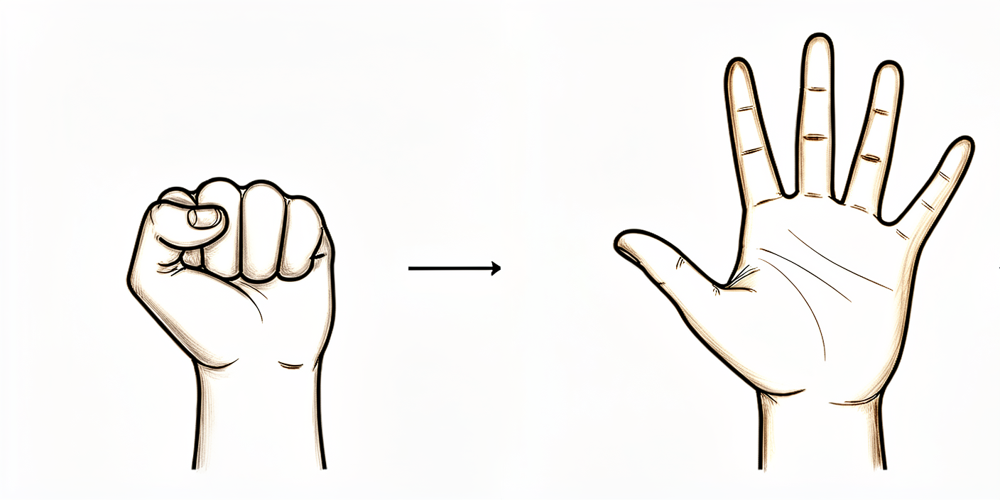
从手术当天起,在手腕本身于石膏或支具中保持静止的同时,请持续活动您的手指和拇指。先将所有手指完全伸直,然后握紧完整的拳头,接着依次用拇指触碰每个手指的指尖。这可以防止手部僵硬,并在腕骨愈合期间保持肌腱的滑动。请勿活动手腕本身。
每项10次,每天数次,从一开始起每天进行
Kieran Hirpara 4.0
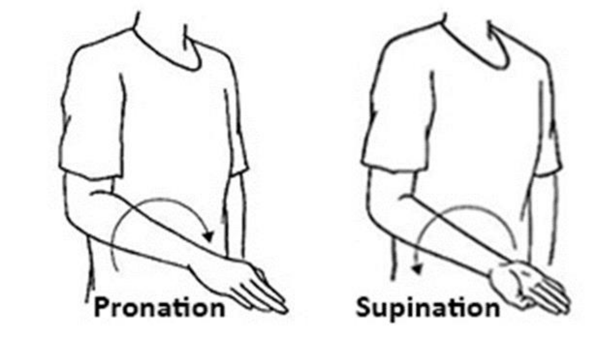
前臂旋转(掌心向上 / 掌心向下)
将肘部紧贴身体一侧并屈曲成直角,轻轻将手掌向上翻转以朝向天花板,然后向下翻转以朝向地面。确保动作源自前臂,而非手腕。这有助于保持前臂的柔韧性,且不会干扰愈合中的骨融合,因此可立即开始进行。
每个方向10次,每天2–3次
Kieran Hirpara 4.0
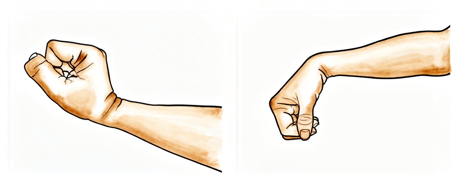
腕部活动(愈合后)
后期练习——仅在融合愈合且您的外科医生和手部治疗师确认腕部活动无碍后方可进行(通常在术后约六至八周)。轻轻将腕部向上(向后)和向下(向前)弯曲,然后向拇指侧和小指侧倾斜,活动范围以舒适为限。请记住,腕部融合部位不会活动——您正在恢复的是来自保留关节的活动,因此最终的活动范围会比之前小,这是预期内的情况。
每个方向10次,每天2–3次,以舒适为度(愈合后)
Kieran Hirpara 4.0
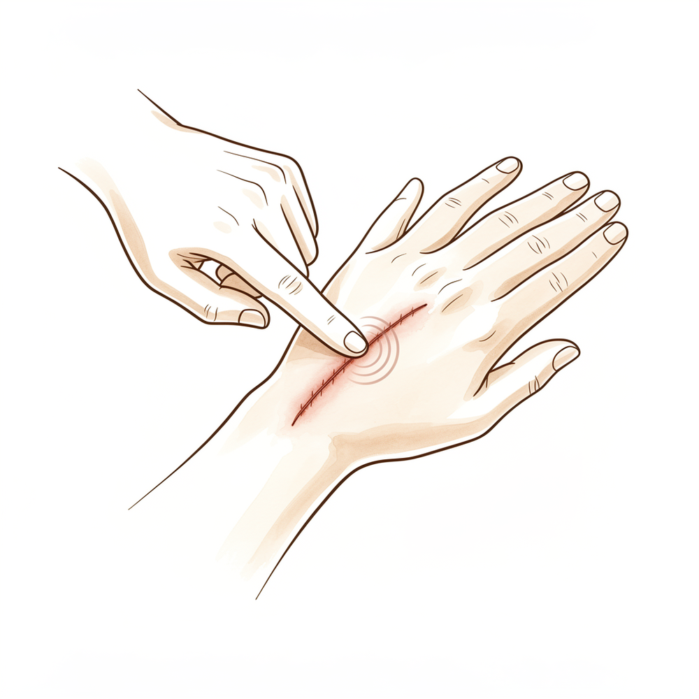
瘢痕护理
待伤口完全愈合且干燥后,取少量无香型润肤霜按摩瘢痕,用对侧拇指以小而有力的圆圈动作按摩数分钟。此举可软化瘢痕并降低敏感度。请勿在伤口未闭合且治疗师未确认前开始操作。
2–3 分钟,每日两次,待伤口愈合后
Kieran Hirpara 4.0
握力强化
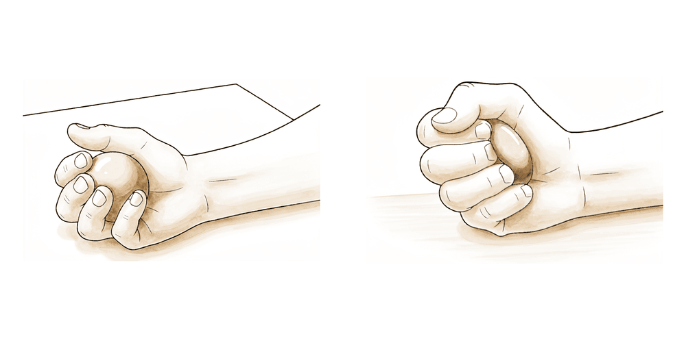
后期练习——仅在融合部位牢固且获准进行强化训练后方可开始(通常从约十二周起)。在手掌中挤压软球或治疗用橡皮泥,短暂保持,然后松开。在数周内逐步增加用力程度。在您的外科医生确认骨骼完全愈合之前,暂不进行用力抓握、提举重物及经手腕负重。
10–15 挤压,2–3 次/天,逐步增加(后期)
这些是您的手册中列出的练习。请仅在Hirpara医生和您的手部治疗师的指导下开始,并严格保持在您被允许的范围内和限制内。早期的练习旨在保持手指、拇指和前臂的活动,同时不干扰愈合中的融合部位;手腕本身需保持静止,置于石膏或支具中。手腕活动和握力强化属于后期阶段,在您的外科医生确认骨骼已愈合之前,不应开始。停止任何在手腕处引起剧烈疼痛的动作。
您的临床方案
本页其余部分为部分腕关节融合术(月骨-头状骨融合 ± 舟骨切除术;四角融合术适用相同原则)后康复的分阶段临床方案。本节内容应提供给您的手部治疗师,每个阶段均以通俗易懂的语言解释正在发生的情况。与肌腱修复不同,此处的结构是骨性的,且进展取决于融合部位的影像学愈合,而非固定的时间表。在外科医生确认愈合之前,腕关节需保持固定,仅对手指、拇指和前臂进行活动;此后恢复腕关节活动范围,然后恢复负重,现实的上限约为对侧屈伸活动度的50–65%以及约70–80%的握力。
在治疗前,请查阅患者的手术报告,并确认所使用的固定方式(环形/背侧钢板、无头加压螺钉、骨钉或克氏针)以及是否切除了舟骨。在治疗外科医生确认影像学愈合之前(通常为6–8周,若使用螺钉或骨钉固定则可能需要更长时间),切勿开始腕关节活动。从首次就诊起,即向患者说明,目标是实现无痛、功能正常的腕关节,其活动范围有意减少,而非完全的活动度。
第一阶段——直至骨性愈合的制动保护期(第0周至约6–8周)
融合部位的愈合过程类似于骨折愈合,因此在骨骼连接期间需保持腕部静止。手部和前臂应保持完全活动,以防止僵硬和肌腱粘连,但腕部不得活动。
致您的手部治疗师:
健康教育与注意事项 - 腕部需通过石膏或支具制动,直至外科医生确认影像学上的骨性愈合(通常为6–8周) - 在此阶段禁止任何主动或被动腕部活动 - 禁止通过手术侧腕部进行抓握、提举、推、拉或负重 - 尽早设定预期:最终活动范围将减少(桡月关节得以保留;中腕关节已融合)
管理措施 - 伤口:按医嘱使用外科敷料;厚敷料/支具固定约10–14天,随后更换为短臂石膏或热塑性支具;监测感染迹象 - 水肿:抬高患肢至心脏水平以上,进行轻柔的手部泵血运动,必要时冰敷 - 练习:手指、拇指及掌指/指间关节的完全主动活动范围;前臂旋前/旋后;轻柔的肩部和肘部活动范围练习;禁止腕部活动
进入下一阶段的指征 - 外科医生确认影像学骨性愈合(切勿仅依据日历时间推进);伤口愈合;肿胀得到控制
第二阶段——恢复腕部活动度(自骨性愈合起,约第6–8周至第12周)
一旦外科医生确认融合牢固,腕部即可脱离石膏,开始轻柔的腕部活动。进展是渐进的;需提醒患者,融合部位将不再活动,且可达到的活动范围小于术前。
致您的手部治疗师:
评估 - 腕部主动和被动活动度(屈曲/伸展,桡偏/尺偏)、前臂旋转、握力基线、疼痛与肿胀、伤口/瘢痕检查
宣教与注意事项 - 在舒适范围内开始腕部主动及主动辅助活动度训练;在两次治疗之间,过渡至可拆卸腕部支具以提供舒适/保护 - 在获得强化训练许可前,继续避免通过腕部进行重握、负重提举及负重 - 强化活动度降低的预期(目标约为对侧屈曲-伸展活动范围的50–65%)
管理 - 练习:腕部主动/主动辅助屈曲、伸展、桡偏和尺偏;伤口完全愈合后开始瘢痕按摩与脱敏;继续全手指/拇指活动度及前臂旋转;按需进行水肿管理
晋级标准 - 在预期的受限范围内,腕部活动弧舒适且可控;疼痛趋于稳定;外科医生许可进行强化训练
第三阶段——强化与功能恢复(约12周起)
随着融合牢固且活动度恢复至其有效上限,开始进行强化训练和分级负重,并在数周内逐步增加强度。恢复手工劳动和体育运动需基于标准评估。
致您的手部治疗师:
评估 - 握力和捏力与对侧对比;负重时的疼痛/肿胀反应;任务及工作相关的功能性测试
教育与注意事项 - 一旦获得许可,开始渐进性握力和腕部强化训练(使用治疗泥、球,随后进行分级阻力训练) - 逐步引入负重和承重任务;应在数周内逐步增加强度,而非一次性完成 - 预期终点:握力恢复至对侧的约70–80%,且活动范围有用、无痛且有所减小
管理 - 练习:渐进性抓握和前臂/腕部强化;分级功能性及工作模拟训练;持续的瘢痕管理和关节活动度(ROM)维持 - 若出现伸腕时持续性背侧腕部疼痛(可能为背侧撞击)、疑似骨不连或恢复停滞,需警惕并转回外科医生处 - 一旦力量和功能足以满足患者的日常及职业需求,可考虑出院
恢复负重/工作的标准 - 融合牢固愈合,在恢复的活动范围内无痛,握力足以应对任务;重体力手工需求需推迟至约4–6个月,并逐步增加强度
恢复工作与活动
鼓励从初期开始进行轻度日常手部使用(如进食、书写、轻度自我护理),以舒适为限,前提是不要对腕关节施加负荷或扭转。由于在腕部佩戴石膏或无法安全控制方向盘期间不得驾驶,请在早期几周内安排交通协助;待拆除石膏且能自信地控制车辆后(经复查确认),方可恢复驾驶。
抓握、提举及通过腕部承重需等待融合确认牢固(通常在约六至八周后),随后逐步增加强度。从事办公室工作或轻体力劳动者通常在约三个月时返回工作岗位;较重的体力劳动通常在四至六个月左右恢复,并分阶段重新引入。在整个过程中,请记住,预期结果是腕关节舒适且功能可用,但活动范围有所减少;评估标准应基于腕关节的感觉与功能,由Hirpara医生及您的手部治疗师指导恢复节奏,而非仅依据日历时间。
您的方案之后
本方案与诊所的总体康复建议配合使用:请参阅术后疼痛管理、伤口护理和疤痕管理。上述分阶段计划反映了部分腕关节融合术后已发表的康复指南,您的持续康复将由Hirpara医生和您的手部治疗师根据您腕部的愈合和进展情况,进行个体化指导。
Evidence & references
This is the clinical evidence summary written for health professionals. It is technical, and it lists the research this page was built from. You do not need to read it to understand your treatment or to make a decision about it.
Topic scope: post-operative rehabilitation after midcarpal partial wrist arthrodesis for scapholunate advanced collapse (SLAC) or scaphoid nonunion advanced collapse (SNAC). The index procedure here is a capitolunate fusion (the capitate fused to the lunate, usually with excision of the worn scaphoid); the closely related four-corner fusion (lunate–capitate– triquetrum–hamate) follows the same rehabilitation principles. This is a bony arthrodesis, so the rehabilitation is a union-gated pathway: the mid-wrist is immobilised until the fusion consolidates, after which a deliberately reduced wrist arc and grip are restored.
Defining principle of the rehab here: a partial wrist fusion is a motion-preserving salvage. The arthritic midcarpal surfaces are fused to abolish pain, while the radiolunate joint is intentionally preserved to retain movement. The construct that needs protecting is healing bone (plate, headless screws, staples or K-wires across the fusion), not a soft-tissue repair — so progression is gated by radiographic union, not a calendar. Counselling the patient that the planned end-point is a pain-free, functional wrist with a reduced arc (~50–65% of normal flexion–extension, ~70–80% grip) is itself part of the treatment: the reduced motion is the intended trade-off, not a failure. The two principal branch points are the fixation method (which sets the immobilisation window and union risk) and whether scaphoid excision was performed (standard in the SLAC/SNAC setting).
A. PROCEDURE OUTCOMES (capitolunate / four-corner fusion for SLAC–SNAC)
Partial wrist fusion is a well-established, durable salvage for the SLAC/SNAC wrist. The dominant debate is which motion-preserving salvage (four-corner / capitolunate fusion vs proximal row carpectomy), not whether to operate; both reliably relieve pain at the cost of some motion.
- Reliable pain relief with a useful but reduced arc. Across series, midcarpal fusion abolishes the painful midcarpal arthritis while preserving radiolunate motion. Typical results are roughly 50–65% of contralateral flexion–extension and ~70–80% of contralateral grip strength, with high rates of pain relief and return to work — the expected, planned trade-off of a partial fusion [Enna Hand Clin 2005; Strauch J Hand Surg Am 2011; Merrell J Hand Surg Am 2008; long-term series J Wrist Surg 2015]. Moderate (cohort/consensus).
- Capitolunate fusion (± scaphoid excision) performs comparably to full four-corner fusion while fusing fewer joints, simplifying the construct. A systematic review of capitolunate arthrodesis and comparative work report comparable motion, grip and union to four-corner fusion, supporting it as a sound index choice [Dunn J Hand Surg Am 2020 (systematic review); lunocapitate series J Hand Surg Eur 2009; J Chin Med Assoc 2017]. Moderate.
- A meta-analysis of two-, three- and four-corner constructs finds the number of fused columns does not materially change motion, grip, union or complications — biomechanically consistent with the radiolunate joint being the motion-determining segment [Hundepool J Hand Surg Am 2025 (SR/meta-analysis); Hernandez-Soria J Hand Surg Am 2016 (capitate-position biomechanics)]. Moderate (SR) + mechanistic.
- Modern fixation is forgiving but union is not guaranteed. Circular dorsal plates, headless compression screws and nitinol staples all achieve consolidation in the great majority, with nonunion and dorsal impingement the characteristic failures to watch for [Merrell J Hand Surg Am 2008 (circular plate); Ahmady J Hand Surg Glob Online 2025 (nitinol staples)]. Moderate.
B. REHABILITATION / THERAPY EVIDENCE
The rehab questions are (1) how long to immobilise, (2) what gates the start of wrist motion, and (3) how to set the patient's expectation of reduced motion. The evidence and published surgeon protocols converge on a union-gated sequence: immobilise the wrist (mobilise the hand and forearm) for ~6–8 weeks, then restore a reduced arc, then strengthen.
- Immobilise until radiographic union, not by the calendar. Because the construct is bony, wrist motion is withheld until the surgeon confirms consolidation — typically 6–8 weeks, longer with some screw/staple fixations. Published institutional protocols use a bulky dressing/splint for ~10–14 days, a short-arm cast to ~4–6 weeks, then a removable splint as motion begins. Moderate (consensus protocols).
- Keep the hand and forearm fully mobile from day one. Immediate active finger, thumb and forearm rotation prevents stiffness and tendon adhesion without disturbing the fusion — the same glide-preserving logic used across hand rehab. Consensus.
- Restore motion gradually after union, against a realistic ceiling. Once united, active and active-assisted wrist ROM is introduced; patients should be counselled that the fused midcarpal segment will not move and the achievable arc is smaller than pre-operatively (the radiolunate joint alone supplies wrist motion). Consensus + mechanistic (capitate-position biomechanics, Hernandez-Soria 2016).
- Strengthen and load only once the fusion is solid. Grip and loaded/weight-bearing work begins after union and is built up gradually; heavy manual demands are deferred to ~4–6 months. Consensus.
Recovery trajectory (expected, evidence-anchored)
| Phase | Window | Restraint | Hand use / therapy focus | Strength / load | Notes |
|---|---|---|---|---|---|
| I — Immobilisation (union) | Week 0 to ~6–8 | Wrist immobilised (cast/splint) | Full active finger, thumb & forearm motion; oedema control, elevation; no wrist motion | None through the wrist | Fusion knits like a fracture; progress is gated by radiographic union, not the calendar |
| II — Restoring wrist motion | From union (~wk 6–8) to 12 | Heavy-load avoidance; removable splint | Begin active/active-assisted wrist flexion/extension & deviation; scar massage once healed | No loaded grip yet | Counsel the reduced-arc expectation (~50–65% of normal flexion–extension) |
| III — Strengthening & return | From ~12 weeks (post-union) | Restrictions lifted progressively | Progressive grip/wrist strengthening; graded job simulation | Grip recovers toward ~70–80% of the other side | Desk work ~3 months; heavy manual ~4–6 months. Watch for dorsal impingement / nonunion |
(Phase windows mirror the precautions in the patient protocol and published surgeon protocols; they are typical guides anchored to union, not trial-derived deadlines.)
C. KEY CONTROVERSIES / EVIDENCE QUALITY
- Capitolunate vs four-corner vs proximal row carpectomy (PRC). All three are accepted motion-preserving salvages for SLAC/SNAC. PRC tends to give a slightly larger arc and avoids fusion-specific complications (nonunion, dorsal impingement, hardware), while fusion may give marginally better grip and is preferred where capitate-head or proximal-capitate cartilage is compromised. Systematic reviews and a meta-analysis find no consistent superiority of one over the other; choice is individualised [Mulford J Hand Surg Eur 2009; J Hand Surg Am 2024 meta-analysis; Strauch J Hand Surg Am 2011]. Moderate (SR/meta-analysis of mostly observational data).
- Capitolunate vs full four-corner construct. Fusing fewer columns (capitolunate) simplifies the construct without clearly compromising motion, grip or union versus four-corner — consistent with the radiolunate joint being the motion-determining segment [Dunn 2020 SR; Hundepool 2025 meta-analysis; Hernandez-Soria 2016]. Moderate.
- When can wrist motion safely start? Union timing varies with fixation, and protocols differ on exact cast duration. The defensible position is surgeon-confirmed radiographic union gates wrist ROM rather than a fixed week number. Weak–moderate (consensus; protocol variation).
- Reduced motion is the plan, not a complication. The preserved radiolunate joint supplies a smaller arc by design; mislabelling the expected ~50–65% range as a poor result drives unnecessary dissatisfaction. Strong mechanistically; cohort-supported.
- Characteristic failures: nonunion and dorsal impingement. Both are recognised, fixation-related complications; persistent dorsal wrist pain on extension or a non-progressing fusion warrants surgical review rather than more therapy [Merrell 2008; Ahmady 2025; long-term series 2015]. Moderate.
D. EVIDENCE STRENGTH FLAGS (summary)
- STRONG (mechanistic / well-supported): the reduced final arc is the planned consequence of preserving the radiolunate joint while fusing the midcarpal segment (capitate-position biomechanics); reliable pain relief from the fusion.
- MODERATE: typical outcome envelope (~50–65% flexion–extension, ~70–80% grip); equivalence of capitolunate and four-corner constructs; no consistent superiority of fusion vs PRC (SR/meta-analysis of largely observational data); modern fixation achieves high union with defined nonunion / dorsal impingement risk.
- WEAK / CONSENSUS: the specific union-gated immobilise → restore-motion → strengthen therapy sequence and exact phase timings (institutional/surgeon protocols, anchored to radiographic union rather than trial-derived).
CITATIONS
RAG corpus (180,000+ Orthopaedic articles)
- Enna M, Hoepfner P, Weiss AC. Scaphoid excision with four-corner fusion. Hand Clin. 2005. DOI: 10.1016/j.hcl.2005.08.012
- Dunn JC, Polmear MM, Scanaliato JP, et al. Capitolunate arthrodesis: a systematic review. J Hand Surg Am. 2020. DOI: 10.1016/j.jhsa.2019.10.007
- Hundepool CA, Duraku LS, Quanjel TJ, et al. Two-, three-, or four-corner arthrodesis for midcarpal osteoarthritis: a systematic review and meta-analysis. J Hand Surg Am. 2025. DOI: 10.1016/j.jhsa.2023.04.018
- Strauch RJ. Scapholunate advanced collapse and scaphoid nonunion advanced collapse arthritis — update on evaluation and treatment. J Hand Surg Am. 2011. DOI: 10.1016/j.jhsa.2011.01.018
- Merrell GA, McDermott EM, Weiss AC. Four-corner arthrodesis using a circular plate and distal radius bone grafting: a consecutive case series. J Hand Surg Am. 2008. DOI: 10.1016/j.jhsa.2008.02.001
- Hernandez-Soria A, Das De S, Model Z, et al. The effect of capitate position on coronal plane wrist motion after simulated 4-corner arthrodesis. J Hand Surg Am. 2016. DOI: 10.1016/j.jhsa.2016.07.101
- Ahmady AA, Zalzaleh M, Riedel BB. Midterm outcomes of four-corner fusion surgery using nitinol staples. J Hand Surg Glob Online. 2025. DOI: 10.1016/j.jhsg.2025.100805
- Mulford JS, Ceulemans LJ, Nam D, Axelrod TS. Proximal row carpectomy vs four corner fusion for scapholunate (SLAC) or scaphoid nonunion advanced collapse (SNAC) wrists: a systematic review of outcomes. J Hand Surg Eur Vol. 2009. DOI: 10.1177/1753193408100954
- Four-corner fusion versus proximal row carpectomy for scapholunate advanced collapse and scaphoid nonunion advanced collapse wrist: a systematic review and meta-analysis. J Hand Surg Am. 2024. DOI: 10.1016/j.jhsa.2024.01.011
- Long-term results of lunocapitate arthrodesis with scaphoid excision for SLAC and SNAC wrists. J Hand Surg Eur Vol. 2009. DOI: 10.1177/1753193409105683
- Lunocapitate fusion with scaphoid excision for the treatment of scaphoid nonunion advanced collapse or scapholunate advanced collapse wrist. J Chin Med Assoc. 2017. DOI: 10.1016/j.jcma.2016.10.001
- The long-term outcome of four-corner fusion. J Wrist Surg. 2015. DOI: 10.1055/s-0035-1549277
Partial wrist fusion rehabilitation literature (protocols & guidance — basis for the union-gated phase structure)
- University of Virginia Department of Orthopaedic Surgery. Wrist Partial Fusion (4-Corner) Rehabilitation Guidelines. https://med.virginia.edu/orthopaedic-surgery/wp-content/uploads/sites/242/2024/09/Wrist-Partial-Fusion-4-corner.pdf
- Oregon Health & Science University (OHSU). Four-Corner Partial Wrist Fusion — Surgery Guide. https://www.ohsu.edu/sites/default/files/2020-12/Four-Corner%20Partial%20Wrist%20Fusion.pdf
- Alaska Orthopedic Specialists. Four-Corner Fusion with Scaphoid Excision — Post-operative Protocol. https://www.akortho.com/wp-content/uploads/Four-Corner-Fusion-with-Scaphoid-Excision-1.pdf
- Twin Cities Orthopedics (Olson). Scaphoid Excision 4-Corner Fusion Post-operative Protocol. https://www.toportho.com/wp-content/uploads/2024/10/olson-_4-Corner-Fusion-Protocol.pdf
- The long-term outcome of four-corner fusion (open-access full text). PMC. https://pmc.ncbi.nlm.nih.gov/articles/PMC4408128/





