

肩峰下减压 资料
本方案适用于在 Mater Private Hospital Rockhampton 由 Kieran Hirpara 医生实施的孤立性关节镜下肩峰下减压术(肩峰成形术,伴或不伴炎性滑囊切除)术后的康复。请在首次物理治疗就诊时携带本页面或其 PDF 版本,以确保您的康复过程协调一致。您的物理治疗师将根据您的肩部恢复情况,通过以下阶段个体化地推进您的康复进程。
重要提示: 肩峰下减压术常与肩袖修复术联合进行。本方案仅适用于孤立性肩峰下减压术。如果您的手术还包含肩袖修复术,请遵循肩袖修复术方案;修复后的肌腱决定了更慢的恢复节奏。
如果您对术后伤口有任何疑虑,请联系诊室。拍摄伤口照片并通过电子邮件发送以供审阅通常很有帮助。
预期情况
肩峰下减压术通过磨除肩峰(肩部的骨性屋顶)下表面并切除发炎的滑囊,为肩袖肌腱腾出更多空间。由于没有需要保护的修复部位,早期康复的重点在于活动而非休息:从第一天起肩部即可安全活动,早期活动有助于防止其在恢复过程中僵硬。
您醒来时佩戴的悬吊带仅用于提供舒适感,并非用于保护任何部位。请尽可能长时间地取下悬吊带,并尽早停止使用:大多数人会在最初几天内取下悬吊带,已发表的方案最迟要求在两周内完全停用。任何肩部手术后,至少六周内请勿驾驶,即使悬吊带已取下;您的外科医生通常会允许您在六周复查时恢复驾驶。
您的锻炼计划包含三种类型的运动,物理治疗师将指导您在每个阶段适用哪些:
- 主动活动范围: 允许在无辅助或帮助的情况下进行运动。
- 主动辅助活动范围: 使用另一只手臂或物体辅助移动手臂。
- 被动活动范围: 完全放松,由另一只手臂或外力完成 100% 的工作。
康复进程概览:
- 第一阶段 — 早期活动 — 大约前两周
- 第二阶段 — 恢复活动范围并开始力量训练 — 第 2–6 周
- 第三阶段 — 力量强化 — 第 6–12 周
- 第四阶段 — 恢复完全活动 — 第 12 周起
在早期几周,肩部通常表现为疼痛而非脆弱,有些人可能会发现不适感持续长达六周。已发表的指南建议,大多数人在约三个月时疼痛会有明显改善,症状可能在长达一年的时间内持续改善。以下周数范围是典型值而非固定值;您的物理治疗师将根据肩部的活动情况而非日历来推进您的康复进程。
第一阶段 — 早期活动(第 0–2 周)
Kieran Hirpara 4.0
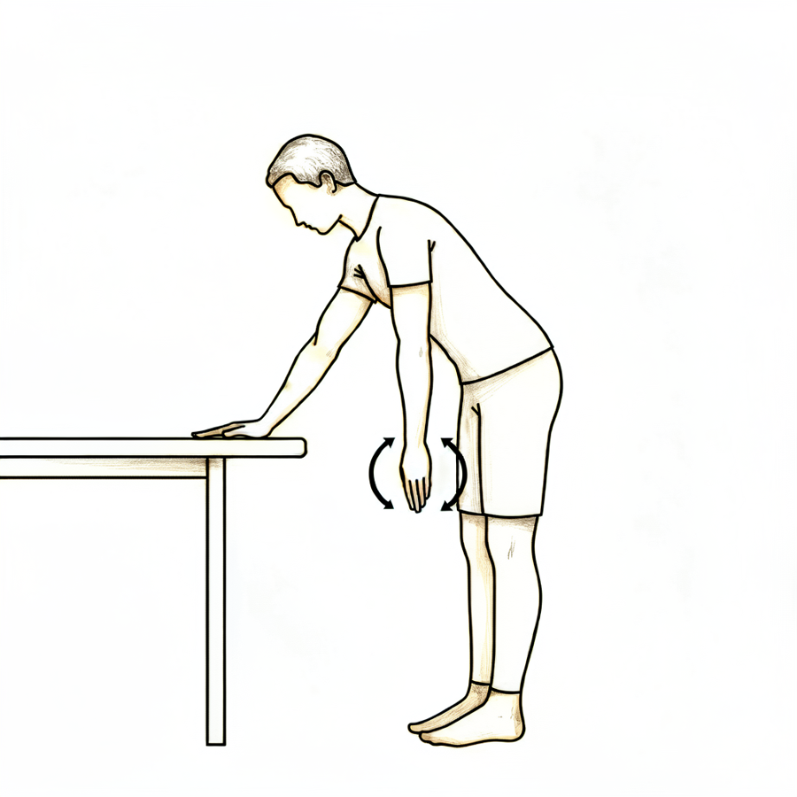
钟摆运动
腰部前屈,让手术侧手臂自然下垂,远离身体。让手臂做小而放松的圆周摆动——动作应来自躯干,而非肩部肌肉。在舒适范围内活动;以疼痛为指引。
在您的物理治疗师指导下
Kieran Hirpara 4.0
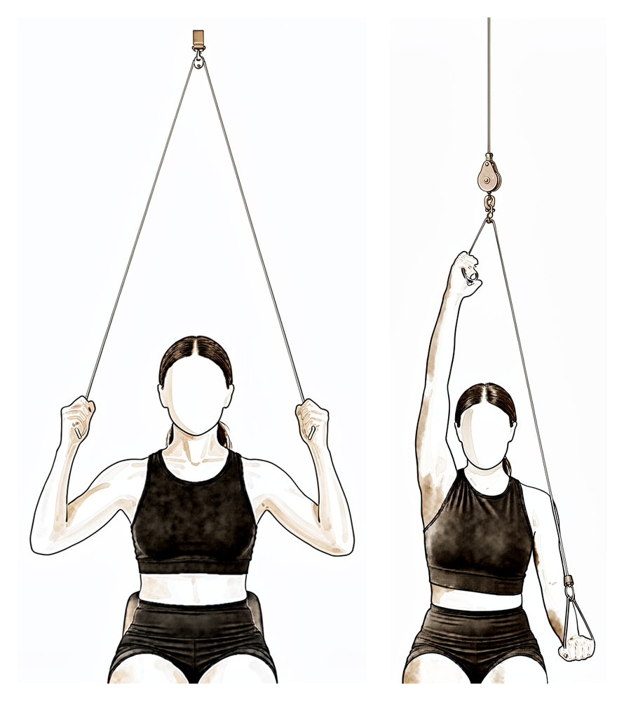
滑轮辅助抬高
坐在门上滑轮装置下方,双手各握一个手柄。用健侧手臂向下拉,使手术侧手臂向上并略向身体前方抬起,保持该手臂放松,然后有控制地放下。切勿强行或过度拉伸——在舒适的活动范围内进行。
在您的物理治疗师指导下
Kieran Hirpara 4.0
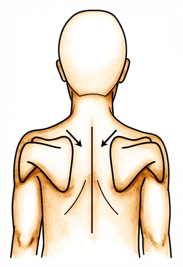
肩胛骨定位练习
坐直,轻轻将肩胛骨向后、向下收拢,使其远离耳朵。保持几秒钟,然后放松。全天注意保持良好姿势,配合此练习。
在您的物理治疗师指导下
Kieran Hirpara 4.0
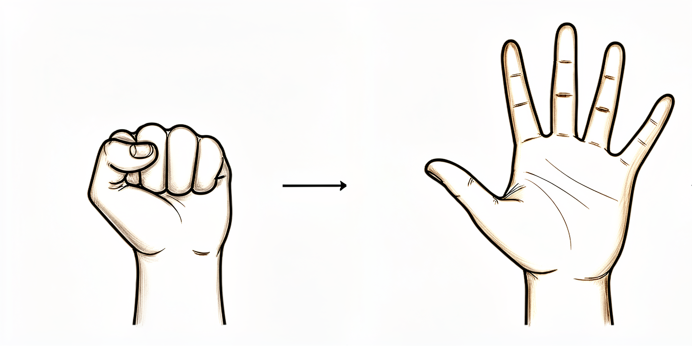
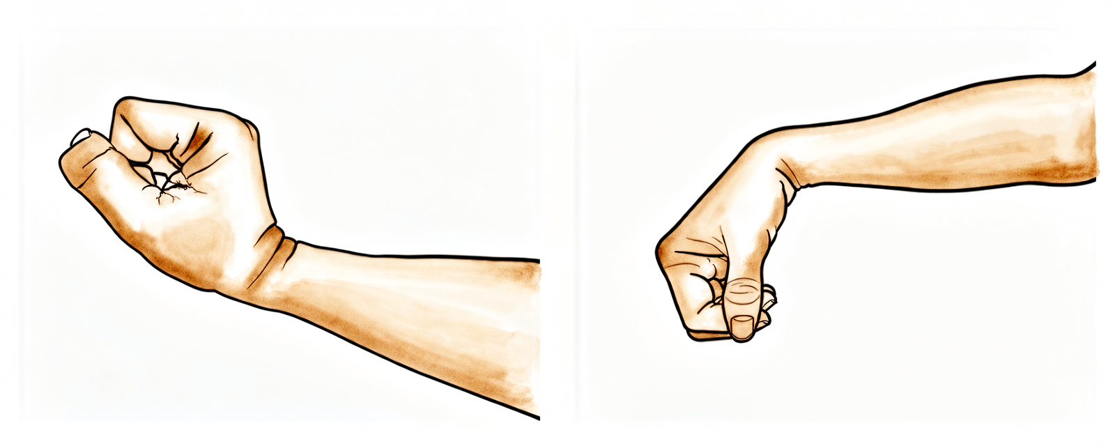
张开和握紧手
通过反复张开和握紧手部及手指,或挤压软球来保持手部和手指的活动。请立即开始此练习,以便在肩部逐渐恢复稳定的过程中维持手部和手臂的功能。
10 次,每日三次
Kieran Hirpara 4.0
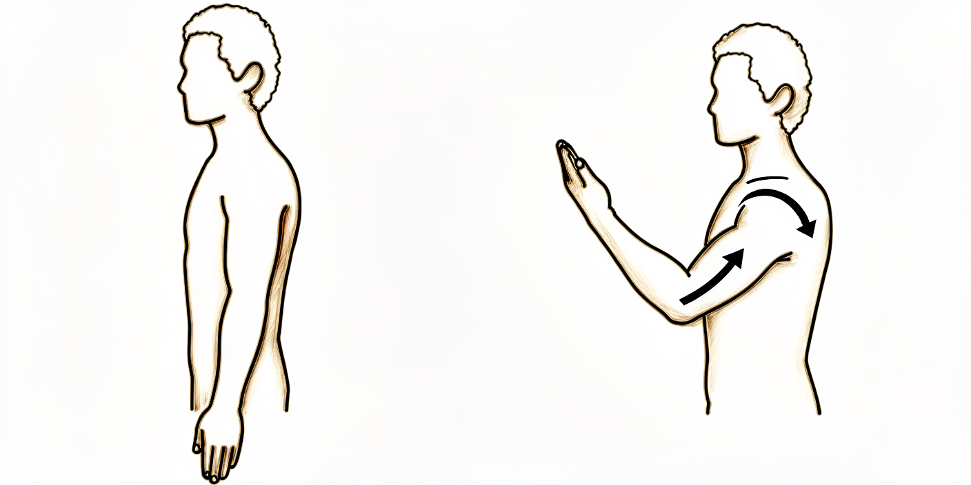
腕部运动
通过向前、向后及左右侧屈腕部来保持手腕活动。从第一天起就进行此练习,以便在肩部恢复期间保持手臂功能。
10 次,每日三次
Kieran Hirpara 4.0
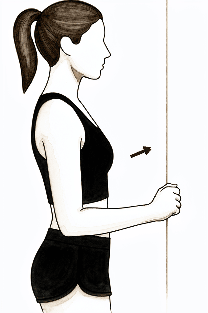
肘部弯曲
上臂自然垂放于体侧,将肘关节弯曲至舒适的最大范围,然后完全伸直。仅肘部活动——保持肩部放松。从第一天开始进行此练习。
10 次,每日三次
Kieran Hirpara 4.0
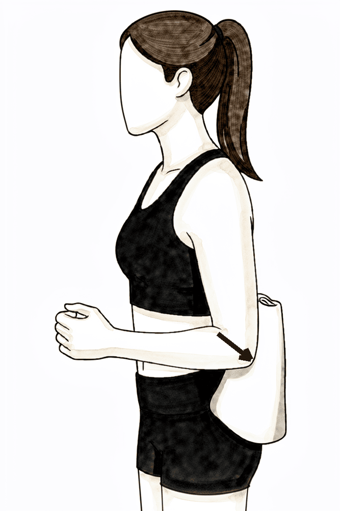
等长屈曲
面向墙壁站立,肘部弯曲并贴紧身体一侧,拳面前部抵住墙壁。轻轻向前推墙,同时保持手臂不动,保持数秒,然后放松。这能让肌肉在肩部逐渐稳定的过程中持续工作而不产生运动。
保持数秒,重复数次;请遵循物理治疗师的指导
Kieran Hirpara 4.0
等长伸展
站立,肘部置于体侧,上臂后侧贴靠墙面。轻轻将手臂向后压向墙面,保持不动,维持数秒,然后放松。这是一种在早期保持肌肉活跃且无需产生动作的方法。
保持数秒,重复数次;请遵循物理治疗师的指导
Kieran Hirpara 4.0
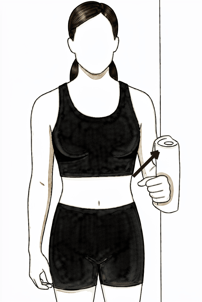
等长外展
侧身站立,上臂外侧靠近墙壁,肘部贴于体侧。将手臂轻轻向外推抵墙壁,保持其不动,维持数秒,然后放松。
保持数秒,重复数次;请遵循物理治疗师的指导
Kieran Hirpara 4.0
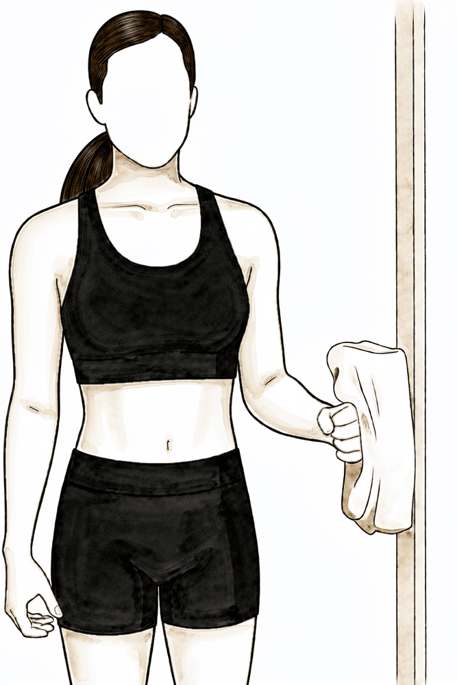
等长外旋
将肘部紧贴体侧并屈曲成直角,手背贴靠墙壁或门框。轻轻向外推压,保持手臂不动,维持数秒,然后放松。此动作可在无肢体运动的情况下锻炼肩袖肌群。
保持数秒,重复数次;请遵循物理治疗师的指导
Kieran Hirpara 4.0
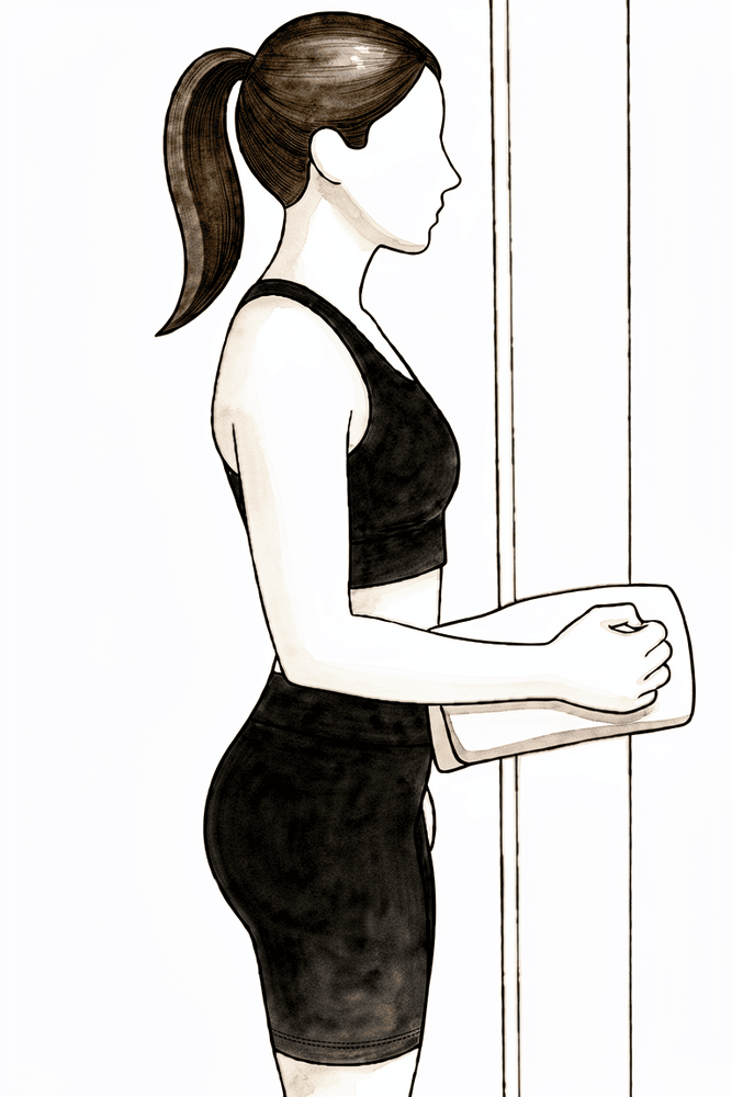
等长内旋
将肘部紧贴体侧并屈曲至直角,手掌抵住墙壁或门框。轻轻向内推压,同时保持手臂不动,维持数秒后放松。这是一种温和的启动肩袖肌群的方法。
保持数秒,重复数次;请遵循物理治疗师的指导
手术期间通常会使用神经阻滞,因此术后数小时内手臂可能会感到麻木;请在麻木感消退前开始服用止痛药。最初两周的目标很简单:控制疼痛和肿胀,并在舒适范围内恢复肩部活动。经常使用冰敷以缓解疼痛。立即开始活动手部、腕部和肘部,随着舒适度允许,逐步加入钟摆练习和辅助手臂活动,并将手臂用于正常的轻度日常活动,如洗漱、穿衣和进食。在练习和物理治疗预约前服用止痛药。这是微创手术,通常通过两个或三个覆盖防水敷料的小穿刺伤口进行;您可以从第 1 天开始淋浴(不要浸泡或用力擦洗),敷料需保留至 10–14 天时的伤口复查。从事办公室工作的人通常在最初两周内即可返回工作岗位;驾驶需等待外科医生批准,通常在六周复查时进行。
致您的物理治疗师:
目标
- 控制疼痛和肿胀
- 重建无痛活动范围(从主动辅助运动过渡到主动运动)
- 预防肌肉萎缩并开始重建动态稳定性
- 能够独立进行轻度日常生活活动
管理
- 经常进行冷疗以缓解疼痛和肿胀;在练习和治疗前使用镇痛药
- 从第 1 天开始进行肘部、腕部、手部、颈部和胸椎的活动范围练习
- 钟摆练习;在肩胛骨平面内进行滑轮或棍棒辅助的主动辅助抬举;外旋和内旋从外展 30–45° 开始
- 随着舒适度允许,过渡到主动活动范围
- 坐姿下的肩胛骨定位;姿势意识
- 次最大等长收缩(屈曲、伸展、外展、外旋和内旋)和轻柔的节奏性稳定训练
- 从第 2 周开始:如有帮助,练习前进行热敷;将旋转训练进展至外展 90°;手臂置于体侧,使用弹力带进行外旋和内旋
注意事项
- 在舒适范围内活动:不要强行或过度拉伸;以疼痛为指引
- 吊带仅用于舒适;在最初几天内逐步停用,并在两周内完全弃用
- 六周内禁止驾驶(此规定适用于任何肩部手术)
- 禁止重物搬运,禁止过头举重,禁止猛烈动作
- 最初六周内避免通过手术侧手臂从椅子或床上撑起身体:肩峰已被磨薄,在重塑期间应避免强力负荷
进展标准
- 疼痛通过简单镇痛药得到良好控制
- 肿胀消退,伤口愈合或正在愈合且无异常
- 主动辅助运动舒适,且肩部高度以下的主动运动正在恢复
第二阶段——恢复活动范围并开始力量训练(第 2–6 周)
Kieran Hirpara 4.0
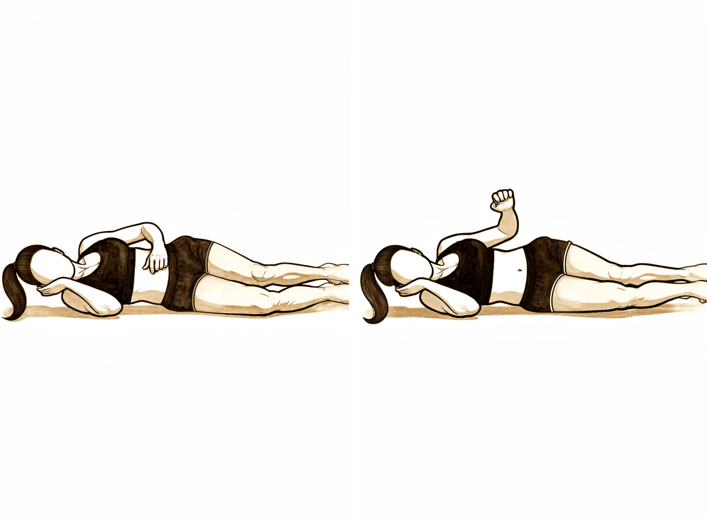
侧卧位外旋
侧卧于非手术侧,上方肘关节屈曲至直角并紧贴身体。保持肘部位于体侧,将前臂向上(朝向天花板)旋转,然后有控制地放下。初始阶段不加负重。
在无痛且控制良好的重复练习一周后,增加轻重量(0.5–1 千克)
Kieran Hirpara 4.0
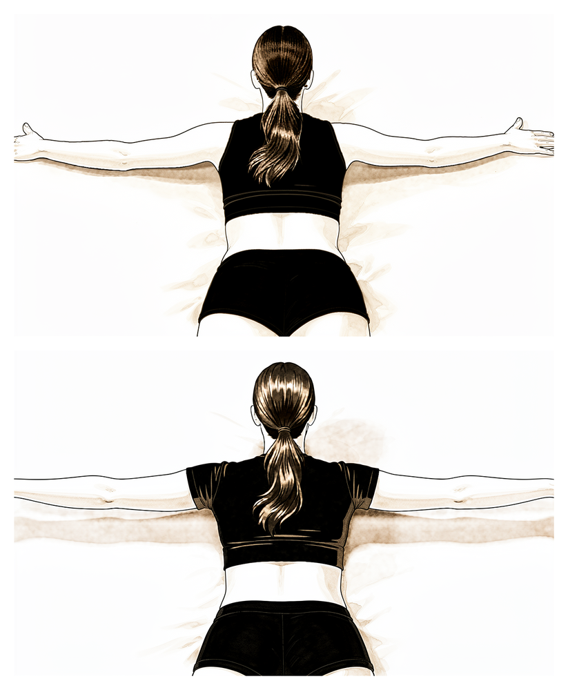
俯卧水平外展(俯卧“T”位)
俯卧,手臂自然下垂朝向地面。将肩胛骨向脊柱方向内收,然后将手臂向侧方抬起至肩部水平,拇指领先,随后缓慢放下。初始阶段不加负重;当重复动作无痛且控制良好时,可添加轻重量。
在您的物理治疗师指导下
Kieran Hirpara 4.0
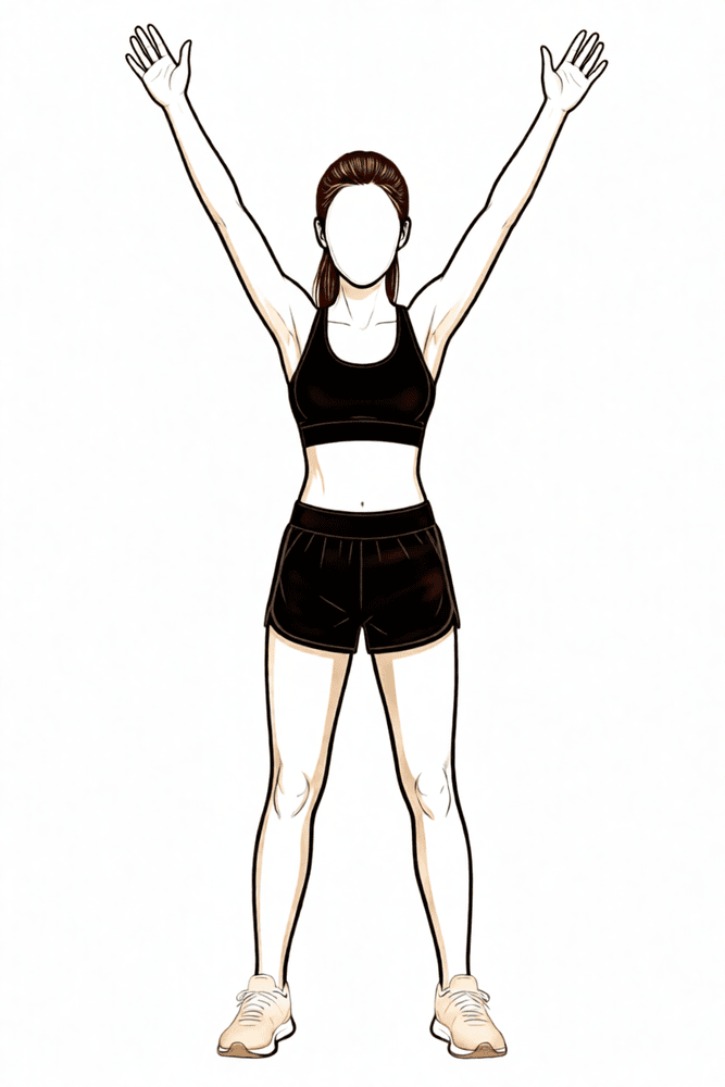
下斜方肌再训练
俯卧于床或桌上,手臂自然放松垂于边缘。将肩胛骨向下并向脊柱方向收拢,随着肩胛骨移动让手臂抬起——发力点来自肩胛骨下方,而非耸肩。缓慢放下并重复。
在您的物理治疗师指导下
Kieran Hirpara 4.0
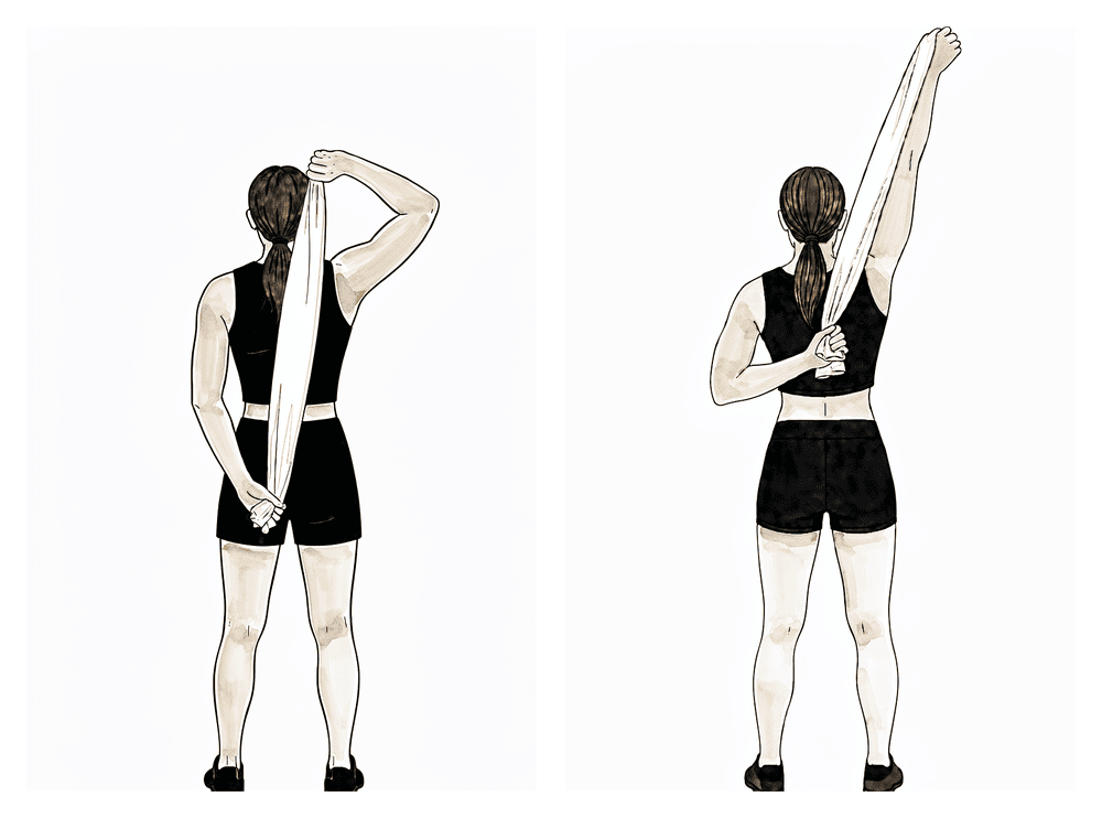
背后伸展
将毛巾置于背后,手术侧手位置较低,健侧手位置较高。用健侧手臂轻轻将手术侧手沿背部向上牵引,直至感到拉伸,短暂保持,然后放下。目标是肩部后方产生轻微拉伸感——切勿强行用力。
在您的物理治疗师指导下
Kieran Hirpara 4.0
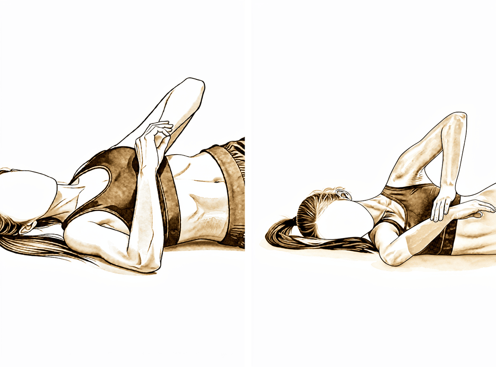
睡眠者拉伸(后关节囊)
侧卧于手术侧,肩关节和肘关节均屈曲至直角。用另一只手轻轻将前臂向下压向床面,直至感到肩后部有牵拉感,然后放松。这是您的物理治疗师将用于恢复旋转功能的后关节囊牵伸——动作务必轻柔,避免产生锐痛。
保持 20–30 秒,重复数次;请遵循物理治疗师的指导
Kieran Hirpara 4.0
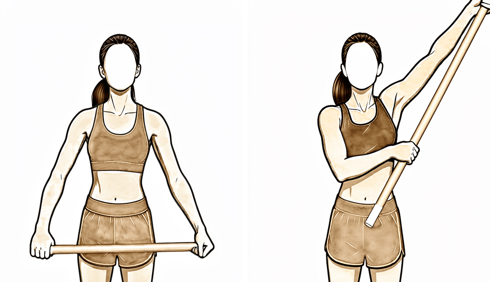
使用棍棒辅助外展
仰卧,双手握住一根棍棒。用健侧手臂引导患侧手臂向侧方抬起至肩部水平,然后有控制地放下。随着活动范围恢复,此动作有助于恢复外展至90度。
在您的物理治疗师指导下
Kieran Hirpara 4.0
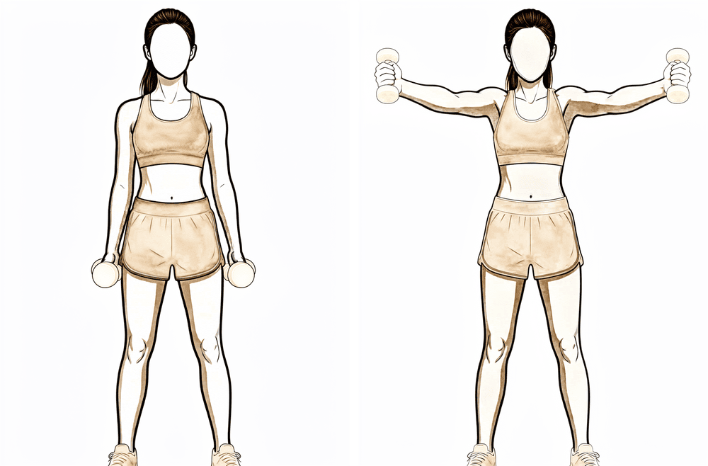
肩胛平面上举(满罐式上举)
手臂垂于体侧,拇指朝上站立。将手臂向前抬起,角度介于正前方与侧方之间,高度以舒适为限,然后缓慢放下。起始时不持重物;保持肩胛骨稳定,避免耸肩。
开始进行无负重训练;当重复动作无痛且控制良好时,再增加轻重量
Kieran Hirpara 4.0
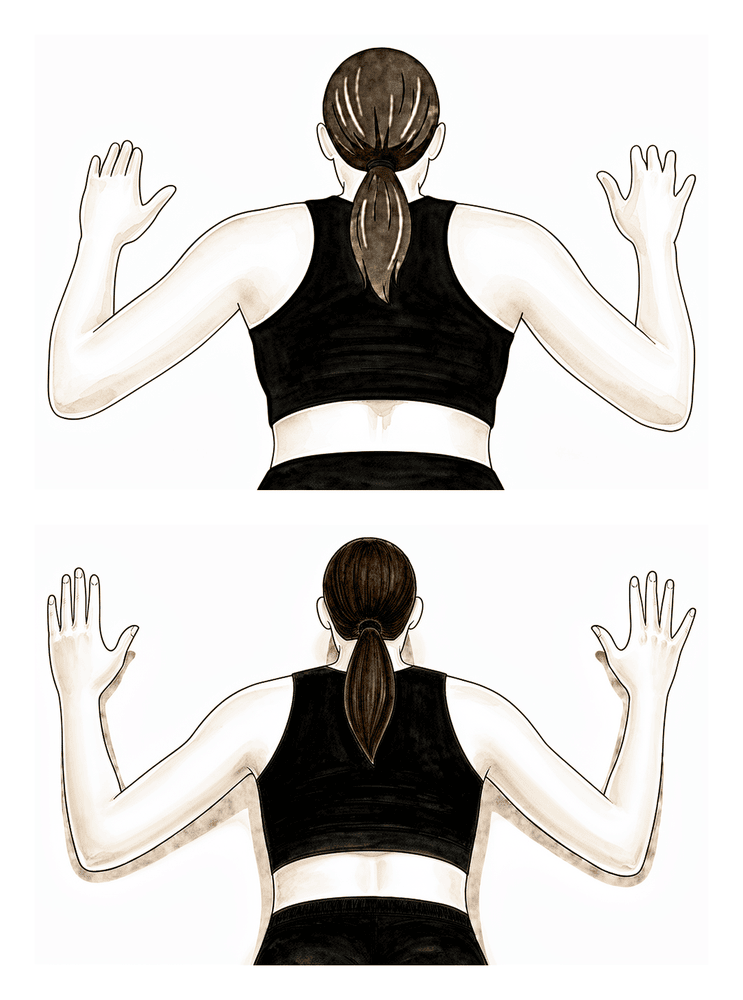
俯卧划船
俯卧,双臂自然下垂指向地面。将肩胛骨相互挤压靠拢,肘部向后下方牵引,使双臂呈“W”形,然后缓慢放下。初始阶段不加负重;当重复动作无痛且控制良好时,可添加轻重量。
从无负重开始;在无痛重复练习一周后,增加轻重量(约 0.5–1 公斤)
本阶段旨在恢复其余的活动范围并开始重建力量。您的锻炼将从辅助运动逐步过渡到在所有方向上主动活动手臂,大多数已发表的方案预计在六到八周左右达到完全或接近完全的活动范围。力量训练从温和开始:首先进行无负重训练,随后针对肩袖和肩胛骨肌肉使用弹力带和极轻的重量。许多人发现运动前热敷和运动后冰敷有帮助。中度活动(肩部高度以下的轻物搬运)通常在本阶段变得可行,具体需由您的物理治疗师指导。
致您的物理治疗师:
目标
- 在约第 6–8 周时,在所有平面上达到完全或接近完全的主动活动范围
- 恢复并改善肩袖和肩胛骨力量
- 使肩肱节律和神经肌肉控制恢复正常
- 继续缓解疼痛
管理措施
- 在所有平面上逐步推进活动范围,包括背后的内旋,并配合温和的后关节囊拉伸
- 根据需要进行盂肱关节松动术(下、后和前向滑动)
- 等长训练从无负重开始(肩部抬举、俯卧划船、俯卧水平外展、俯卧伸展至中立位、侧卧外旋、外展至 90°),在一周无痛且控制良好的重复动作后,增加轻重量(约 0.5–1 公斤)
- 使用弹力带进行外旋和内旋;随着舒适度提高,逐步过渡到更高位置下的旋转训练
- 肩胛骨神经肌肉控制和下斜方肌训练;躯干、核心及下肢体能训练
- 上肢耐力训练;根据偏好,在训练前热敷,训练后冰敷
注意事项
- 避免在引起疼痛的撞击范围内进行负重训练;锻炼可能具有挑战性,但不应重现术前疼痛
- 在冈上肌或中三角肌负荷训练无痛之前,不要针对这些肌肉进行负荷训练;如果引发静息痛或夜间痛,则应避免此类训练
- 继续避免通过手术侧手臂向上推举,以及重物或过头举重,直至术后六周
晋级标准
- 活动范围完全且无痛,压痛极轻微
- 手动测试显示肩袖力量约为 4/5 级,且肩胛骨控制良好
第三阶段 — 强化(第 6–12 周)
Kieran Hirpara 4.0
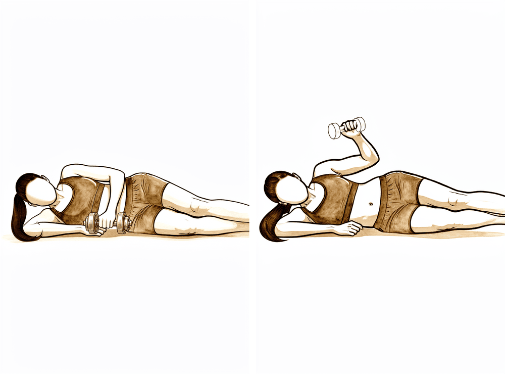
侧卧位负重外旋
侧卧于非手术侧,上方肘关节屈曲成直角并紧贴身体。手持轻重量,将前臂向上旋转朝向天花板,然后缓慢放下。练习应感觉有负荷但基本无痛——若出现静息痛或夜间痛,应减轻强度。
2–3 组,每组 8–12 次
Kieran Hirpara 4.0
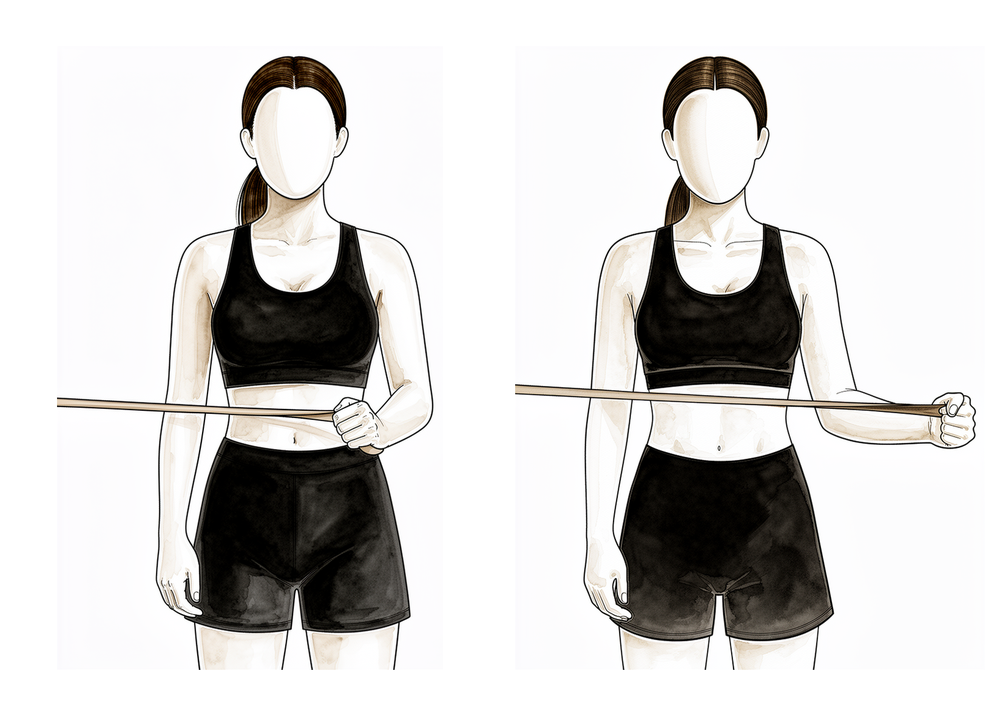
弹力带外旋
将肘部贴紧体侧并屈曲至直角,手持一根固定在腰部高度的弹力带。保持肘部贴于体侧,对抗弹力带阻力将前臂向外旋转,然后缓慢复位。此动作是在早期等长及无负重训练基础上,进一步锻炼肩袖肌群。
2–3 组,每组 8–12 次
Kieran Hirpara 4.0
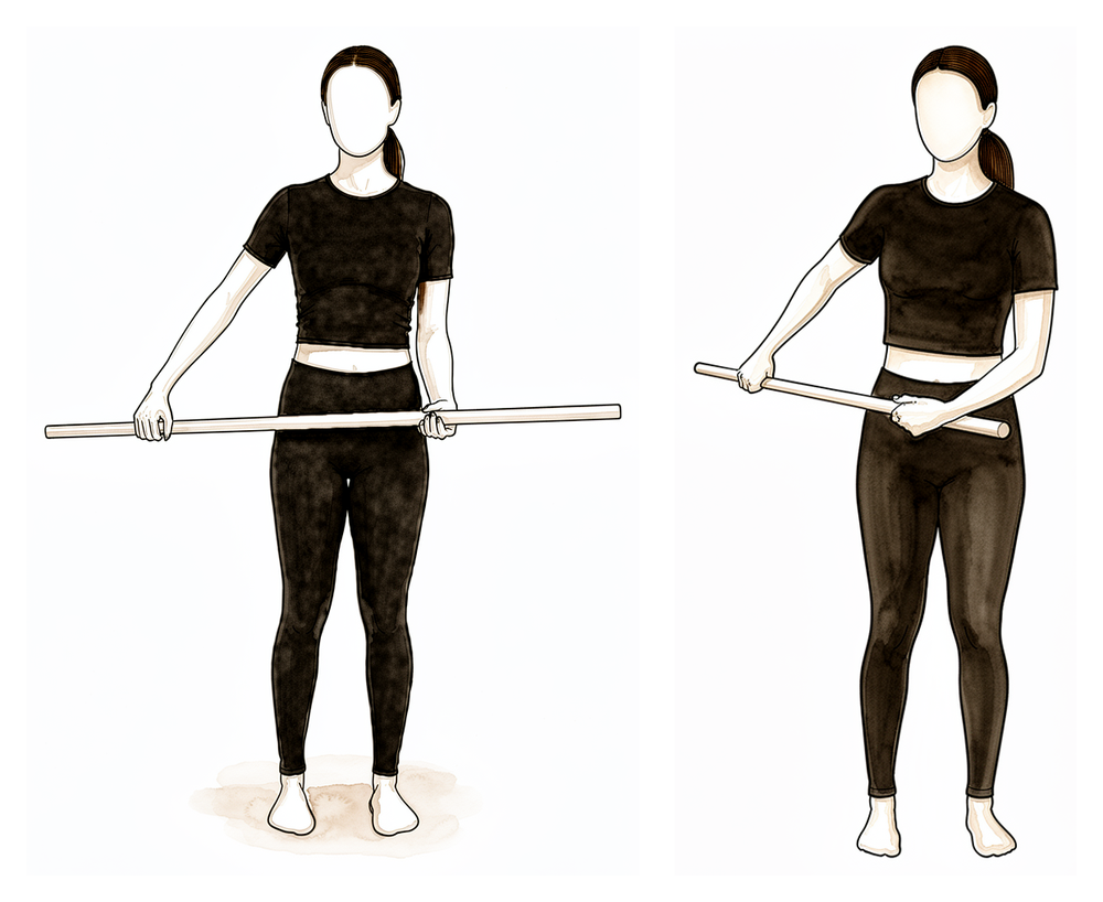
弹力带内旋
将肘部紧贴体侧,握住固定在一侧的弹力带,将前臂向内旋转并横过身体以对抗弹力带,然后缓慢复位。随着力量增强,逐步增加阻力。
2–3 组 8–12 次
Kieran Hirpara 4.0
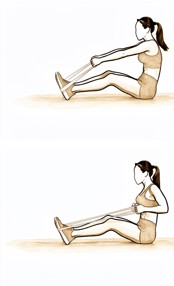
站立位划船
将一条弹力带固定在身前腰部高度。保持手臂相对伸直,向后下方拉向髋部,同时收缩肩胛骨使其向下向后移动,然后缓慢还原。此动作可锻炼支撑肩袖的肩胛骨肌肉。
2–3 组 8–12 次
Kieran Hirpara 4.0
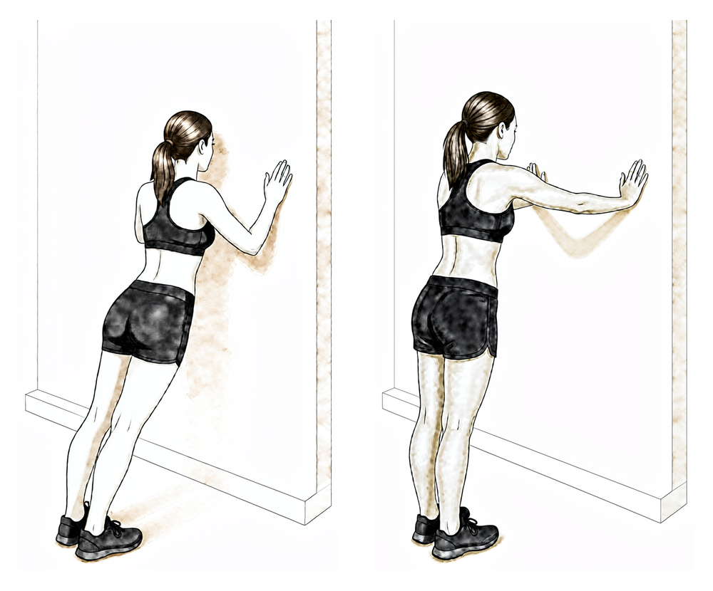
靠墙俯卧撑加强
面向墙壁站立,双手平贴于墙面,高度与肩部齐平。屈肘向墙壁方向前倾,随后向后推回,并在动作末端通过双手施加轻柔推力,以展开肩胛骨。这是一种用于建立控制能力的闭链运动——请在物理治疗师的指导下进行。
在您的物理治疗师指导下
随着活动度的恢复,重点转向力量、耐力和控制。抗阻训练从弹力带过渡到负重,练习变得更加动态,包括针对重返运动人群,开始受控的增强式训练和循序渐进的训练回归。游泳通常从大约六周开始恢复(先蛙泳,舒适后自由泳),中等强度工作(肩部高度以下的轻负荷提举)通常从六周起可管理。间歇性重返运动计划通常在满足力量标准时于第 10–12 周开始。
致您的物理治疗师:
目标
- 改善肩关节复合体的力量、爆发力和耐力
- 优化神经肌肉控制、本体感觉和运动模式(无肩部耸起)
- 为循序渐进地重返运动和更重的工作做准备
管理
- 推进等长训练方案:等长收缩 → 弹力带 → 负重;针对肩袖、三角肌和肩胛骨稳定肌,进行 2–3 组、每组 8–12 次
- 引入离心抗阻训练、闭链练习和节律性稳定训练;本体感觉训练包括负重和四点跪位
- 增强式活动:从双手练习(胸前传球、左右投掷)过渡到阶段末期的单手练习(墙壁运球、投掷练习)
- 若满足标准,在第 10–12 周开始间歇性运动计划
- 继续核心和体能训练;必要时进行冷疗
注意事项
- 练习应具有一定难度但基本无痛;若任何练习引发静息痛或夜间痛,应降低强度
- 重负荷提举和持续的过头负荷需等待至约三个月
晋级标准
- 完全、无痛的主动活动范围(大致与对侧相等)
- 力量接近对侧(已发表的标准范围从进入动态训练的70%,到重返运动的90%)
- 渐进负荷下无疼痛或压痛
第四阶段 — 恢复完全活动(第12周起)
最后阶段是逐步恢复重体力劳动、过头动作和体育运动。重负荷或重复性举重以及持续高于肩部的作业通常从约三个月后开始恢复。恢复竞技体育(特别是过头运动)基于达到标准而非日历时间:完全无痛的活动、力量接近对侧水平,以及对运动专项训练的信心。肩部通常在此阶段之后仍会持续改善;已发表的指南描述改善可持续长达一年。
致您的物理治疗师:
目标
- 逐步恢复重体力劳动、过头活动和体育运动
- 长期维持关节活动度、力量和控制力
管理
- 继续强化训练计划,在可耐受范围内推进健身房及运动专项训练
- 继续间歇性运动计划,分阶段恢复投掷及其他过头运动
- 根据需要进行自我管理的关节囊拉伸和维持练习
注意事项
- 进展仍以症状为导向;如果疼痛复发,应减少负荷,恢复舒适的活动度并重建
进展标准
- 完全且无痛的关节活动度
- 力量和功能性测试令人满意(在已发表的恢复运动标准中约为对侧的90%)
- 临床复查令人满意
您的康复方案之后
上述各阶段改编自已发表的肩关节镜下肩峰下减压术康复方案及患者指导,来源包括:OrthoIndy、Sports Surgery New York、Gundersen Health System Sports Medicine、Twin Cities Orthopedics、牛津大学医院 NHS 基金会信托及皇家伯克郡 NHS 基金会信托。周数范围通常为典型值而非固定值,您的持续康复将由您的物理治疗师根据肩关节恢复情况,在诊所的配合下为您个体化指导。本页面与诊所的一般恢复建议相辅相成;请参阅术后疼痛管理和伤口护理。关于手术本身及其治疗的病症,请参阅肩峰下减压术。本方案背后的证据(包括其与肩峰下手术试验证据的契合度)在证据部分进行了总结,可从本页面顶部获取 PDF 文件。
Evidence & references
This is the clinical evidence summary written for health professionals. It is technical, and it lists the research this page was built from. You do not need to read it to understand your treatment or to make a decision about it.
Topic scope: Post-operative rehabilitation after isolated arthroscopic subacromial decompression (ASD / acromioplasty ± bursectomy). When a decompression is performed together with a rotator cuff repair, the repaired tendon sets the (slower) pace and the rotator-cuff-repair protocol takes priority — this page is for the isolated decompression.
Defining principle of this rehab: a subacromial decompression shaves bone and clears bursa — it repairs nothing that needs protecting. So (like a debridement, and unlike a cuff repair or stabilisation) the rehab is an early-movement pathway: a short sling for comfort only, weaned within days, motion and normal light use from day one, and a rapid return of range and function. The aim is to settle the post-operative flare and keep the shoulder moving while it quiets down — rehabilitation, not rest, does the work.
A. THE PROCEDURE & ITS EVIDENCE CONTEXT (important)
Arthroscopic subacromial decompression removes the subacromial bursa and shaves the under-surface of the acromion to "make room" for the rotator cuff, on the impingement model of subacromial pain.
The efficacy of the bony decompression itself is one of the most debated questions in shoulder surgery, and the rehabilitation context cannot be stated honestly without it:
- CSAW (Beard et al, Lancet 2018; n=313, 3-arm) — a placebo-controlled surgical RCT. Decompression gave no clinically important benefit over arthroscopy-only (placebo) surgery, and both surgical arms were only marginally better than no treatment — a difference below the pre-specified minimal clinically important threshold.
- FIMPACT (Paavola et al, BMJ 2018) — a second placebo-controlled RCT: no benefit of ASD over diagnostic arthroscopy, and neither was superior to a structured exercise programme at 2 years.
- Cochrane review (Karjalainen et al, 2019) — high-certainty evidence that subacromial decompression provides little or no clinically important benefit over placebo for pain, function, or quality of life.
The practical consequence is that structured exercise/physiotherapy is first-line for subacromial pain, and ASD is now a selective operation — reserved for patients who have failed an adequate non-operative programme or who have a specific mechanical lesion. This does not make a well-selected decompression valueless, but it explains why the post-operative emphasis is on early movement and rehabilitation, which is what reliably drives recovery.
B. POST-OPERATIVE PHASED TIMELINE (isolated decompression)
A nerve block is commonly used (numb arm for some hours — start analgesia before it wears off). Keyhole wounds; desk-based work commonly resumes within ~2 weeks.
| Phase | Window | Sling | ROM / use | Strengthening | Notes |
|---|---|---|---|---|---|
| I — Early movement | Week 0–2 | Comfort only, off within days (gone by ≤ 2 wk) | Hand/elbow immediately; pendulums + assisted motion as comfort allows; normal light daily use from day 1 | — | Settle pain/swelling; ice; analgesia before exercise. No driving while in sling (typically back ~1–3 wk once out of sling + safe emergency stop) |
| II — Restore range / start strength | Week 2–6 | Off | Progress to full active ROM all planes | Begin gentle cuff + scapular strengthening | Range comfortable below shoulder height; most daily activity resumed |
| III — Strengthening | Week 6–12 | Off | Full | Progressive cuff/scapular loading, band → light weight | Heavier and overhead loading built gradually |
| IV — Return to full activity | Week 12 + | Off | Full | Advanced/sport-specific | Full unrestricted activity typically ~3 months |
There is no construct-protection branch in the isolated decompression — the only branch is if a rotator cuff repair was also done, which converts recovery to the protected cuff-repair pathway.
C. KEY CONTROVERSIES / EVIDENCE QUALITY
- Does the bony decompression add anything? Two placebo-controlled RCTs (CSAW, FIMPACT) and a Cochrane review say it adds little or nothing over placebo or exercise for subacromial pain. Strong (RCT/SR). → exercise-first, selective surgery.
- Decompression added to a cuff repair — multiple RCTs show no added benefit of routine acromioplasty at the time of arthroscopic cuff repair. Moderate–strong.
- The post-operative rehab protocol itself — consensus/expert (institutional protocols), no defining rehab RCT; phase timings are typical, not trial-derived. Weak/consensus.
(Patient-facing note: the efficacy debate belongs in this clinician evidence section, not the patient protocol page — surfaced here for the surgeon's awareness.)
D. EVIDENCE STRENGTH FLAGS (summary)
- STRONG (RCT / SR): ASD provides little/no benefit over placebo surgery or exercise (CSAW Lancet 2018; FIMPACT BMJ 2018; Cochrane Karjalainen 2019); no benefit of added acromioplasty at cuff repair.
- MODERATE: early-motion rehabilitation after isolated decompression (institutional cohorts / consensus protocols, consistent ~3-month recovery).
- WEAK / CONSENSUS: the specific phase/timeline structure of the post-op protocol.
CITATIONS
RAG corpus (180,000+ Orthopaedic articles)
- Is acromioplasty justifiable? Orthop Traumatol Surg Res. 2019. DOI: 10.1016/j.otsr.2019.10.002
- Indications for Arthroscopic Subacromial Decompression: a Level V evidence clinical guideline. Arthroscopy. 2019. DOI: 10.1016/j.arthro.2019.06.012
- The role of subacromial decompression in patients undergoing arthroscopic repair of full-thickness rotator cuff tears. Arthroscopy. 2012. DOI: 10.1016/j.arthro.2011.11.022
- Arthroscopic repair of full-thickness rotator cuff tears with and without acromioplasty (RCT). Am J Sports Med. 2014. DOI: 10.1177/0363546514529091
- A comparative study of arthroscopic débridement versus repair for Ellman grade I partial cuff tears. J Shoulder Elbow Surg. 2020. DOI: 10.1016/j.jse.2020.03.006
Landmark trials / reviews (URLs)
- Beard DJ, et al. Arthroscopic subacromial decompression for subacromial shoulder pain (CSAW): placebo-controlled 3-group RCT. Lancet. 2018. https://www.thelancet.com/journals/lancet/article/PIIS0140-6736(17)32457-1/fulltext
- Paavola M, et al. (FIMPACT) Subacromial decompression versus diagnostic arthroscopy for shoulder impingement: randomised, placebo surgery controlled trial. BMJ. 2018;362:k2860. https://www.bmj.com/content/362/bmj.k2860
- Karjalainen TV, et al. Subacromial decompression surgery for rotator cuff disease. Cochrane Database Syst Rev. 2019. https://www.cochranelibrary.com/cdsr/doi/10.1002/14651858.CD005619.pub3/full
Published rehab protocols (basis for the phase structure)
- Kendall C. Arthroscopic Subacromial Decompression Physical Therapy Protocol. OrthoIndy. https://www.orthoindy.com/wp-content/uploads/Arthroscopic-Subacromial-Decompression.pdf
- Strauss EJ. Rehabilitation Protocol: Arthroscopic Subacromial Decompression / Distal Clavicle Excision. Sports Surgery New York. https://www.sportssurgerynewyork.com/pdf/arthroscopic-subacromial-decompression-distal-clavicle-excision-rehab-protocol.pdf
- Gundersen Health System Sports Medicine. Subacromial Decompression / Acromioplasty Rehabilitation Program. https://www.gundersenhealth.org/sites/default/files/2022-06/Sports-Medicine-Protocol-Subacromial-Decompression-Acromioplasty.pdf
- Meisterling RC. Arthroscopic Subacromial Decompression Rehabilitation. Twin Cities Orthopedics. https://tcomn.com/wp-content/uploads/2016/06/Arthroscopic-Subacromial-Decompression-Rehabilitation.pdf
- Scott-Dempster C, Harper J. Outpatient Post-operative Physiotherapy Guidelines: Sub-Acromial Decompression. Oxford University Hospitals NHS FT. https://www.ouh.nhs.uk/media/zidppie2/sub-acromial-decompression.pdf
- Royal Berkshire NHS FT. Arthroscopic subacromial decompression and/or ACJ excision: discharge advice. https://www.royalberkshire.nhs.uk/media/smegtn3c/arthroscopic-subacromial-decompression-and-or-acromioclavicular-joint-excision-discharge-advice_jul24.pdf





